Introduction
Remission rates following antidepressant first-line treatment in major depression with agents such as selective serotonin reuptake inhibitors (SSRIs) range between 30–45% (Carvalho et al. Reference Carvalho, Cavalcante, Castelo and Lima2007) and only ⩽50% of patients achieve full remission (Rush et al. Reference Rush, Trivedi, Wisniewski, Nierenberg, Stewart, Warden, Niederehe, Thase, Lavori and Lebowitz2006). There is a well-known delayed onset in response following antidepressant treatment attributed to presynaptic and postsynaptic adaptive mechanisms (Celada et al. Reference Celada, Puig, Amargós-Bosch, Adell and Artigas2004) which affects the time available to clinicians to make decisions about next step treatments. Hence clinical guidelines often offer pragmatic advice on switching to a different antidepressant (e.g. different class), after 4–8 weeks of continued treatment with a therapeutic dose (APA, Reference APA2010; Bauer et al. Reference Bauer, Pfennig, Severus, Whybrow, Angst and Möller2013). The possibility of identifying response/remission to a given antidepressant early in treatment history confers the great advantage of reducing disease burden by giving the opportunity to consider alternative treatments sooner if necessary. Clinical research has already indicated that it is possible to detect susceptibility to response to treatment within the first 2 weeks of treatment by identifying individuals unlikely to benefit from a given antidepressant regimen (Kim et al. Reference Kim, Kim, Stewart, Yoo, Bae, Jung, Lee, Yim and Jun2011). Furthermore, there is evidence that antidepressant response might supervene within a few days of treatment (Taylor et al. Reference Taylor, Freemantle, Geddes and Bhagwagar2006). It has therefore become increasingly evident that identification of early predictors of antidepressant response at brain level is an essential step to more effectively personalize treatment in major depression.
Traditional antidepressants (i.e. SSRIs), have acute direct effects of augmentation of extracellular 5-HT in serotonergic networks (Artigas, Reference Artigas1993). Preliminary evidence suggests that functional magnetic resonance imaging (fMRI) offers the potential to investigate acute treatment effects at brain level following antidepressant treatment (Kraus et al. Reference Kraus, Ganger, Losak, Hahn, Savli, Kranz, Baldinger, Windischberger, Kasper and Lanzenberger2014). A single oral dose of citalopram has been demonstrated to influence task-negative processes during goal-directed behavior in the frontal cortex in healthy controls (Klomp et al. Reference Klomp, van Wingen, de Ruiter, Caan, Denys and Reneman2013). Augmenting serotonin neurotransmission with acute citalopram can modulate emotional expression in healthy male participants (Labuschagne et al. Reference Labuschagne, Croft, Phan and Nathan2010). Compared to viewing neutral faces, intravenous citalopram pretreatment also enhanced different blood oxygen-level dependent (BOLD) response of brain regions in subjects with depression, including left anterior cingulate to happy faces, right posterior insula and right lateral orbitofrontal responses to sad faces, and reduced bilateral amygdala responses to fearful faces (Anderson et al. Reference Anderson, Juhasz, Thomas, Downey, McKie, Deakin and Elliott2011). It has also been demonstrated that duration of treatment influences neural responses in the prefrontal cortex (PFC), resulting in decreased activity in the left dorsomedial prefrontal cortex (DMPFC) following 8-week SSRI administration (Wang et al. 2014a), and increased activity in the right dorsolateral prefrontal cortex (DLPFC) after 6 months administration (Heller et al. Reference Heller, Johnstone, Peterson, Kolden, Kalin and Davidson2013). An interesting technical development in functional brain MRI is the possibility to investigate brain networks ‘at rest’ in the absence of overt stimuli. Biswal and others (Biswal et al. Reference Biswal, Zerrin Yetkin, Haughton and Hyde1995) demonstrated that ‘at rest’ low-frequency fluctuation is equivalent to self-representation of highly synchronous spontaneous neuronal activity (Mantini et al. Reference Mantini, Perrucci, Del Gratta, Romani and Corbetta2007). As the ruminative cognitive style underlying self-generated thoughts in major depression is believed to be important in the generation of symptoms and their maintenance (Perkins et al. Reference Perkins, Arnone, Smallwood and Mobbs2015), resting-state fMRI might be optimally designed to investigate the ‘neural predisposition’ underlying the neuro-pathology of depressive disorders (Sheline et al. Reference Sheline, Barch, Price, Rundle, Vaishnavi, Snyder, Mintun, Wang, Coalson and Raichle2009). Preliminary evidence suggests that resting-state fMRI can guide treatment response in depressive disorders (Dichter et al. Reference Dichter, Gibbs and Smoski2015). For example, a 10-week treatment study with duloxetine showed an increase in baseline connectivity in the default mode network (DMN) in dysthymic individuals with clinical improvement (Posner et al. Reference Posner, Hellerstein, Gat, Mechling, Klahr, Wang, McGrath, Stewart and Peterson2013).
Conventional resting-state approaches take advantage of functional connectivity analysis, which examine the inter-regional temporal correlation between predefined seed regions and functionally related brain circuitry. In this work we apply a novel technique named ‘amplitude of low-frequency fluctuation (ALFF)’ developed by Zang and others (Yu-Feng et al. Reference Yu-Feng, Yong, Chao-Zhe, Qing-Jiu, Man-Qiu, Meng, Li-Xia, Tian-Zi and Yu-Feng2007) to explore the amplitude of intra-regional brain activity at rest with fMRI. This technique minimizes the risk of overlooking the functional segregation or regional specialization of the brain functional integrity likely to occur with pre-hypothesized regions of interest (ROI), approaches typical of conventional resting-state techniques (Zhou et al. Reference Zhou, Wang, Liu, Song, Song and Jiang2010). The validity and reliability of ALFF has already been tested in major depression (Liu et al. Reference Liu, Ren, Womer, Wang, Fan, Jiang, Blumberg, Tang, Xu and Wang2014) suggesting sensitivity to detect differences between cases and controls, correlations with antidepressant treatment response (Yamamura et al. Reference Yamamura, Okamoto, Okada, Takaishi, Takamura, Mantani, Kurata, Otagaki, Yamashita and Yamawaki2016), high temporal stability (Küblböck et al. Reference Küblböck, Woletz, Höflich, Sladky, Kranz, Hoffmann, Lanzenberger and Windischberger2014), a correlation with conventional functional connectivity methods (Liu et al. Reference Liu, Ren, Womer, Wang, Fan, Jiang, Blumberg, Tang, Xu and Wang2014) and consistent sensitivity and specificity in detecting spontaneous brain activities (Zou et al. Reference Zou, Zhu, Yang, Zuo, Long, Cao, Wang and Zang2008).
The aim of the present study was to investigate treatment effects in depression measured with fMRI ALFF. We were particularly interested in detecting early neurobiological predictors of treatment efficacy after a single dose of escitalopram by identifying initial changes in resting state especially in brain regions involved in mood regulation, and correlations with treatment effects following a single dose of antidepressant treatment.
Materials and method
Participants and baseline assessment
Seventy-nine first-episode, treatment-naive, right-handed Han Chinese individuals, aged 18–50 years, meeting DSM-IV diagnostic criteria for moderate to severe major depression (First et al. Reference First, Spitzer, Gibbon and Williams1996) and 78 sex-, age- and handedness-matched healthy controls were recruited from the purpose of this study. Depressed participants, clinically assessed by a board-certified psychiatrist, were outpatients recruited from the Department of Psychiatry, First Affiliated Hospital, Kunming Medical University, China, between 2008 and 2011. The study received approval from the local University ethics committee (Kunming Medical University, Clinical registration no. http// www.ClinicalTrials.gov, NCT00703742) and only participants who were able to consent in writing were included in the research. Recruited patients scored ⩾17 on the Hamilton Depression Rating Scale (HAMD-17; Hamilton, Reference Hamilton1960), <28 on the Hamilton Anxiety Scale (HAMA; Hamilton, Reference Hamilton1959) and had experienced symptoms of depression for <2 years. Exclusion criteria for both groups included: (1) present or previous history of Axis I and Axis II disorders (including substance misuse and anxiety disorder) aside major depression for patients, neurological and/or significant physical morbidity, (2) previous systemic psychiatric treatment and overt suicidal ideation/behavior for patients, and (4) inability to undergo MRI scans including pregnancy. The MRI examination was performed within 3 days of the clinical evaluation and prior to treatment initiation.
Longitudinal assessment
Patients
A subset of 48 patients participated to the 8-week longitudinal component which included pharmacological treatment and regular clinical reviews at weeks 2 and 4 with a final evaluation at week 8. The highly selective SSRI escitalopram (Owens & Rosenbaum, Reference Owens and Rosenbaum2002) was the only treatment administered at a fixed oral dose of 10 mg/day to minimize possible dose-related effects on brain activity. Neuroimaging data were acquired at baseline, 5 h (5H) after the first dose (T max of escitalopram, 5H), and at the end of week 4 (4W) and week 8 (8W). In this study, response was conventionally defined as a reduction of ⩾50% in depression scores at endpoint. In order to measure treatment efficacy, clinical remission at the end of the 8 weeks was used as main clinical outcome and it was conservatively defined as HAMD score of ⩽7.
Controls
A cohort of 16 healthy controls received a single dose of placebo and received the baseline and the 5H MRI scan to control for placebo effects. Additionally 12 healthy controls underwent MRI at baseline and at 8W without any treatment, to control for the time-drift effect on the stability of the resting-state signal.
Imaging acquisition and data processing
All subjects received MRI scans with a 1.5-T clinical GE MRI scanner (Twinspeed, GE, USA) using a birdcage head coil. Obvious structural abnormalities were excluded. Resting-state MRI data were obtained using an echo-planar imaging sequence: repetition time (TR) = 2000 ms, echo time (TE) = 40 ms, flip angle = 90°, 24 axial slices, thickness/skip = 5/1 mm, matrix = 64 × 64, field of view = 240 × 240 mm2. Image preprocessing was conducted using the statistical parametric mapping software (SPM8, http://www.fil.ion.ucl.ac.uk/spm). The first 10 volumes of each functional time-series were discarded, and the remaining 150 volumes of fMRI images were employed in subsequent preprocessing for slice timing. Section-timing adjustment and realignment for head motion correction were performed. The group differences in translation and rotation of head motion were evaluated (Qi et al. Reference Qi, Zhang, Wu, Zhong, Zhang, Zhong, Ni, Zhang, Li and Jiao2012) and no group differences were found comparing the four time points. The data were spatially normalized into MNI space and resampled to 3 × 3 × 3 mm cubic voxels and were spatially smoothed using a 6-mm full-width half-maximum (FWHM) Gaussian kernel. After smoothing, the imaging data were temporally filtered (band pass, 0.01–0.08 Hz) to remove the effects of low-frequency drift and high-frequency noise.
Functional amplitude of low-frequency fluctuation calculation
Functional ‘fractional amplitude of low-frequency fluctuation’ (fALFF) was calculated using REST software (http://resting-fmri.sourceforge.net) (Supplementary material). To explore the within-group whole-brain fALFF patterns, one-sample t tests were performed on the individual fALFF maps in a voxelwise manner for each group. The fALFF difference between patients and controls and the difference between remitted and non-remitted patients were compared using two-sample t tests.
Statistical analysis
Using SPM8 software, one-way repeated-measures ANOVA analysis was used to identify overall changes in the fALFF at the four time-points. To reveal fALFF differences between the baseline and three post-treatment time-points separately, paired t tests in SPM8 were also used. All results were considered statistically significant at p < 0.005 and a cluster size >22 voxels, which corresponded to a corrected p < 0.05 (corrected by using the AlphaSim program, http://afni.nih.gov/afni/docpdf/AlphaSim.pdf). Small volume correction was applied by using a gray-matter mask made from the SPM template.
Functional fALFF in relation to treatment efficacy and clinical improvement
Functional fALFF differences between measurements at baseline and 5 h after treatment were calculated using the ‘ImCalc’ function in SPM8, and difference value maps (Dmap = fALFF5H-map– fALFFbase-map) were obtained for each individual. The SSRI treatment efficacy was evaluated using the reducing rate of HAMD scores:
 $$\left(\Delta {\rm HAMD} = \displaystyle{{({\rm HAMD}_{{\rm base}} - {\rm HAMD}_{8{\rm W}} )} \over {{\rm HAMD}_{{\rm base}}}} \times 100{\rm \%} \right).$$
Correlation analyses were subsequently performed between the fALFF Dmap and HAMD using SPM8 (p < 0.05, FEW-corrected).
As an exploratory analysis we used the subtraction in the fALFF value at 5H and baseline in the ROIs to predict clinical response at 8W. We subsequently calculated sensitivity, specificity, positive and negative predictive values and receiver-operating characteristics for fALFF5H (see Supplementary material).
Results
Demographic and clinical data
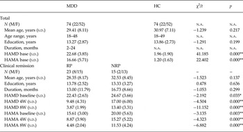
Seventy-four patients and 74 healthy controls were included in the analyses. Table 1 shows that patients and controls were well matched. Five patients and four controls had to be excluded due to motion artifacts. In the longitudinal component 40 of the 48 patients completed the study and two participants had to be excluded due to motion artifacts (N = 38, 79%). Details of the patients and reasons for discontinuation are provided in Supplementary Table S1 and Supplementary Fig. S1. Twenty-three of the 38 patients remitted (61%, mean HAMD score at endpoint: 3.87 ± 1.99), and 15 patients were non-remitters (39%, mean HAMD score at endpoint: 13.40 ± 3.31). The total reduction of HAMD for the remitters was 82.85%, while for the non-remitters it was 44.75% so that responders largely achieved remission (see also Table 1 and Supplementary Fig. S2). There were no significant difference of baseline clinical data between patients who dropped out and completers.
Demographic and clinical data

MDD, Major depressive disorder; HC, healthy controls; HAMD, Hamilton Depression Rating Scale; HAMA, Hamilton Anxiety Scale; M, male; F, female; RP, remitted patient; NRP, non-remitted patient; n.a., not applicable.
* p < 0.01, ** p < 0.001.
State effects in major depression
Baseline evaluation of the fALFF signal suggested that depressed patients exhibited a decreased signal compared to healthy controls in the DLPFC, DMPFC, precuneus, and right angular gyrus. Conversely an increase in fALFF signal was detected bilaterally in the temporal lobe (see Supplementary Table S2 and Supplementary Fig. S3 for details).
Pharmacological effects and clinical improvement
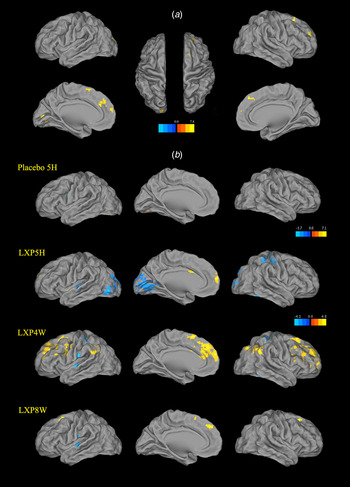
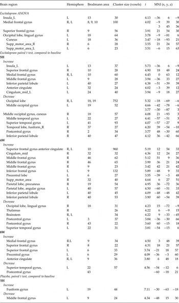
One-way repeated-measures ANOVA analysis among the four time-points identified a group × time interaction in regions of the DLPFC, anterior cingulate cortex (ACC), and occipital cortex (Table 2 and Fig. 1). Individual t tests indicated that following acute (5H) and sub-chronic (4W and 8W) treatment administration the fALFF signal decreased in the bilateral post-central gyrus and left superior temporal gyrus (STG) while it increased in a vast area of the PFC incorporating dorsomedial and dorsolateral regions. The acute effect of escitalopram appeared to prominently decrease the activity of the occipital cortex, whereas after 4 weeks of treatment an increase in the activity of the PFC and ACC was evident. We were not able to detect fALFF and resting-state time effects in the cohort of healthy controls during the 8-week period at the corrected significance level (p < 0.05), supporting test–retest reliability of the procedures. The alteration of fALFF after a single dose of placebo in the controls demonstrated only a slight effect of increased temporal lobe activity, whereas escitalopram resulted in obvious changes in major depression compared to baseline.
The figure shows resting-state changes over the course of treatment and in relation to placebo. (a) One-way repeated-measures ANOVA analysis among the four time-points of baseline, 5 h (5H), 4 weeks (4W) and 8 weeks (8W). (b) Dynamic regulation of fALFF after acute and sub-chronic antidepressant treatment in major depression. The alteration of fALFF after escitalopram treatment at different time-points showed different patterns. LXP, escitalopram.

Brain regions where differences were measured between major depression (MDD) and healthy controls (HC) during the course of treatment and in the placebo experiment

Differences between remitters and non-remitters
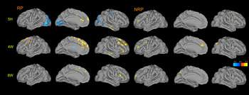
Differences were detectable at 5H with decreased activity bilaterally in the orbitofrontal cortex, left STG, right supra-marginal gyrus and post-central gyrus and increased activities in ACC, mid cingulate cortex and right STG in remitters (Fig. 2 and Supplementary Table S3). At 4W changes resulted in increased activities in bilateral PFC [including the DMPFC and ventrolateral prefrontal cortex (VLPFC), mid cingulate cortex, and inferior temporal gyrus] and decreased activity in the midbrain, thalamus and occipital lobe. At 8W increased activity was noted in the left VLPFC and DMPFC and decreased activity in the left STG. However, for the non-remitters, fewer regions with increased activities were identified at all three post-medication time-points (Supplementary Table S3). Only a very small region of the occipital cortex with decreased activity was identified at 4W in the non-remitters. The non-remitters appeared to have a delayed and restricted response to the SSRI at weeks 4 and 8. The comparison between remitters and non-remitters also identified a significantly different resting-state response to escitalopram at different time-points (Supplementary Table S4 and Supplementary Fig. S4). Responders showed higher activity in precuneus, inferior temporal gyrus and occipital cortex, while lower activity in right parahippocampal gyrus, hippocampus and temporal lobe than non-remitters at baseline. At 5H, remitters had higher activity in the ACC, mid cingulate cortex, supplementary motor area, and middle frontal gyrus, but lower activity in occipital cortex, and postcentral gyrus than non-remitters. However, during the sub-chronic treatment, remitters showed lower activity in midbrain (4W) and left parahippocampal gyrus, including amygdala, and hippocampus, than non-remitters (8W) (Fig. 3). Both acute (5H) and sub-chronic (4W) treatment increased more activity on supplementary motor area in remitters than in non-remitters.
Brain regions active in remitted patients during the course of the pharmacological study. Compared to baseline, remitted patients (RP) displayed a tendency towards more active responses to the SSRI than non-remitters (NRP), especially at 5 h (5H) and 4 weeks (4W) after treatment. At 5H, significant lower activity was found in the bilateral occipital lobe, left superior temporal gyrus, right supramarginal gyrus and post-central gyrus, while higher activity was found in the ACC, MCC and right superior temporal gyrus in remitters. At 4W, significantly higher activity was found in bilateral prefrontal cortex (including DLPFC, DMPFC and VLPFC), MCC, and inferior temporal gyrus, while lower activity was found in left thalamus in remitters. As for 8 weeks (8W), relatively higher activity was found in the left VLPFC and DMPFC and lower activity in left superior temporal gyrus in remitters. Only fewer regions in the frontal lobe, supramarginal gyrus, ACC and occipital gyrus with higher activity were found in non-remitters at 5H, 4W and 8W. Except for a small region in the occipital gyrus at 4W, there were almost no regions with deceased activity found in non-remitters at all the three time-points compared to baseline. ACC, Anterior cingulate cortex; MCC, mid cingulate cortex; DLPFC, dorsolateral prefrontal cortex; DMPFC, dorsomedial prefrontal cortex; VLPFC, ventral lateral prefrontal cortex.

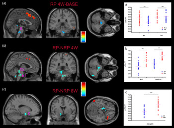
Escitalopram elicited more prominent responses in remitted patients (RP) than non-remitted patients (NRP) in midbrain and amygdala at 4 (4W) and 8 (8W) weeks. (a) fALFF reduction of RP in pons (a) at 4W compared to baseline; (b) fALFF of pons (b1) and midbrain (b2) decreased in RP v. NRP at 4W; (c) fALFF of amygdala (c, cluster of left amygdala) decreased in RP v. NRP at 8W (** p < 0.001).

Exploratory correlation of functional fALFF with clinical response
Exploratory correlation analyses identified five clusters where fALFF5H alteration correlated with HAMD (Supplementary Table S5). Negative correlations were measured in two clusters in the bilateral occipital cortices and one cluster in the right temporal lobe. The fALFF alterations of clusters in the left caudate and the right middle temporal gyrus positively correlated with HAMD (Supplementary Fig. S5, A-E).
In all five ROIs, fALFF5H was highly sensitive (70–85%) and moderately specific (60–75%) for the prediction of the later response (Supplementary Table S6). In addition, the positive predictive values for all treatment outcomes (76–82%) were higher than the negative predictive values (59–75%). The area under the curve (AUC) of fALFF5H for all five ROIs was >0.8 (0.80–0.89) (see Supplementary Figs S5F, S6 and Supplementary Table S6 for cut-off values of fALFF5H of each ROI).
Discussion
In this study, dynamic changes in resting-state activity after acute/sub-chronic escitalopram administration were found and became manifest after a single dose of oral antidepressant medication. After 5 h the antidepressant induced a significant increase in the DLPFC and DMPFC and middle cingulate cortex. Furthermore, while remitters demonstrated more obvious changes following treatment, these were more modest in non-responders suggesting possible tonic and dynamic differences in the serotonergic system. Changes after 5 h in the caudate, occipital and temporal cortices were the best predictor of clinical remission at endpoint. We also found a significant association between acute responses to antidepressant 5 h after administration and delayed (8W) clinical effects of the SSRI.
Consistent with previous reports (Liu et al. Reference Liu, Guo, Liu, Long, Ma, Xue, Wang, Li, Hu and Zhang2013), abnormal resting-state activity indexed by fALFF in depressed patients was detected at baseline, including reduction of fALFF at DLPFC, middle cingulate cortex, posterior cingulate cortex, and right angular gyrus, while increased fALFF was seen in the bilateral temporal lobe. These abnormalities, especially at the DLPFC, were partially reversed by escitalopram after 4 weeks of treatment and might explain adaptive changes which appeared related to the insurgence of a clinical response. Regions where resting-state activity increased over the 4 weeks of treatment included prefrontal and cingulate cortices, and occipital regions, whereas neural activity decreased in the temporal lobe during the same time period. In contrast the acute administration (5H) of an SSRI decreased resting-state neural activity in the occipital cortex. These reversions appeared dampened at the end of the treatment phase (8 weeks), and may be related to a continuous process of synaptic adaption to prolonged pharmacological treatment. Although a single dose of placebo slightly changed resting-state neural activity in healthy controls, these alterations were topographically different from the alterations induced by acute escitalopram in depression. Unfortunately we could not include a placebo arm in the study to investigate placebo effects in major depression because of ethical considerations. Results from a positron imaging study, however, supports independent drug-specific effects induced by 6 weeks’ treatment with the SSRI fluoxetine compared to placebo (Mayberg et al. Reference Mayberg, Silva, Brannan, Tekell, Mahurin, McGinnis and Jerabek2002).
The most impressive changes induced by escitalopram in our study are the increased activity in prefrontal networks including DMPFC, DLPFC and cingulate cortex at week 4 accompanied by decreased activity in the occipital cortex at 5H. A recent study reported that an acute administration of ketamine leads to reductions in the resting-state functional connectivity of subgenual ACC (sgACC) with other regions (Wong et al. Reference Wong, O'Daly, Mehta, Young and Stone2016). Other studies found the fMRI change of PFC and cingulate cortex following antidepressant treatment at a task condition. For example, a single dose of mirtazapine can attenuate responses to self-referential processing task in the medial PFC and the ACC (Komulainen et al. Reference Komulainen, Heikkila, Meskanen, Raij, Nummenmaa, Lahti, Jylha, Melartin, Harmer, Isometsa and Ekelund2016). Another study also found a different effect of agomelatine on brain activities after short-term and long-term use. Seven days agomelatine treatment significantly deactivated the VLPFC during an emotional self-referential task, while significantly increasing the activation of the ventral ACC after 7 weeks in major depression (Delaveau et al. Reference Delaveau, Jabourian, Lemogne, Allaili, Choucha, Girault, Lehericy, Laredo and Fossati2016). The occipital cortex is part of resting-state networks (Beckmann et al. Reference Beckmann, DeLuca, Devlin and Smith2005; Fransson et al. Reference Fransson, Skiöld, Horsch, Nordell, Blennow, Lagercrantz and Åden2007) and visual recognition circuits (i.e. containing the lingual gyrus, middle occipital gyrus, fusiform gyrus and cuneus) were also implicated in two studies of early SSRI response (Wang et al. Reference Wang, Li, Zhang, Zeng, Dai, Su, Wang, Tan, Jin and Yu2014a , Reference Wang, Kuang, Xu, Lei and Yang b ). A growing body of evidence indicates the importance of the PFC in the pathogenesis of mood disorders (Price & Drevets, Reference Price and Drevets2010; Wise et al. Reference Wise, Radua, Via, Cardoner, Abe, Adams, Amico, Cheng, Cole, de Azevedo Marques Perico, Dickstein, Farrow, Frodl, Wagner, Gotlib, Gruber, Ham, Job, Kempton, Kim, Koolschijn, Malhi, Mataix-Cols, McIntosh, Nugent, O'Brien, Pezzoli, Phillips, Sachdev, Salvadore, Selvaraj, Stanfield, Thomas, van Tol, van der Wee, Veltman, Young, Fu, Cleare and Arnonein press) including neuropsychological homeostasis and cognitive control. PFC and cingulate cortex are particular relevant in exercising cognitive control (Rive et al. Reference Rive, van Rooijen, Veltman, Phillips, Schene and Ruhe2013), including the tendency often seen in depression to generate negative valenced cognitions (Quirk & Beer, Reference Quirk and Beer2006). Gray-matter reduction shown in the PFC (Arnone et al. Reference Arnone, Job, Selvaraj, Abe, Amico, Cheng, Colloby, O'Brien, Frodl, Gotlib, Ham, Kim, Koolschijn, Perico, Salvadore, Thomas, Van Tol, van der Wee, Veltman, Wagner and McIntosh2016) might relate to the decreased baseline activities revealed in this study. A recent study found that sensory experience within the occipital cortex can modulate visual cortical circuitry by modifying neurotransmission and synaptic connectivity (Larsen et al. Reference Larsen, Smith, Miriyala, Han, Corlew, Smith and Philpot2014). Increased activity in the PFC and decreased activity in the occipital cortex suggest that SSRI treatment might play a role in dynamically regulating these regions by setting a different balance in the resting-state network potentially linked with clinical response.
The exact mechanism of how escitalopram affects resting-state activity remains unclear. A recent study demonstrated that escitalopram decreased intrinsic DMN regional connectivity, including ACC, posterior cingulate cortex, hippocampal complex and lateral parietal regions, suggesting that the serotonergic system plays an important role in default mode connectivity and its contribution to cognition (van de Ven et al. Reference van de Ven, Wingen, Kuypers, Ramaekers and Formisano2013). There are reports that the neurophysiological basis of the fMRI signal may originate more extensively from synaptic activity (Logothetis et al. Reference Logothetis, Pauls, Augath, Trinath and Oeltermann2001) or be driven by neurotransmitter-related signaling (Rauch et al. Reference Rauch, Rainer and Logothetis2008). In the central nervous system, of all the serotonergic (5-HT) receptors, the 5-HT1A receptor is the most extensively distributed (Pytliak et al. Reference Pytliak, Vargova, Mechirova and Felsoci2011), present in high density in the cortex, hippocampus, septum, and raphe nuclei, and in low density in the basal ganglia and thalamus (el Mestikawy et al. Reference el Mestikawy, Fargin, Raymond, Gozlan and Hnatowich1991). The 5-HT1A receptors in the raphe nuclei are largely somatodendritic autoreceptors, whereas those in other areas are postsynaptic receptors. The acute response for SSRI decreases the serotonergic output via the negative feedback of autoreceptors in the raphe nuclei. It is believed that chronic use of SSRI can induce the increase of serotonergic output following 5-HT1A autoreceptor desensitization. The desensitization of 5-HT1A autoreceptor and increased postsynaptic activation via a general increase in serotonin levels has been shown to be a major mediator in the therapeutic benefits of SSRIs. Reduced resting-state activity at 5 h with increased activity at 4 and 8 weeks in our results were consistent with putative receptor-level alterations.
Receptor mapping studies in rodents have demonstrated that serotonergic afferents project to multiple regions of the limbic system, including the anterior and posterior cingulate cortices (Bozkurt et al. Reference Bozkurt, Zilles, Schleicher, Kamper, Arigita, Uylings and Kötter2005). Serotonergic projection may inhibit the ‘aversive amplification’ circuit, including the ‘prefrontal-amygdala circuit’, and might contribute to the negative affective bias shown in mood disorders (Robinson et al. Reference Robinson, Overstreet, Allen, Letkiewicz, Vytal, Pine and Grillon2013). Neurophysiological studies in animals support the notion that functional results emerging from neuroimaging studies might result from changes in metabolic demands as a consequence of altered serotonin levels (McBean et al. Reference McBean, Ritchie, Olverman and Kelly1999; Schwarz et al. Reference Schwarz, Gozzi, Reese and Bifone2007). Thus, the global component of the fMRI fluctuations measured during resting-state appears tightly coupled with underlying neural activities (Schölvinck et al. Reference Schölvinck, Maier, Frank, Duyn and Leopold2010).
Remitters to treatment showed a significant tendency to modify resting-state activity in the occipital cortex where the signal decreased while neural activity increased in regions such as the cingulate cortex and furthermore decreased in areas such as the post-central gyrus at week 4 of treatment. Conversely, non-remitters showed much lower levels of significant changes. This would support the notion that a relatively silent network might reflect the persistence of pathological mechanisms and unchanged cognitive function. We also noted that the remitters had increased activities in pre- and post-central gyri but decreased activity levels in the midbrain at week 4 and in the hippocampal gyrus, amygdala and subgenual ACC after 8 weeks of treatment compared to non-responders. Speculatively it might be postulated that increased neural activity in the midbrain, being located in proximity of the dorsal raphe nuclei might indicate a higher responsiveness of the serotonin system in remitters. The limbic system is vital to affective regulation circuits in depression, and increased connectivity has been demonstrated in limbic regions such as the insula, amygdala (Connolly et al. Reference Connolly, Wu, Ho, Hoeft, Wolkowitz, Eisendrath, Frank, Hendren, Max and Paulus2013), and PFC (Späti et al. Reference Späti, Hänggi, Doerig, Ernst, Sambataro, Brakowski, Jäncke, grosse Holtforth, Seifritz and Spinelli2015). Furthermore, abnormalities in functional connectivity between limbic and brain areas such as the subgenual cingulate cortex have also been found in both emotionally triggered and resting conditions (Palmer et al. Reference Palmer, Crewther and Carey2015). A possible interpretation of the lower activity detected at endpoint in non-responders in this study might reflect a delayed clinical response in some individuals or indicate a lower sensitivity to serotonin reuptake inhibition in the brain areas where neural activity was measured in this experiment (van de Ven et al. Reference van de Ven, Wingen, Kuypers, Ramaekers and Formisano2013).
The finding of resting-state activity correlating with later remission to escitalopram treatment is of interest. The observation of decreased reactivity at 5H in the occipital lobe combined with increased activity in the caudate and medium temporal gyrus in response to a single dose of escitalopram suggested a better clinical outcome. The caudate and temporal lobe are important in the emotion pathway (Mayberg, Reference Mayberg1997; Phillips et al. Reference Phillips, Drevets, Rauch and Lane2003).
Although explorative in nature, the fALFF5H for all five ROIs was significantly sensitive and specific in predicting the later response based on the change of the resting-state activity at 5H. If confirmed, these data suggest that acute neural changes in specific brain regions might provide potentially useful biomarker data for the identification of patients favorably responding to SSRIs at week 8.
Limitations of this study include the relatively small sample size, the use of a fixed-dose regimen for the antidepressant of choice, which although in the therapeutic range might have restricted remission rate in some clinical cases where higher doses were required, and the relatively milder severity of depression. Future studies could confirm the generalizability of the findings in a larger sample (1) with more severe depressive features, (2) with/without co-morbid anxiety symptoms, (3) clinically treated with variable doses of antidepressant medication and (4) with confirmatory blood levels for efficacy. Furthermore, subjective responses to the acute effects of antidepressants could be better assessed in future studies with rating scales evaluating state effects. Alternative methods to model nonlinear data may be better suited than the technics used in this work.
In conclusion this work identified resting-state dynamic alterations in major depression following pharmacological treatment and in relation to clinical improvement. Our results may provide potentially useful information to further understand the neurobiological basis of treatment and clinical response which might help clinicians to make decisions in relation to medication.
Supplementary material
The supplementary material for this article can be found at http://dx.doi.org/10.1017/S0033291716002440
Acknowledgments
We thank all the volunteers participated in this study. This work was supported by grants from the National Natural Science Foundation of China (81160379, 81160171, 81460256, 81560233), the Funding of Yunnan Provincial Health Science and Technology Plan (2014NS171, 2014NS172). Middle and Young Aged Academic and Technology Leaders Reserve Personnel Foundation of Yunnan Province (2015HB071). Danilo Arnone is supported by the Academy of Medical Sciences (AMS-SGCL8). Yuqui Cheng and Xiufeng Xu had full access to all of the data in the study and take responsibility for the integrity of the data and the accuracy of the data analysis.
Declaration of Interest
Danilo Arnone has received travel grants from Servier and Jannsen.