Introduction
Imaging plays an important role in aiding diagnosis and planning surgery for middle-ear cholesteatoma. Computed tomography (CT) is the cornerstone for imaging primary (pre-operative) cholesteatoma. It shows delineation of the bony anatomy of the middle-ear cleft and aids diagnostic assessment of disease by showing disease extent and any complications, notably bony and ossicular chain erosion.Reference Khemani, Singh, Lingam and Kalan1 The diagnostic capability of CT is unfortunately limited by its inability to differentiate cholesteatoma from co-existing inflammatory tissue and fluid.Reference Muzaffar, Metcalfe, Colley and Coulson2 Diffusion-weighted magnetic resonance imaging (MRI), specifically non-echoplanar diffusion-weighted MRI, can reliably characterise cholesteatoma and differentiate it from soft tissue and fluid within the middle-ear cleft.Reference Khemani, Singh, Lingam and Kalan1–Reference Majithia, Lingam, Nash, Khemani, Kalan and Singh3 This is because cholesteatoma, by virtue of its keratin content, has high signal intensity on diffusion-weighted MRI images (b-value of 800 or 1000) due to a combination of restricted diffusion and T2 shine-through effect.
Given the high diagnostic performance of non-echoplanar diffusion-weighted MRI, it is currently the modality of choice in detecting post-operative cholesteatoma and has been applied as an adjunct to CT in the assessment of primary cholesteatoma.Reference Majithia, Lingam, Nash, Khemani, Kalan and Singh3 Diffusion-weighted MRI also performs well in depicting the size and extent of cholesteatoma.Reference Khemani, Lingam, Kalan and Singh4–Reference Lingam and Bassett5 However, bony anatomy is lacking on diffusion-weighted MRI, meaning precise disease localisation is challenging when planning surgery.
Modern post-processing fusion imaging techniques allow high resolution CT and diffusion-weighted MRI of the temporal bone to be superimposed and viewed simultaneously. This harnesses the capabilities of the two types of scans and depicts the precise location and extent of disease in relation to the detailed temporal bone anatomy.Reference Plouin-Gaudon, Bossard, Ayari-Khalfallah and Froehlich6–Reference Alzahrani, Alhazmi, Bélair and Saliba10 This arguably provides a more useful map for the surgeon who is better acquainted with CT when planning middle-ear surgery. The fused map allows the surgeon to plan an optimal approach and technique to clear the disease. We discuss our experience in using the CT and diffusion-weighted MRI fusion technique to practically assist the otologist in assessment and treatment of both primary and post-operative cholesteatoma. We illustrate the practical implications of the technique through a series of cases.
Ethical considerations
Software was used to utilise existing scans performed as part of routine clinical care. No additional imaging was performed in this case series study.
Materials and methods
Computed tomography was performed using a 16-row multi-detector CT scanner (Phillips Brilliance; Phillips, Amsterdam, The Netherlands). Magnetic resonance imaging was performed using a 1.5-T superconductive unit (Magnetom Avanto; Siemens Medical Solutions, Erlangen, Germany) using standard head matrix coil. Coronal 2-mm thick turbo spin-echo T2-weighted images were performed. In all patients, a 2-mm thick non-echoplanar half-Fourier acquisition single-shot turbo spin-echo diffusion-weighted sequence was acquired in the coronal plane.
Following acquisition, fusion of the CT and diffusion-weighted MRI b1000 images was performed using TeraRecon (Durham, North Carolina, USA) Aquarious software (version 4.4) on the picture archiving and communication system (Sectra, Linkoping, Sweden). Axial CT images and the coronal b1000 images were selected for fusion. Automatic multiplanar registration of the images was performed by the software followed by manual fine-tuning by the radiologist, taking approximately twenty minutes per scan. The fused CT and diffusion-weighted MRI images were saved onto the picture archiving and communication system for review.
Assessment of primary cholesteatoma
Case 1
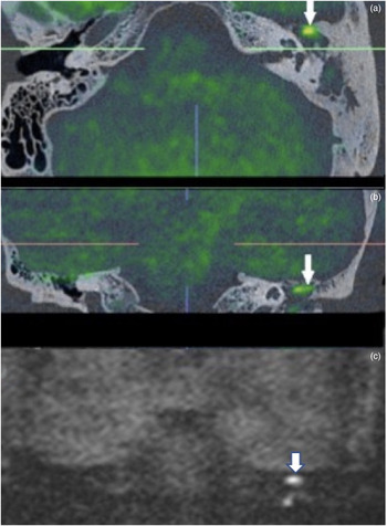
A 47-year-old man presented with ongoing foul-smelling ear discharge and was diagnosed with cholesteatoma clinically. His high-resolution CT scan of temporal bone demonstrated complete soft tissue opacification of the entire left middle-ear cleft. Diffusion-weighted MRI was performed to characterise and define the extent of cholesteatoma within the opacified middle-ear cleft and was also fused with the CT. This was helpful as the fused images clearly showed that the disease was confined to the epi-mesotympanum without intruding into the mastoid antrum (Figure 1a–c). This level of definition provided information about the extent, size and precise location of disease. This made it possible to have a clear discussion with the patient regarding technique, approach, duration and expected recovery from surgery, empowering the consenting process. The improved pre-operative planning allowed us to tailor the surgery with recognition that opening the mastoid was not required.
Cholesteatoma anatomically localised to the anterior epi-mesotympanum on fusion imaging (axial (a) and coronal (b) images), as indicated by arrows, in comparison to diffusion-weighted technique only (coronal b1000 image (c)).

Case 2
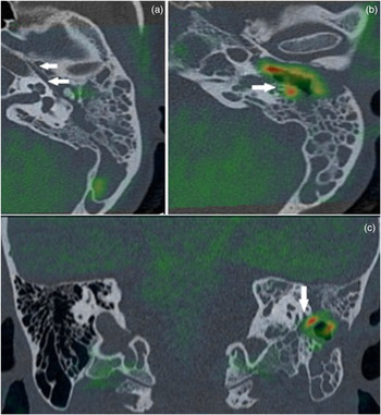
A 34-year-old woman presented with a discharging left ear and left side grade III facial nerve palsy. A clinical diagnosis of cholesteatoma was made on otoscopic examination. High-resolution CT showed abnormal soft tissue in the bony external auditory canal and middle-ear cleft associated with bony destruction of the canal wall, notably the posterior canal wall. However, diffusion-weighted MRI showed abnormal restricted diffusion of cholesteatoma primarily within the bony external auditory canal extending into the mastoid. These findings helped ascertain that given the degree and location of bone erosion, a canal wall down procedure was required to ensure adequate disease clearance. Fusion imaging mapped the abnormal diffusion-weighted MRI signal of cholesteatoma onto the CT images and showed the bony relations of the cholesteatoma in the region and specifically where it eroded the facial nerve canal (Figure 2). This gave the operating surgeon greater pre-operative awareness of the areas where the nerve was exposed and at particular risk in the dissection of disease. Post-operatively, the patient recovered to a House–Brackmann grade I with a well lined epithelialised cavity.
Left facial nerve and its intact tympanic canal (axial fusion image (a)) with an area of erosion at its mastoid segment from ear canal cholesteatoma (axial (b) and coronal (c) fusion images) shown.

Assessment of post-operative cholesteatoma
Case 3
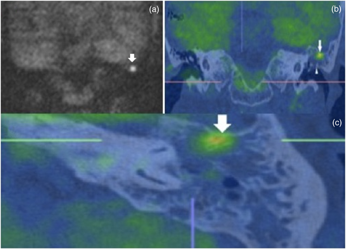
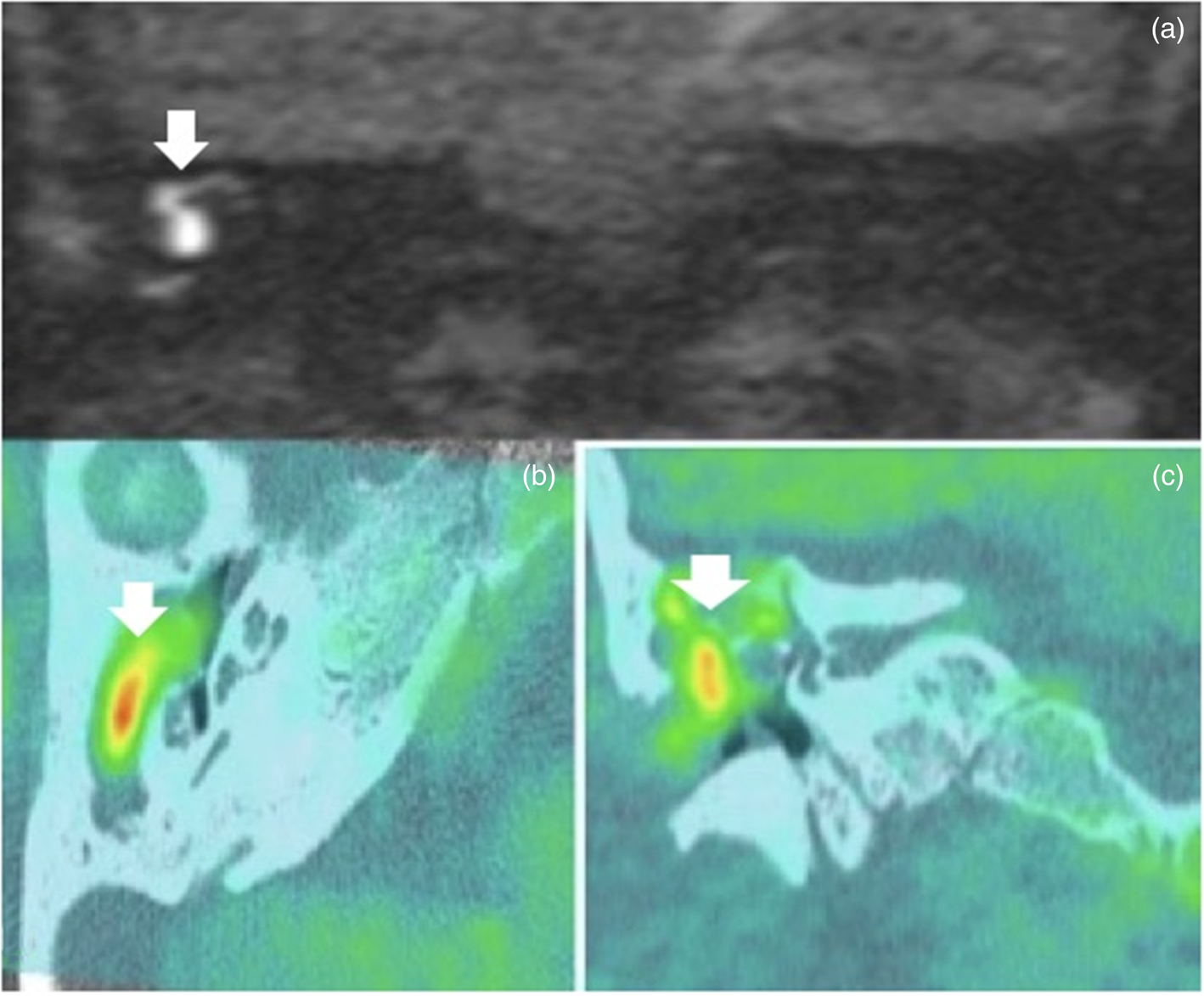
A 64-year-old woman underwent a left-hand side combined approach tympanomastoidectomy (canal preserving) procedure for cholesteatoma. During primary surgery, extensive cholesteatoma was located medial to the neck of the malleus and filling the epitympanum. Despite being asymptomatic, a follow-up diffusion-weighted MRI scan at 12 months demonstrated a 3-mm residual cholesteatoma in the region of the lateral epitympanum although the exact anatomical location was unclear on the diffusion-weighted MRI (Figure 3). In order to avoid radiation exposure from performing another CT scan, fusion of the diffusion-weighted MRI was performed with the original pre-operative CT scan. This was an option as the key bony landmarks would remain unchanged as the images are registered automatically by the software and then adjusted manually by the observer on fixed bony anatomical registration points such as the clivus and skull base.Reference Alzahrani, Alhazmi, Bélair and Saliba10 The fused CT and diffusion-weighted MRI images (Figure 3) show improved localisation of disease, not in the epitympanum but in the pneumatised bone just superior to the bony external auditory canal, which communicates posteriorly with the mastoid. A combined approach tympanomastoidectomy (second look) confirmed the disease as being isolated to this specific area and was removed. Correlation of the initial intra-operative findings and subsequent fusion images can help to educate us in the evaluation of how and where recurrent or residual disease may arise.
Localisation of cholesteatoma superior to the left bony external auditory canal communicating posteriorly with the mastoid in (a) coronal diffusion-weighted MRI only and (b (coronal) and c (axial)) fusion imaging.

Case 4
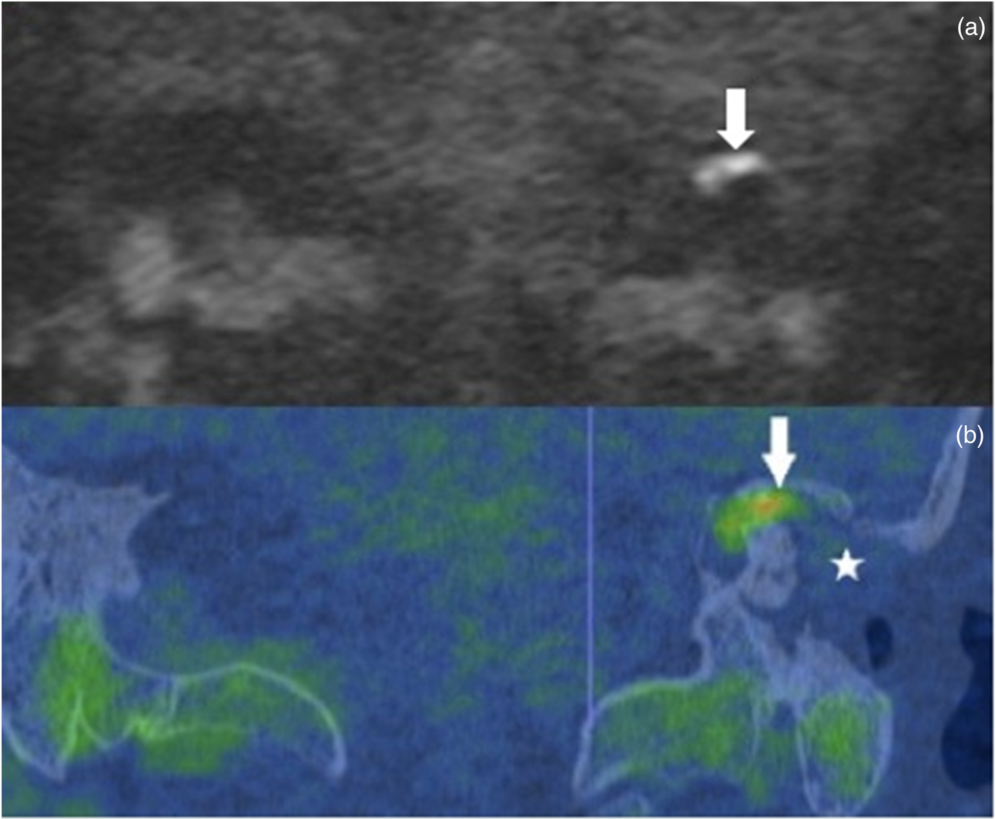
A 38-year-old man underwent multiple preceding surgical procedures for recurrent cholesteatoma, most recently a canal wall down procedure. He displayed ongoing otological symptoms of discharge and imbalance. Diffusion-weighted MRI is not routinely used in the detection of recurrent disease in canal wall down mastoidectomy because the open cavity can be easily inspected clinically, and any disease can be easily microsuctioned in clinic. However, in this case, microscopic and otoendoscopic evaluation showed a clean and epithelialised mastoid cavity making it diagnostically difficult to explain the patient's recurring symptoms which were suspicious for recurrent disease. Though not typically indicated for canal wall down mastoidectomy, diffusion-weighted MRI was subsequently performed on the basis of persisting symptoms. This detected cholesteatoma within the altered middle-ear cleft, but it was not possible to precisely identify this within a canal wall down mastoid. A CT temporal bone scan was performed to depict the bony anatomy and fused with the diffusion-weighted MRI to precisely depict the extent of disease within the bone (Figure 4). This clearly identified residual cholesteatoma medial to the superior semicircular canal in the petrous bone. Normally, the clinician can visualise the mastoid cavity in canal wall down cases and manage the patient without having to image them. However, this case highlights the diagnostic difficulty that diffusion-weighted MRI presents in localising disease when there is only a cavity. Fusion imaging elegantly rectifies this problem. Subsequently, the patient underwent a labyrinthectomy for control of his aggressive cholesteatoma.
(a) Coronal b1000 diffusion weighted image (b) coronal fusion image: Residual cholesteatoma identified medial to the superior semicircular canal (arrow) in left canal wall down cavity (depicted with a star).

Case 5
A 26-year-old woman presented with significant ear discharge and pain with a large ear canal polyp occluding the entire mastoid cavity, hindering clinical examination. She had previous mastoid surgery at another institution prior to her referral. It was clinically unclear whether this was a mucosal or squamosal process and imaging was requested. A high-resolution CT scan of the temporal bones helped to identify bony changes consistent with previous mastoid surgery, but the middle-ear cleft was completely opacified. Diffusion-weighted MRI confirmed the presence of underlying cholesteatoma but given the altered anatomy it was difficult to localise. Fusing the two modalities clearly depicted the precise location of the cholesteatoma in relation to the previous surgical changes (Figure 5). Aural polyps often arise due to mucosal disease and are treated topically or are examined under anaesthesia for biopsy. In a case that was proving to be a clinical dilemma, imaging helped to inform the diagnosis and facilitate early surgical planning by providing accurate localisation and information on the extent of disease. In this case, imaging helped to prevent delay in definitive revision surgery.
Identification of cholesteatoma medial to an aural polyp with anatomical localisation compared from (a) coronal b1000 diffusion-weighted MRI and (b) axial and (c) coronal fusion images.

Discussion
Our series highlights the visibility of disease relative to the underlying bony anatomy and therefore provides the otologist with an enhanced map for surgical planning. Use of fusion imaging for assessing cholesteatoma has recently been advocated in providing these detailed navigational maps to allow less invasive approaches such as transcanal endoscopic surgery. In a recent US study, blinded investigators found the diagnostic sensitivity and localisation accuracy to be improved using the fusion technique, with an accuracy of 90 per cent.Reference Locketz, Li, Fischbein, Holdsworth and Blevins8 Similarly, a recent prospective study of 55 patients from Japan found positive and negative predictive values of over 90 per cent for all areas of the middle ear.Reference Watanabe, Ito, Furukawa, Futai, Kubota and Kanoto9 The precise localisation of cholesteatoma has also been demonstrated within the paediatric population, in primary and revision cases.Reference Plouin-Gaudon, Bossard, Ayari-Khalfallah and Froehlich6,Reference Alzahrani, Alhazmi, Bélair and Saliba10
The limitations to use of fusion imaging include the costs of obtaining the fusion software package and the radiologist's or otologist's time in fusing and interpreting the images. Acquiring additional post-operative CT scans for fusion in the assessment of post-operative cholesteatoma would incur radiation exposure; however, this may be avoided by using the original pre-operative CT scan instead.Reference Alzahrani, Alhazmi, Bélair and Saliba10 It is important to recognise the limitations of detection (3 mm) remain as with stand-alone diffusion-weighted MRI and false positive b1000 diffusion weighted signal from various causes.
Our series highlights the value of CT and diffusion-weighted MRI fusion as a problem-solving tool in the clinical management of primary and recurrent cholesteatoma and its associated complications. Within our recent practice we found fusion imaging can also aid in the patient counselling process with respect to outlining potential complications and also in teaching and training. We demonstrate its practical relevance to operating otologists, where it provides a greater understanding of the anatomical structure of the disease, which can help to guide optimal surgical outcomes rather than simply indicating presence or absence of disease.
Competing interests
None declared