Introduction
The multi-step process of in vitro embryo production (IVP) has faced many difficulties since the birth of the first calf on 1982 after transfer of an in vitro-matured, in vitro-fertilized and in vitro-cultured embryo into a recipient cow (Brackett et al., Reference Brackett, Bousquet, Boice, Donawick, Evans and Dressel1982). During the last decades, the whole process has been improved, attempting to mimic the natural system and, nowadays, in vitro fertilization (IVF) has become a well-established assisted reproductive technology in both animals and humans (Wrenzycki et al., Reference Wrenzycki, Herrmann, Korsawe, Hadeler and Niemann2000). Protocols of IVF are successfully used in many areas of applied sciences, for example for research investigations, for treating infertility, for the conservation of biology in endangered animals and also for enhancing the productivity of food animals (Bavister, Reference Bavister2002). This fact is thanks to the possibility of providing a large number of viable embryos, which, after transfer into recipient females, are able to progress into normal healthy offspring. In order to apply IVP technology on an intensive commercial level, particularly for bovine species, a series of problems have to be dealt, focused and investigated to reach a significant and fully available point for applicability. Applications of such technology, combined with the modern molecular tools, can open wide horizons for different procedures and techniques in which gametes and embryos are involved (Gordon & Lu, Reference Gordon and Lu1990). These therefore possess strategic importance to enhance the bovine reproductive and genetic advances. A widespread commercialization of such technology would also provide a new future goal for the production of sexed embryos and transgenic animals (Brackett & Zuelke, Reference Brackett and Zuelke1993). The preimplantation period of embryo development, during which embryos can be manipulated, is essential to improve their quality through the IVP techniques in connection with genetic approaches like gene expression, nuclear transfer, gene insertion, cloning and stem cell biology. The establishment of such molecular approaches in farm animals not only will provide a new knowledge about the mammalian embryonic development (Kanka, Reference Kanka2003), but will also aid in the achievement of genomic advances within the livestock. Embryo development is characterized by a minor and a major form of genome activation, during which genes are expressed in a stage-specific manner (Niemann & Wrenzycki, Reference Niemann and Wrenzycki2000). The expression pattern of specific genes can be usefully used as molecular marker during the preimplantation period (Wrenzycki et al., Reference Wrenzycki, Herrmann, Lucas-Hahn, Lemme, Korsawe and Niemann2004). Effect of the in vitro system has been reflected by the occurrence of clinically important phenomena, such as the large offspring syndrome (LOS) (Lazzari et al., Reference Lazzari, Wrenzycki, Herrmann, Duchi, Kruip, Niemann and Galli2002) and apoptosis (Yang & Rajamahendran, Reference Yang and Rajamahendran2002). While studies of gene expression in the oocytes are more advanced (Dalbies-Tran & Mermillod, Reference Dalbies-Tran and Mermillod2003), there is still a defect of information about the mechanisms that can affect such patterns of expression in relation to changes in oocyte environmental conditions (Lonergan et al., Reference Lonergan, Rizos, Gutierrez-Adan, Fair and Boland2003a). In vitro maturation (IVM) is an important phase that may have an influence on oocyte transcripts abundance (Wrenzycki et al., Reference Wrenzycki, Herrmann, Lemme, Eckert, Carnwath and Niemann1997; Bettegowda et al., Reference Bettegowda, Patel, Ireland and Smith2006). In particular, mRNA expression pattern can differ among the various IVM media and chemical supplementation (Watson et al., Reference Watson, De Sousa, Caveney, Barcroft, Natale, Urquhart and Westhusin2000). In a study of Knijn et al., (Reference Knijn, Wrenzycki, Hendriksen, Vos, Herrmann, Van Der Weijden, Niemann and Dieleman2002)), no significant difference in the expression pattern of a series of genes in blastocysts deriving from oocytes matured under different IVM regimens was reported, suggesting that an altered gene expression pattern cannot be related necessarily to prematuration–maturation environment, but more likely with post-IVM and IVF environment. Oocyte quality of different classes (based on cumulus cell investment and ooplasm homogeneity) was reported to have no significant effect on the transcriptional activity either in oocytes (Bilodeau-Goeseels, Reference Bilodeau-Goeseels2003) or in in vitro-produced bovine embryos (Bilodeau-Goeseels & Panich, Reference Bilodeau-Goeseels and Panich2002). Conversely, the expression of specific pro-apoptotic and anti-apoptotic transcripts was significantly affected among different quality bovine oocytes and embryos (Yang & Rajamahendran, Reference Yang and Rajamahendran2002). Despite the oocyte origin and its intrinsic quality represent a major factor affecting the bovine blastocyst yield, the period of postfertilization culture environment can have great impact on mRNA abundance (Lonergan et al., Reference Lonergan, Rizos, Gutierrez-Adan, Fair and Boland2003a) and on the pattern of gene expression of genes responsible for a correct embryo development. This could be a determining factor for a series of quality parameters of blastocyst, such as cryotolerance (Lonergan et al., Reference Lonergan, Rizos, Kanka, Nemcova, Mbaye, Kingston, Wade, Duffy and Boland2003b) and clinical normality of embryos. Effect of the in vitro culture (IVC) has been studied in relation to the phenomenon of apoptosis (Rizos et al., Reference Rizos, Lonergan, Boland, Arroyo-Garcia, Pintado, de la Fuente and Gutierrez-Adan2002) that characterizes the in vitro-produced bovine embryos largely (Gjorret et al., Reference Gjorret, Knijn, Dieleman, Avery, Larsson and Maddox-Hyttel2003). Improving the quality of the in vitro-produced blastocysts in order to equalize their in vivo-deriving counterparts, will depend on the possibility to induce specific changes in the gene expression patterns during oocyte maturation and embryo culture. Definitely, this would have beneficial effects not only in increasing the in vitro production of blastocysts but also in the production of higher quality blastocysts (Lonergan et al., Reference Lonergan, Rizos, Gutierrez-Adan, Fair and Boland2003d).
This review focuses on the morphogenetic embryo development and gene expression profile in the in vitro-produced bovine embryos with special reference to those parameters that may alter the gene expression pattern during the critical period of IVC. Effect of the IVP system, as reflected by some clinically important events like apoptosis, is also reviewed.
Development from immature oocyte to blastocyst
The multi-step process of in vitro embryo production begins with the recovery of immature oocytes from the ovaries. These oocytes are then exposed to in vitro maturation, during which the oocyte passes through different nuclear and cytoplasmic changes to become mature. Oocytes are then in vitro fertilized and zygotes are cultured to the morula or blastocyst (period of preimplantation) stages. At the blastocyst stage, the in vitro-produced embryos can be either transferred to synchronized recipient females or cryopreserved and stored indefinitely (Gasparrini, Reference Gasparrini2002; Neglia et al., Reference Neglia, Gasparrini, di Brienza, Di Palo, Campanile, Presicce and Zicarelli2003).
Oocyte maturation
Mammalian oocytes are arrested at the diplotene of the first meiotic prophase, the germinal vesicle stage (GV) (Motlik & Kubelka, Reference Motlik and Kubelka1990). In vitro, resumption of meiosis is observed in fully grown oocytes when they are released from the inhibitory influence of their follicles (Edwards, Reference Edwards1965) and cultured in suitable conditions, during which the oocyte passes both nuclear and cytoplasmic changes (Gasparrini, Reference Gasparrini2002). Final target of oocyte maturation is the production of a haploid secondary oocyte that is fully equipped with the cell biological machinery necessary for successful fertilization and embryonic development (Hyttel et al., Reference Hyttel, Fair, Callesen and Greve1997).
Nuclear maturation
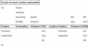
During the 18 to 24 h of maturation, the oocyte nucleus passes several developmental changes: the resumption of the meiotic arrest is characterized by the germinal vesicle (4N) breakdown (GVBD), chromosome condensation and spindle formation. Thereafter, the first meiotic division occurs and the first polar body containing half of the genome (2N) is extruded, while the remaining diploid condensed part of chromatin proceeds to MII. Meiosis is arrested again at this stage until oocytes are activated by the fertilising spermatozoa or by an artificial stimulus (parthenogenetic activation) (Fig. 1). After fertilization, the second polar body (1N) is extruded and the remaining part of chromatin (1N) forms the female pronucleus (Chan et al., Reference Chan, Homan, Ballou, Burns and Bremel1998). This process, which is accompanied by the formation of the male pronucleus, is followed by the fusion of maternal and paternal pronuclei, as haploid sets of DNA, to form a zygote through the union of chromosomes (syngamy) that restores the diploid state (Bavister, Reference Bavister2002).
A summary of major events during the oocyte meiotic cycle, classified according to their dependence on (+) or independence from (–) the requirement for a new protein synthesis in the 4 h preceding each meiotic event (adapted from Zhang et al., Reference Zhang, Lee and Moor1998).

Cytoplasmic maturation
Oocyte cytoplasmic maturation is characterized by a series of ultrastructural and molecular changes. In immature oocytes, the metabolic activity appears to be high as reflected by a large number of organelles in the ooplasm such as mitochondrial clusters, smooth endoplasmic reticulum and lipid vacuoles with a peripheral localization, closed to the cumulus cells–oocyte coupled gap junctions, related to the intensive exchange of activity at this level (Gasparrini, Reference Gasparrini2002). In mature oocytes (MII), cumulus–oocyte junctions disappeared, the cumulus projections cannot be longer detected, the lipid droplets increase and mitochondria and smooth endoplasmic reticulum are rearranged towards a more central location in the ooplasm. Golgi complexes are decreased or can be even absent, while the cortical granules (produced by Golgi complex) are localized peripherally, close to the membrane, forming a structural background for the block against polyspermy (Hyttel et al., Reference Hyttel, Fair, Callesen and Greve1997).
Morphogenetic embryo development
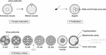
Regarding the molecular and morphological events contributing the bovine embryo development, three important morphogenetic events take place during the period of preimplantation: morula compaction, that begins at the 32-cell stage, blastocyst formation, which starts between the 64- to 128-cell stages (Betteridge & Flechon, Reference Betteridge and Flechon1988; Van Soom Reference Van Soom, Boerjan, Bols, Vanroose, Lein, Coryn and de Kruifet al., 1997a) and blastocyst hatching. The intercellular communications, which are the base for the morphological and transcriptional event mediated by cellular adhesions of morula compaction, play an important role during the embryonic development. Boni et al. (Reference Boni, Tosti, Roviello and Dale1999) accounted a low expression of the intercellular communication device transcripts as one of the causes of the low developmental potential associated with the in vitro-produced bovine embryos. The process of embryo compaction is followed by the cavitation, characterized by the formation of a fluid filled cavity (blastocoele) accompanied by the differentiation of an outer epithelial trophectoderm and an inner cell mass, within the forming blastocyst (Fig. 2). These two morphogenetic events are mediated by a specific expression profile of a number of gene families encoding for cellular processes and growth factor gene products (Watson, Reference Watson1992). The regulation of the expression during these stages is important for obtaining good quality blastocysts and for the maintenance of pregnancy (Watson et al., Reference Watson, Natale and Barcroft2004).
Development phases of bovine embryo. Phase I: oocyte maturation from germinal vesicle stage of metaphase II stage, extrusion of first polar body. Phase II: fertilization and zygote formation; extrusion of second polar body. Phase III: cleavage and divisions of blastomeres, forming cell–cell junctions at the compact morula stage and differentiating into trophectoderm and inner cell mass at the blastocyst stage. Before the zygotic genome activation (ZGA), embryonic protein synthesis depends mainly on the maternal transcripts (mRNA and proteins) stored in the oocyte (adapted from Young et al., Reference Young, Sinclair and Wilmut1998).

Embryonic transcriptional activity
Mammalian developmental stages include the accumulation of maternal RNA and proteins during the growth and development of the oocyte. Thus, the earliest stages of embryogenesis are supported by these maternally inherited components stored within the oocyte (Pavlok et al., Reference Pavlok, Kopecny, Lucas-Hahn and Niemann1993) and play an important role for the following development of the oocyte up to the blastocyst stage (Pavlok et al., Reference Pavlok, Lucas-Hahn and Niemann1992; Lonergan et al., Reference Lonergan, Monaghan, Rizos, Boland and Gordon1994; Sirard & Blondin, Reference Sirard and Blondin1996; De Sousa et al., Reference De Sousa, Caveney, Westhusin and Watson1998; Meirelles et al., Reference Meirelles, Caetano, Watanabe, Ripamonte, Carambula, Merighe and Garcia2004). With further development, the oogenetic maternally inherited informational molecules begin to decrease and the embryonic development becomes dependent on the expression of embryonic genome. This switching from maternal to embryonic control, includes loss of mRNA molecules of maternal origin, activation of transcription of embryonic genome, developmental arrest in the presence of transcriptional inhibitors and a marked change in the pattern of protein synthesis (Telford et al., Reference Telford, Watson and Schultz1990). This transition is essential for the activation of a large number of genes and for the following pattern of gene expression that is responsible for successful embryonic differentiation and development (Lequarre et al., Reference Lequarre, Marchandise, Moreau, Massip and Donnay2003; Lonergan et al., Reference Lonergan, Rizos, Gutierrez-Adan, Moreira, Pintado and de la Fuente2003c).
Embryonic genome activation
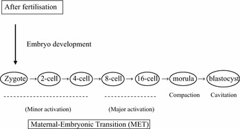
With activation of embryonic genome, the maternal transcripts are replaced by the embryonically expressed ones (Kanka, Reference Kanka2003). Embryonic genome activation is characterized by two different minor and major forms in which the embryo passes a complicated pattern of activation (Fig. 3).
Stages of embryo development, with regard to the minor and major embryonic genome activation and the importance of maternal transcripts and proteins to initiate the early embryonic development until the fourth cell cycle (adapted from Dielman et al., 2002).

Minor genome activation
Minor bovine embryonic genome activation has been observed between the 1-cell to 4-cell stages, suggesting that zygotes and early embryos are transcriptionally and translationally active (Barnes & Eyestone, Reference Barnes and Eyestone1990; Memili et al., Reference Memili, Dominko and First1998; Memili & First, Reference Memili and First1999). This finding was confirmed by Hyttel et al. (Reference Hyttel, Viuff, Avery, Laurincik and Greve1996) and Viuff et al. (Reference Viuff, Avery, Greve, King and Hyttel1996), who demonstrated the presence of a transcriptional activity of bovine embryos at the second cell cycle by incubating the embryos with [3H]-uridine and using autoradiography. Thus, the successful embryonic development requires an adjusted embryonic physiological pattern in response to maternal regulatory signals and the surrounding environment, as also suggested by Chandolia et al. (Reference Chandolia, Peltier, Tian and Hansen1999) who reported an increased embryonic gene expression pattern of specific heat shock protein 70 in 2-cell bovine embryos cultured at elevated temperatures.
Major genome activation, the maternal–embryonic transition (MET)
Time of switching from maternal to embryonic control of mRNA synthesis, is termed MET. In bovine, the major activation of embryonic genome occurs at the 8- to 16-cell stages and includes an increase of mRNA transcribed from the embryonic genome together with a marked change in the pattern of protein synthesis (Memili & First, Reference Memili and First1998, Reference Memili and First2000). Maternal transcripts and proteins accumulated within the oocyte are essential to sustain the first few cell cycles of bovine embryos until the major activation of embryonic genome takes place (Dieleman et al., Reference Dieleman, Hendriksen, Viuff, Thomsen, Hyttel, Knijn, Wrenzycki, Kruip, Niemann, Gadella, Bevers and Vos2002), as confirmed by a work of Barnes & First (Reference Barnes and First1991), which revealed that bovine embryos cultured in the presence of a transcriptional inhibitor (α-amanitin) were able to undergo cleavage and to progress up to the 8- to 16-cell stages. Hyttel et al. (Reference Hyttel, Laurincik, Viuff, Fair, Zakhartchenko, Rosenkranz, Avery, Rath, Niemann, Thomsen, Schellander, Callesen, Wolf, Ochs and Greve2000) showed that the transcriptional activity of the ribosomal RNA genes starts in the nucleolus of bovine embryos during the fourth cell cycle postfertilization, evidencing at this time, an activation of the embryonic genome at major cascade. Moreover, a proper activation of these ribosomal genes is important for maintenance of the subsequent protein synthesis for both housekeeping functions and embryonic cells specialization. Meirelles et al. (Reference Meirelles, Caetano, Watanabe, Ripamonte, Carambula, Merighe and Garcia2004), studied the phenomenon of embryo developmental arrest that is generally observed in bovine embryos at the 8- to 16-cell stages and correlated this process with the quality of cytoplasmic factors and proteins within the oocyte and with the failure of transcription of the embryonic genome during MET.
Implications and final developmental patterns of the IVP system
Obtaining high quality, clinically normal blastocysts and healthy offspring remains the basic target of IVP and can be influenced by several factors and quality parameters:
In vitro-produced bovine embryos show low developmental potential
Although in vitro-matured and in vitro-fertilized bovine oocytes may reach high cleavage rates (about 80%), no more than 50% of the zygotes are able to progress to blastocyst stage (Gordon & Lu, Reference Gordon and Lu1990; Lonergan et al., Reference Lonergan, Monaghan, Rizos, Boland and Gordon1994; Boni et al., Reference Boni, Tosti, Roviello and Dale1999; Dieleman et al., Reference Dieleman, Hendriksen, Viuff, Thomsen, Hyttel, Knijn, Wrenzycki, Kruip, Niemann, Gadella, Bevers and Vos2002; Lequarre et al., Reference Lequarre, Marchandise, Moreau, Massip and Donnay2003). This suggests the culture period and culture conditions to be a critical phase for determining blastocyst yield and quality (Lonergan et al., Reference Lonergan, Rizos, Gutierrez-Adan, Fair and Boland2003a).
An impaired embryo quality in vitro
The quality of in vitro-produced bovine embryos usually lags behind those produced in vivo (Rizos et al., Reference Rizos, Lonergan, Boland, Arroyo-Garcia, Pintado, de la Fuente and Gutierrez-Adan2002; Lonergan et al., Reference Lonergan, Rizos, Kanka, Nemcova, Mbaye, Kingston, Wade, Duffy and Boland2003b). Moreover, there is evidence of transcriptional differences in the relative abundance of physiologically and clinically important genes among postfertilization developmental stages between in vitro- and in vivo-deriving bovine embryos (Tesfaye et al., Reference Tesfaye, Ponsuksili, Wimmers, Gilles and Schellander2004). Although the efficiency of the in vitro systems is progressing, until now the final developmental potential of IVP, in terms of production of good quality blastocyst and healthy offspring free from clinical abnormalities, is not able to equalize the ideal ‘gold standard’ represented by the in vivo system. Embryo quality can be expressed according to different parameters, such as morphology, cryotolerance, metabolism and gene expression. With regard to this, the IVP system has reflected morphogenetic alterations of quality parameters associated with the embryos, such as the different cell number and inner cell mass allocation when compared to their in vivo counterparts (Van Soom Reference Van Soom, Ysebaert and de Kruifet al., 1997b), the different morphology and developmental kinetics between embryos cultured in vivo or in vitro (Holm et al., Reference Holm, Booth and Callesen2002; Gutierrez-Adan et al., Reference Gutierrez-Adan, Rizos, Fair, Moreira, Pintado, De La Fuente, Boland and Lonergan2004), the energy and glucose metabolism (Khurana & Niemann, Reference Khurana and Niemann2000), embryo cryosurvival (Enright et al., Reference Enright, Lonergan, Dinnyes, Fair, Ward, Yang and Boland2000; Rizos et al., Reference Rizos, Gutierrez-Adan, Perez-Garnelo, de la Fuente, Boland and Lonergan2003) and the incidence of chromosomal abnormalities associated with the in vitro- produced embryos (Hyttel et al., Reference Hyttel, Viuff, Fair, Laurincik, Thomsen, Callesen, Vos, Hendriksen, Dieleman, Schellander, Besenfelder and Greve2001; Viuff et al., Reference Viuff, Hendriksen, Vos, Dieleman, Bibby, Greve, Hyttel and Thomsen2001). The mRNA expression pattern of several candidate genes regulating embryo development is a relevant parameter determining embryo quality (Wrenzycki et al., Reference Wrenzycki, Herrmann, Carnwath and Niemann1999; Farin et al., Reference Farin, Farin and Piedrahita2004; Dode et al., Reference Dode, Dufort, Massicotte and Sirard2006) and when this is altered, an impaired embryo status may result. These alterations may lead to deviations affecting embryo development and the occurrence of pathological phenomena that make the in vitro-produced bovine embryos lacking the perfect potential before or after cryopreservation, transfer and implantation (Wrenzycki et al., Reference Wrenzycki, Herrmann, Carnwath and Niemann1996, Reference Wrenzycki, Herrmann, Carnwath and Niemann1999, Reference Wrenzycki, Herrmann, Keskintepe, Martins, Sirisathien, Brackett and Niemann2001, Reference Wrenzycki, Herrmann, Lucas-Hahn, Lemme, Korsawe and Niemann2004; Niemann & Wrenzycki, Reference Niemann and Wrenzycki2000; Yang & Rajamahendran, Reference Yang and Rajamahendran2002). To this regard, LOS is a molecularly related phenomenon, reported occasionally in the in vitro-produced embryos, that is characterized by an increased birth weight of the calves as compared with the normal and a number of related problems which frequently lead to abortion or an unhealthy status (Lazzari et al., Reference Lazzari, Wrenzycki, Herrmann, Duchi, Kruip, Niemann and Galli2002). This impaired consequence of the IVP systems has been related to cellular and genetic alterations and would require to be more handled by cellular and molecular focusing (Galli & Lazzari, Reference Galli and Lazzari1996; Young et al., Reference Young, Sinclair and Wilmut1998). Apoptosis, another pathologically important alteration, was found to occur in the in vitro-derived bovine embryos more frequently than in the in vivo (Gjorret et al., Reference Gjorret, Knijn, Dieleman, Avery, Larsson and Maddox-Hyttel2003) and was related to suboptimal culture environment. Such phenomenon was associated with molecular alterations in the gene expression pattern of the pro- and anti-apoptotic transcripts (Yang & Rajamahendran, Reference Yang and Rajamahendran2002).
Gene expression and bovine preimplantation embryonic development
A number of 60 to 70 different genes have been associated with about 15 physiological functions controlling bovine preimplantation development, whereas, genes are expressed in a stage-specific manner and show two expression patterns: the first starts after the activation of the embryonic genome at the 8- to 16-cell stages and depends completely on embryonic activity and the second starts before and after embryonic genome activation indicating dependence on both maternal and embryonic transcriptional patterns (Niemann & Wrenzycki, Reference Niemann and Wrenzycki2000). More recently, the expression pattern of about 100 genes (Wrenzycki et al., Reference Wrenzycki, Herrmann, Lucas-Hahn, Lemme, Korsawe and Niemann2004), that could be potentially used as molecular markers for embryo quality, viability and pre and postimplantation progress, has been estimated and compared between the in vitro and the in vivo system. The recognition of different genes and characterization of their expression pattern during the preimplantation stages will provide useful information about the genes involved during the development and about genes whose expression is altered during abnormal development. Furthermore, the relationship between phenotype and gene expression profile would help to determine the clinical effect of particular genes and to elucidate the expression pattern of grouped genes required to ensure a desirable development (Ponsuksili et al., Reference Ponsuksili, Wimmers, Adjaye and Schellander2002).
Gene expression profile and IVP system of bovine embryos
As already mentioned, mRNA phenotyping pattern could be a useful tool to evaluate the quality and clinical normality of the in vitro-produced embryos and to optimize the culture conditions in order to minimize adverse effects in such in vitro-produced embryos (Corcoran et al., Reference Corcoran, Fair and Lonergan2005; Hall et al., Reference Hall, Ruddock and French2005). In fact, particular in vitro conditions or the presence of different factors in the IVM, IVF and IVC media, have been suggested to have a role in the occurrence of clinical abnormalities during preimplantation period or after transfer, determining an altered expression pattern of genes linked to morphological or functional differences. For this reason, identification of these genes would provide information necessary to adjust the existing protocols towards the more suitable conditions and to clarify the molecular basis of the observed preimplantation and postimplantation molecularly related abnormalities. For this aim, understanding of the relationship between an altered phenotype and an alteration in mRNA or protein expression profile within the IVP system is required and remains essential. When preimplantation bovine embryos are exposed to adverse culture conditions, they undergo an enormous resiliency (certain degree of flexibility) to adapt themselves to the environment or to adjust their developmental programme to the suboptimal culture conditions (Niemann & Wrenzycki, Reference Niemann and Wrenzycki2000; Wrenzycki et al., Reference Wrenzycki, Herrmann, Lucas-Hahn, Lemme, Korsawe and Niemann2004). However, when the capacity for compensation is overloaded, development is compromized and may result in pathological abnormalities (Young et al., Reference Young, Sinclair and Wilmut1998). Perturbations and an aberrant expression profile of some physiologically and developmentally important genes essential for vital processes, like placental development (Hall et al., Reference Hall, Ruddock and French2005) and subsequently preimplantation of embryos, were detected among bovine embryos deriving from different systems. This, reflected the existence of different gene expression patterns among embryos produced either in vitro, in vivo or even by nuclear transfer (Niemann et al., Reference Niemann, Wrenzycki, Lucas-Hahn, Brambrink, Kues and Carnwath2002) and these alterations may affect the physiological preimplantation and postimplantation status. The evolution in the use of sensitive methods like RT-PCR (Watson, Reference Watson1992) for molecular investigation of mammalian preimplantation embryonic development and to characterize the expression profile and the role of the sequenced genes, can facilitate the progress of the embryo culture systems and in the recognition of the genes that may be implicated in the origin of the clinical abnormalities associated to IVP.
Suspected causes of an altered pattern of mRNA gene expression
The expression pattern of mRNA may be altered or exposed to a number of deviations by a series of factors:
An altered mRNA steady state
Steady state levels of specific mRNAs are important indicators for a functional mRNA expression pattern (Niemann & Wrenzycki, Reference Niemann and Wrenzycki2000). As previously mentioned, the maternally derived transcripts undergo gradual degradation during the first few cell cycles until the major activation of the embryonic genome takes place. At this time, these transcripts are mostly decreased and disappeared (De Sousa et al., Reference De Sousa, Caveney, Westhusin and Watson1998; Meirelles et al., Reference Meirelles, Caetano, Watanabe, Ripamonte, Carambula, Merighe and Garcia2004). An altered mRNA stability status could be illustrated with regard to different factors:
• Degradation of the maternal transcripts and proteins is completed at the period of MET, allowing the developmental control to become dependent on the zygotic transcriptional products. In Drosophila embryos, maternal mRNA is destabilized through two pathways: a maternally encoded pathway for mRNA degradation that occurs through maternal specific elements, beside an embryonically dependent pathway, which is activated 2 h after fertilization. The joint augmented action of both pathways ensures the elimination of maternal transcripts just before MET (Bashirullah, Reference Bashirulla, Halsell, Cooperstock, Kloc, Karaiskakis, Fisher, Fu, Hamilton, Etkin and Lipshitz1999; Tadros et al., Reference Tadros, Houston, Bashirullah, Cooperstock, Semotok, Reed and Lipshitz2003). On the same basis, it can be hypothesized the existence of similar pathways in bovine embryos, where the selection of oocytes from relatively small follicles during in vitro production can affect the mRNA degradation by maternal pathway (Knijn, Reference Knijn2004). In addition, suboptimal culture conditions can affect the zygotic pathway for mRNA degradation and subsequently the gene expression pattern (Niemann & Wrenzycki, Reference Niemann and Wrenzycki2000; Knijn, Reference Knijn2004).
• The mRNA molecules stored within the ooplasm are important during oocyte growth phases. The extent of poly(A) tail at the 3′ end of mRNA transcripts is important for determining the stability of mRNA. A shorter poly(A) tail, which evidences an altered mRNA stability, was detected after IVM of oocytes and was correlated with low developmental potential (Brevini-Gandolfi et al., Reference Brevini-Gandolfi, Favetta, Mauri, Luciano, Cillo and Gandolfi1999).
In summary, suboptimal conditions during prematuration and maturation of oocytes can alter the degradation of the maternally derived mRNA, as well as the suboptimal culture conditions of embryos, particularly during the period of MET, would alter the degradation of the mRNA of embryonic derivation (Knijn, Reference Knijn2004).
An affected (perturbed) mRNA expression level
mRNA expression level during the IVP can be affected by several factors:
• Changes of the IVC system or IVC conditions (as supplementation with different compounds) may lead to a disturbance in the level of mRNA expression with regard to a significant gene up-regulation or down-regulation/gene expression or no expression (Niemann & Wrenzycki, Reference Niemann and Wrenzycki2000; Niemann et al., Reference Niemann, Wrenzycki, Lucas-Hahn, Brambrink, Kues and Carnwath2002; Wrenzycki et al., Reference Wrenzycki, Herrmann, Lucas-Hahn, Lemme, Korsawe and Niemann2004).
• Several authors (Young & Fairburn, Reference Young and Fairburn2000; Niemann et al., Reference Niemann, Wrenzycki, Lucas-Hahn, Brambrink, Kues and Carnwath2002; Reik et al., Reference Reik, Santos and Dean2003; Farin et al., Reference Farin, Farin and Piedrahita2004; Wrenzycki et al., Reference Wrenzycki, Herrmann, Lucas-Hahn, Lemme, Korsawe and Niemann2004) showed that alterations in the epigenetic patterns (process of reprogramming with modifications in the chromatin without altering the primary DNA sequence, including DNA methylation, histone modifications and imprinting) may originate from the surrounded environmental conditions and can affect the phenotype. Moreover, alterations of epigenetic patterns of the chromatin of preimplantation embryos due to inadequate IVC conditions may lead to an altered expression profile of imprinted genes that regulate the fetal growth and development. DNA methylation is involved in the phenomenon of imprinting which is the process of parental epigenetic inheritance in which the expression of genes depends on either maternal or paternal inheritance of a specific allele (Surani, Reference Surani1998). This suggests that the occurrence of some abnormalities, that may originate from the IVP systems due to an adverse culture environment, can be related to epigenetic alterations of expression pattern of imprinted genes, showing an altered methylation pattern which would lead to silencing or induction of a specific gene (Niemann & Wrenzycki, Reference Niemann and Wrenzycki2000; Young & Fairburn, Reference Young and Fairburn2000; Farin et al., Reference Farin, Farin and Piedrahita2004).
• IVP system can result in a deviated expression pattern and alterations in dosage compensation of some X-chromosome-linked gene transcripts between male and female bovine embryos that could result in a deviated normal sex ratio (Wrenzycki et al., Reference Wrenzycki, Lucas-Hahn, Herrmann, Lemme, Korsawe and Niemann2002). With regard to dosage compensation, Xist (X inactive-specific transcript) is one of X-linked genes that is predominantly expressed in female embryos (XX) and its function is to inactivate one of the two X chromosomes in order to initiate the process of dosage compensation, while it is already down-regulated in male embryos (XY) (De La Fuente et al., Reference De La Fuente, Hahnel, Basrur and King1999).
IVC environment can affect gene expression profile
Despite the oocyte origin and its intrinsic quality represent a basic factor affecting the blastocyst yield, the IVC environment influences a series of embryonic molecular parameters during preimplantation development. In fact, the expression pattern of many genes resulted to be variable in the in vitro-derived bovine embryos cultured in media with different supplementations like proteins (Wrenzycki et al., Reference Wrenzycki, Herrmann, Korsawe, Hadeler and Niemann2000; Yaseen et al., Reference Yaseen, Wrenzycki, Herrmann, Carnwath and Niemann2001; Rizos et al., Reference Rizos, Lonergan, Boland, Arroyo-Garcia, Pintado, de la Fuente and Gutierrez-Adan2002; Lonergan et al., Reference Lonergan, Rizos, Gutierrez-Adan, Fair and Boland2003a, Reference Lonergan, Rizos, Gutierrez-Adan, Moreira, Pintado and de la Fuentec; Rizos et al., Reference Rizos, Gutierrez-Adan, Perez-Garnelo, de la Fuente, Boland and Lonergan2003; de Oliveira et al., Reference de Oliveira, Lopes and Rodrigues2006) or co-cultured in the presence of somatic cells (Harvey et al., Reference Harvey, Arcellana-Panlilio, Zhang, Schultz and Watson1995; Rief et al., Reference Rief, Sinowatz, Stojkovic, Einspanier, Wolf and Prelle2002). This finding could be a determining factor for a series of blastocyst quality parameters, such as cryotolerance and clinical normality of embryos (Lonergan et al., Reference Lonergan, Rizos, Kanka, Nemcova, Mbaye, Kingston, Wade, Duffy and Boland2003b, Reference Lonergan, Rizos, Gutierrez-Adan, Fair and Bolandd), due to the induction of alterations in the well-orchestrated pattern of expression of developmentally important genes (Wrenzycki et al., Reference Wrenzycki, Herrmann, Carnwath and Niemann1999; Stice & Rzucidlo, Reference Stice and Rzucidlo2001; Farin et al., Reference Farin, Farin and Piedrahita2004, Rizos et al., Reference Rizos, Gutierrez-Adan, Moreira, O'Meara, Fair, Evans, Boland and Lonergan2004; Hall et al., Reference Hall, Ruddock and French2005). In fact, the IVC systems may cause a persistent silencing or an enhanced expression of certain genes, it is supposed through either an altered rate of mRNA degradation thus affects methylation and subsequently the expression or an initiated expression pattern of the cell death genes leading to early embryonic death (Niemann & Wrenzycki, Reference Niemann and Wrenzycki2000). Therefore, it is clear that any progress in the quality of the obtained blastocysts is likely to originate from the modification of the postfertilization culture conditions, as such period represents the critical window (Rizos et al., Reference Rizos, Gutierrez-Adan, Perez-Garnelo, de la Fuente, Boland and Lonergan2003) in which several morphogenetic embryonic developmental events take place, including the time of first cleavage which is associated with embryo developmental potential (Lonergan et al., Reference Lonergan, Khatir, Puimi, Rieger, Humblot and Boland1999), activation of embryonic genome (Memili & First, Reference Memili and First1999), morula compaction and the cellular differentiation of blastocyst (Watson et al., Reference Watson, Natale and Barcroft2004). The process of embryo compaction and cavitation is dependent on a well-orchestrated expression pattern of genes mediating cell adhesions and differentiation (Watson et al., Reference Watson, Westhusin, De Sousa, Betts and Barcroft1999) and perturbations in the expression of the genes involved in these processes have been noticed in bovine morulae and blastocysts produced in vitro (Wrenzycki et al., Reference Wrenzycki, Herrmann, Carnwath and Niemann1996, Reference Wrenzycki, Herrmann, Keskintepe, Martins, Sirisathien, Brackett and Niemann2001). Supporting this point, Wrenzycki et al. (Reference Wrenzycki, Herrmann and Niemann2003) studied the pattern of expression of a series of genes that are important for bovine blastocyst formation and expansion in vitro and demonstrated that the expression pattern of these genes is related to the two cell lineages (either in the inner cell mass or trophectoderm cell layer) and their expression was affected by the time of blastocyst expansion (either day 7 or day 8).
Apoptosis is an important pathological event in preimplantation embryos
Messages and different proteins necessary for cellular cleavage are carried in the ooplasm along with several cell-death gene transcripts, which have a role in directing the oocytes towards cellular death (as in case of atresia). Healthy embryo can direct its genome activation towards developmentally important genes to keep an appropriate balance of death/survival factors and transcripts in the cytoplasm, as the inability to maintain this balance may initiate the apoptotic cascade (Meirelles et al., Reference Meirelles, Caetano, Watanabe, Ripamonte, Carambula, Merighe and Garcia2004). Apoptosis is considered a self-directed cell death controlled by a molecular genetic mechanism that characteristically affects single cells (Hardy, Reference Hardy1997) and can be induced by a variety of external and internal stimuli (Hardy, Reference Hardy1999). The process of apoptosis is characterized by cell shrinkage, cytoplasmic and chromatin condensation, DNA fragmentation and degradation and is accomplished by blebing of the whole cell and fragmenting into membrane bound apoptotic bodies (Brill et al., Reference Brill, Torchinsky, Carp and Toder1999; Betts & King, Reference Betts and King2001). Apoptosis differs from necrosis in its mode of induction, whereas necrosis can be induced by several chemical, physical or biological events and is accompanied by an inflammatory response in the healthy adjacent tissues. Apoptotic cell death is regulated by maternal and embryonic survival and growth factors, together with a molecular regulation by a gene-directed energy-dependent process (Cotter et al., Reference Cotter, Lennon, Glynn and Martin1990). It is characterized by morphologically and biochemically related features triggered by changing in the level of stimuli (Yang & Rajamahendran, Reference Yang and Rajamahendran2002). Apoptosis has been demonstrated to be a pathological event with a potential role in the early embryonic loss. In the in vitro-produced bovine blastocysts, apoptosis has been detected more frequently than in the in vivo-derived ones, with a higher apoptotic pattern detected in the inner cell mass than in the trophectoderm (Gjorret et al., Reference Gjorret, Avery, Larsson, Schellander and Hyttel2001, Reference Gjorret, Knijn, Dieleman, Avery, Larsson and Maddox-Hyttel2003). During early development, the ability of the embryo to respond to different stress factors can be controlled through the induction of a cell death programme or a protective cell programme including stress-induced genes (Betts & King, Reference Betts and King2001).
Apoptosis and embryonic cell death
In preimplantation embryos, apoptosis may support some physiological events thus maintaining the clinical normality. Some of its suspected possible roles can be described as following:
• Apoptosis may selectively eliminate the damaged or abnormal blastomeres (pathologically affected and no longer required) at different stages of development without affecting the remaining embryonic cells in order to adjust the clinical normality of the embryos under sub-optimal environmental conditions (Hardy, Reference Hardy1997, Reference Hardy1999; Betts & King, Reference Betts and King2001).
• Apoptosis provides the elimination of the cells with abnormal nuclei and with chromosomal anomalies, as binucleation, anucleation (Hardy, Reference Hardy1997, Reference Hardy1999) or polyploid cells, particularly in the in vitro-derived embryos (Viuff et al., Reference Viuff, Hendriksen, Vos, Dieleman, Bibby, Greve, Hyttel and Thomsen2001).
• Through apoptosis, embryos can eliminate a part of the inner cell mass cells of the blastocyst, which still have the potential to form trophectoderm, reducing the risk of an inappropriate ectopic trophectoderm expression during the germ cell layer differentiation (Handyside & Hunter, Reference Handyside and Hunter1986; Pierce et al., Reference Pierce, Lewellyn and Parchment1989). Although this idea may hypothesize the cell death through removal of the inner cell mass cells that are able to regenerate to form trophectoderm cells, it does not explain the cellular death observed in the trophectoderm cell layer (Hardy, Reference Hardy1997).
Possible causes of apoptosis in preimplantation embryos
• Suboptimal artificial conditions that characterize IVM, IVF and IVC can influence the occurrence of chromosomal abnormalities like polyploidy (Viuff et al., Reference Viuff, Hendriksen, Vos, Dieleman, Bibby, Greve, Hyttel and Thomsen2001) and together with cellular stress or nutritional imbalance during IVP may influence the process of apoptosis (Leunda-Casi et al., Reference Leunda-Casi, Genicot, Donnay, Pampfer and De Hertogh2002).
• The lack of survival and growth factors (maternal and embryonic) produced during the preimplantation development that may influence embryo survival by regulating cell death (Brison & Schultz, Reference Brison and Schultz1997; Hardy, Reference Hardy1997, Reference Hardy1999).
• Improper activation of the maternal embryonic genome during the IVC may influence the onset of apoptosis (Knijn, Reference Knijn2004), as supported by the detection of apoptosis in the in vitro-produced bovine embryos from the 6- and 8-cell stages onward (Byrne et al., Reference Byrne, Southgate, Brison and Leese1999; Gjorret et al., Reference Gjorret, Knijn, Dieleman, Avery, Larsson and Maddox-Hyttel2003).
Genes regulating cell death
Apoptosis is genetically governed via specific genes involved in the regulation of preimplantation embryo survival and is dependent on a series of pro-apoptotic and anti-apoptotic proteins (Jurisicova & Acton, Reference Jurisicova and Acton2004). Proteins of Bcl-2 family are located in the intracellular membranes and are mainly involved in either triggering or regulating apoptosis through cell specific enzymes. They include pro-apoptotic (Bax, Bcl-XS, Bak, Bad, Bik and Bid) members and anti-apoptotic (Bcl-2, Bcl-XL, Bcl-W, Mcl-1, A1) members (Krajewski et al., Reference Krajewski, Tanaka, Takayama, Schibler, Fenton and Reed1993; Jurisicova & Acton, Reference Jurisicova and Acton2004). The cellular decision towards an apoptotic response is thought to depend on the balance of the cellular survival and cellular death inducers, indicating that the cellular ratio of pro-death and anti-death members of Bcl-2 family plays a crucial role in determining the cellular sensitivity to apoptosis more than the expression level of individual members (Betts & King, Reference Betts and King2001). Under the effect of a specific stimulus, the cell activates or disactivates the genetic cascade that controls the process of cell survival or suicidal mechanisms (Ellis et al., Reference Ellis, Yuan and Horvits1991). Bcl-2 protein, prevents apoptosis by maintaining the cell survival through the release of cytochrome C from mitochondria rather than by altering proliferation (Yang et al., Reference Yang, Liu and Bhalla1997). The Bax gene is the first pro-apoptotic member that when is over-expressed, the cellular apoptotic pathway takes place (Yang & Rajamahendran, Reference Yang and Rajamahendran2002). On contrary, heat shock protein 70 (HSP70) protects the organisms from the different environmental stresses and subsequently from undergoing damage, as supported by the detection of an increased apoptotic level in the in vitro-produced bovine embryos and a lower developmental potential to blastocysts in the presence of anti-HSP70 antibodies in the culture medium (Matwee et al., Reference Matwee, Kamaruddin, Betts, Basrur and King2001).
Apoptosis and gene expression in the IVP system
A significantly higher expression level of the pro-apoptotic Bax gene, was reported in the in vitro-derived bovine blastocysts cultured in SOF medium when compared with in vivo-derived ones or blastocysts co-cultured in the presence of somatic cells (Rizos et al., Reference Rizos, Lonergan, Boland, Arroyo-Garcia, Pintado, de la Fuente and Gutierrez-Adan2002; Lonergan et al., Reference Lonergan, Rizos, Kanka, Nemcova, Mbaye, Kingston, Wade, Duffy and Boland2003b), as well as in blastocysts produced in the presence of serum (Yang & Rajamahendran, Reference Yang and Rajamahendran1999; Rizos et al., Reference Rizos, Gutierrez-Adan, Perez-Garnelo, de la Fuente, Boland and Lonergan2003). This fact reflected a significant effect of the culture system on the incidence of apoptosis and indicated a correspondence between the level of this transcript and the quality and clinical normality of the embryos produced in vitro. Furthermore, apoptosis transcripts were detected earlier (from the 8-cell stage) in the in vitro-derived bovine embryos respect to the in vivo-derived embryos, where these transcripts were detected starting from the 16-cell stage (Lonergan et al., Reference Lonergan, Rizos, Gutierrez-Adan, Moreira, Pintado and de la Fuente2003c). Blastocysts originating from late cleaved zygotes had an increased apoptotic pattern when compared to those that cleaved earlier (Byrne et al., Reference Byrne, Southgate, Brison and Leese1999), reflecting the effect of an incorrect development. The expression of Bax gene was also significantly higher in the low quality oocytes, denuded oocytes and fragmented embryos, while the anti-apoptotic (Bcl-2) gene expression was higher in the good quality oocytes and embryos, suggesting the interaction and the ratio of Bax to Bc12 to be responsible for the susceptibility to apoptotic processes and to the tendency towards survival or death and towards a different developmental competence (Yang & Rajamahendran, Reference Yang and Rajamahendran2002).