




No CrossRef data available.
Article contents
In vitro titration of Theileria parva tick derived stabilates
Published online by Cambridge University Press: 01 March 2004
Abstract
Immunization against the protozoan Theileria parva by infection and treatment has proved to be very efficient for the control of East Coast fever, an acute and often-fatal lymphoproliferative tick-borne disease of cattle in Eastern, Central and Southern Africa. The immunizing dose of live T. parva sporozoites used in this method is usually determined by in vivo titration. An alternative in vitro method of quantification of sporozoites in whole tick-derived stabilates is proposed. The method consists of incubating serially diluted T. parva stabilates with bovine peripheral blood lymphocytes, the host cell that is infected naturally. Allowing the cultures to incubate undisturbed for the full cultivation period (10 days) reduced the variability among replicate titrations. Fungal contaminations were avoided by centrifuging stabilates at 400 g prior to the incubation, which did not precipitate sporozoites significantly. Fungistatics, Nystatin and Flucytosine, did not appear to interfere with the in vitro development of T. parva but their effect on fungal growth was limited. In vitro titration data were compared to in vivo infection data for 2 stabilates. In vitro titration of T. parva sporozoites should allow more ethical and efficient research on the preparation and storage of T. parva tick-derived stabilates.
Information
- Type
- Research Article
- Information
- Copyright
- 2004 Cambridge University Press
INTRODUCTION
Immunization of cattle by infection and treatment is an effective method for East Coast fever control (Uilenberg, 1999). This acute and often fatal protozoan tick-borne disease occurs in many countries of Eastern, Central and Southern Africa where it is a major constraint to livestock improvement. Immunization by infection and treatment (I and T) consists of the simultaneous inoculation of live Theileria parva sporozoites, the cause of East Coast fever, and treatment with oxytetracycline (Radley et al. 1975). Sporozoite suspensions for field immunization are prepared by grinding whole infected ticks and cryopreserving the resulting supernatant at ultra-low temperatures (<−70 °C). The infectivity of stabilates is estimated by dissecting infected ticks and determining the abundance of infected acini in their salivary glands. However, this estimate does not reflect the properties of the final product and these have to be evaluated in vivo. Attempts to relate the quantity of infective particles present in a stabilate to the intensity of clinical reactions (Jarrett, Crighton & Pirie, 1969; Radley et al. 1974; Dolan et al. 1984) have been problematical and are inaccurate, expensive and ethically unacceptable. Additionally, binomial analyses of I and T immunizations (Musisi et al. 1992; Kanhai et al. 1997) require large numbers of animals.
Brown et al. (1973) developed a method for in vitro infection of bovine leucocytes, the natural host cells, with Theileria sporozoites. Infected cells multiply indefinitely as the parasite undergoes schizogony (Ole-Moi Yoi, 1989) and the cultures are therefore easy to maintain. In vitro titrations of Theileria annulata are done routinely (Wilkie, Kirvar & Brown, 2002) while T. parva-infected tick salivary glands have been used in various trials but the in vitro titration of whole-tick derived stabilates of T. parva has never been done comprehensively. One of the constraints is the risk of bacterial and fungal contamination. Antibiotics can control bacteria and Brown et al. (1973) proposed the use of Nystatin to control fungi. The maintenance of the appropriate concentration of Nystatin requires that the culture medium be replaced every 3 days.
The objective of this study was to explore in vitro titration of T. parva whole-tick-derived stabilates in which the cultures are left undisturbed for the full cultivation period, from infection until day 10 (when the detection of schizonts in successfully infected cultures is certain). This was in an attempt to reduce the variability among replicates. Two fungistatics, Nystatin and Flucytosine, were tested and the removal of fungi by centrifugation of the stabilates was evaluated. Once fungal growth was controlled, the optimal number of replicates required to quantify a stabilate was studied and in vitro results were compared to in vivo data for 2 stabilates. Finally, the concentration of sporozoites by centrifugation was assessed as a method for increasing the sensitivity of in vitro titration of stabilates containing low numbers of infective sporozoites.
MATERIALS AND METHODS
Theileria parva tick-derived stabilates
Two T. parva Katete tick-derived sporozoite stabilates (A and B) were produced according to the method described by Marcotty et al. (2001). Rhipicephalus appendiculatus nymphs were infected by feeding on a T. parva-infected bovine. At 3–5 months after engorgement, the resulting adult ticks were fed for 4 days on rabbits to induce sporogony. Infection rates were determined in tick samples by dissecting and staining salivary glands (Büscher & Otim, 1986). The infection parameters of stabilates A and B are presented in Table 1. The tick-derived material was cryopreserved using 0·3 M sucrose as cryoprotectant, dispensed in 1 ml volumes and stored below −70 °C (Cunningham et al. 1973). A third stabilate (C) was produced in a similar manner but appeared to be contaminated with yeast (Stephanoascus ciferrii) and fungi (Penicilium sp. and Acremonium sp.) and was used in Experiment 1 to evaluate the efficiency of the different methods of fungal control.

Preparation of naive peripheral blood lymphocytes
Peripheral blood lymphocytes (PBL) were prepared using a slight modification of the method described by Goddeeris & Morrison (1988). A Friesian bull, free of ticks and tick-borne diseases and housed in tick-proof conditions was used as a lymphocyte donor throughout the study. Blood was collected from the jugular vein into an equal volume of Alsever's solution (Goddeeris & Morrison, 1988) 3 days before in vitro infection attempts. Then 17 ml of the mixture were carefully laid on top of 17 ml of Nycoprep 1077 Animal® (Nycomed Pharma AS, Oslo) and centrifuged at 900 g for 15 min. The cells were aspirated from the interface and washed twice in the cultivation medium of RPMI 1640 with HEPES, newborn calf serum, glutamine, mercaptoethanol and gentamycin (Goddeeris & Morrison, 1988). The concentration was adjusted to 3×10⁶ cells/ml and the suspension was transferred to an incubator containing 5% CO2 in air and maintained at 37 °C. After 24 h, the cell concentration was doubled and the cells were stimulated using concanavalin A (Goddeeris & Morrison, 1988).
Experiment 1: fungal control
Three different methods for control of fungal growth were assessed: (1) Nystatin at 25 U/ml (Brown et al. 1973), (2) Flucytosine at 50 μg/ml (Mäser et al. 2002) and (3) purification of stabilates by centrifugation. One vial of heavily contaminated stabilate (stabilate C) was diluted 10-fold in culture medium. A 2 ml volume was centrifuged at 400 g for 10 min. The supernatant was diluted 4-fold and 200 μl aliquots were transferred into a 96-well culture plate. The remainder of the suspension was diluted 2-fold and 100 μl aliquots were deposited into three 96-well plates. Then 100 μl of either Nystatin (50 U/ml), Flucytosine (100 μg/ml) or cultivation medium were added to each well. The Nystatin and Flucytosine solutions were prepared on the day of use using the cultivation medium as diluent. The plates were kept in a 5% CO2 atmosphere in an incubator at 37 °C and examined after 3 and 6 days for gross and microscopic fungal growth. Exact binary logistic regression (Mehta & Patel, 1995) was used for statistical analysis like in a similar study reported by Dorny et al. (2002).
Experiment 2: the effect of fungal control on the development of T. parva
The methods used in Experiment 1 to control fungi were tested on stabilate A, a non-contaminated stabilate, to assess their effects on T. parva development. Three tests were conducted for which 2 vials of T. parva sporozoite stabilate were thawed and pooled. Part of the suspension, 1·2 ml, was then purified by centrifugation as described above. The supernatant and the non-purified stabilate were diluted serially (11 dilution steps of 1·5) in stabilate diluent containing 0·3 M sucrose. Aliquots of 150 μl of diluted stabilate were transferred to 1·5 ml microtubes with 100 μl of PBL suspension (6 million PBL/ml). The tubes were sealed and incubated for 1 h in thermomixers compact® (Eppendorf, Hamburg) at 37 °C and a shaking at 1000 rpm. The medium was replaced following centrifugation at 210 g for 10 min. The PBL were resuspended in culture medium with or without fungistatics, as appropriate. Microtubes were split into two wells of 96-well plates to give a final concentration of 250000 cells in 200 μl in each well. The cultivation plates were left undisturbed for 10 days at 37 °C in an atmosphere with 5% CO2 in air. On day 10, 100 μl of culture were sampled from each well and cyto-centrifuged onto microscopy slides, stained with May-Grünwald/Giemsa and screened for the presence of T. parva schizonts.
The binary response data were analysed through a stratified logistic regression (StataCorp, 2001) using the test repetitions as strata. The sporozoite dose is expressed in tick equivalents per culture well and the effective dose for 10, 50 and 90% of successes (ED10, 50 and 90 respectively) was calculated.
Experiment 3: effect of high speed centrifugation on sporozoite concentration
Three tests were conducted in assessing the effect of centrifugation on sporozoite infectivity using 2 cryopreserved vials of stabilate A for each. The protocol was similar to that of Experiment 2, except that after the purification centrifugation at 400 g, part of the stabilate was centrifuged further at 3200 g for 10 min. The rest of the stabilate, the supernatant and the resuspended pellet, were diluted serially as above, incubated and cultivated for 10 days, without fungistatics. Again, the data were analysed using a stratified logistic regression. The unit used was the maximal quantity of stabilate that can be incubated with PBL in a well of a 96-well plate (60 μl). This corresponds to 0·6 tick-equivalents for the non-centrifuged stabilate and the supernatant, and to 6 tick-equivalents for the pellet re-suspended in 10% of its initial volume.
Experiment 4: in vitro quantification of stabilate, determination of the optimal number of repetitions and comparison of in vitro titration to in vivo observations
Stabilates A and B were compared in vivo and in vitro.
In vitro.
Stabilate A was titrated in 7 tests of 88 to 96 wells each and stabilate B was tested separately, once in 48 wells and twice in 96 wells. The protocol used was that described in Experiment 2 using 400 g centrifugation to remove fungi. A robust logistic regression was used as a model (StataCorp, 2001). Fieler confidence intervals (95%) of the ED50 (Collet, 1991) of all possible combinations of the 7 tests conducted with Stabilate A were calculated.
In vivo.
Two vials of each of neat stabilates A and B were thawed in a water bath at 37 °C, pooled and diluted serially. Neat (10 tick-equivalents/ml), 1/10 and 1/100 doses were inoculated in 1 ml volumes subcutaneously below the left ear in 1, 2 and 2 susceptible Friesian cattle respectively. The rectal temperature of the animals was recorded every morning and fever was defined as a temperature of 39·5 °C or more. Biopsies were prepared daily from the left parotid lymph node once it was found to be enlarged and until the death or the recovery. Biopsies were stained using May-Grünwald/Giemsa and schizonts were quantified. Post-mortem examinations were conducted on dead animals. The experimental animals, weighing on average 120 kg, originated from a farm free of ticks and tick-borne diseases, and were maintained and treated according to the Guidelines of the Ethical Committee for Experimental Animals of the Institute of Tropical Medicine of Antwerp (Belgian registration number LA 1100120). Survival analysis (Cleves, Gould & Gutierrez, 2002) was performed on the days to fever, the days to schizont detection and the days to death. A logarithmic regression was used as a model for the macroschizont index (Jarrett et al. 1969).
RESULTS
Experiment 1: fungal control
No fungal growth was observed in the cultures initiated with centrifuged (400 g) contaminated T. parva stabilate whereas Nystatin and Flucytosine failed to control fungal development for 3 and 6 days respectively. The number of contaminated wells and the results of the statistical analyses conducted separately for day 3 and day 6 are shown in Table 2.

Experiment 2: the effect of fungal control on the in vitro development of T. parva
None of the methods of fungal control tested had a significant effect on the in vitro development of T. parva when compared to tests without fungal control (Table 3). It is therefore concluded that the methods of fungal control tested neither reduce the number of sporozoites in the suspensions nor interfere with the development of T. parva-infected cells.
Table 3. Non-significant effect of fungal control on the in vitro Theileria parva development (Stabilate A) – stratified logistic regression on dose (287 Observations, 3 test repetitions as strata, F(4,281)=19·46 (P<0·001).)

Experiment 3: effect of high speed centrifugation on sporozoite concentration
High speed centrifugation of the stabilate had a significant effect on the sporozoite concentration; 5 times more sporozoites were detected in the re-suspended pellet (P=0·02) while the supernatant contained 33 times less (P<0·001) when compared to non-centrifuged stabilate. The titration curves of the 3 fractions are displayed with their confidence intervals in Fig. 1.

Fig. 1. In vitro titrations using non-centrifuged Theileria parva sporozoite stabilate A (——), the pellet (---) or the supernatant (–––) of 3200 g centrifuged stabilate A: average proportions of positive wells as function of the dose of stabilate (bold lines) and the 95% confidence intervals (fine lines).
Experiment 4: in vitro titration profiles, determination of the optimal number of repetitions and comparison of in vitro titration to in vivo observations
In vitro quantification of stabilate A and determination of the optimal number of repetitions.
The results of the 7 individual titrations, their average and the confidence intervals are shown in Fig. 2. The ED 10, 50 and 90 of the average were respectively 1/2500, 1/510 and 1/104 tick-equivalents. The evolution of the confidence interval width (upper limit−lower limit) in function of the number of repetitions is presented in Fig. 3. After an initial plateau between 2 and 3 repetitions and a drop between 3 and 4, the average confidence interval width decreased steadily from repetition 4 onwards. The ED50 confidence interval (95%) had a dilution factor of 5 as a width for 4 repetitions and 3·6 for 7.

Fig. 2. Average proportions of positive wells as function of the dose of Theileria parva stabilate used for in vitro titration (stabilate A): curves of 7 individual tests (——), their average (–––) and 95% confidence interval of the average (---).

Fig. 3. Effect of the number of test repetitions on the average width of the 95% confidence interval of the ED50 (c.i. width) in Theileria parva sporozoite in vitro titrations.
Comparison of in vitro and in vivo titrations.
Numerous wells of stabilate B titration tests were contaminated by a multi-antibiotic-resistant Pseudomonas species. Yet, in vitro titrations showed statistical differences between stabilates A and B (Fig. 4). It was estimated that stabilate A contained 10 times more viable sporozoites than stabilate B.

Fig. 4. In vitro comparison of stabilates A (——) and B (---): averages (bold lines) and 95% confidence intervals (fine lines).
In vivo, statistical differences (P<0·05) were found between stabilates A and B. The pre-patent periods (time to fever and time to schizont detection) were shorter for stabilate A (Table 4). Differences were not observed in the schizont index or day of death. There was no significant effect of dose and therefore no indication of the relative quantity of sporozoites per unit volume of stabilate.

DISCUSSION
The in vitro titration method presented allowed the concentration of infective sporozoites in a stabilate to be determined using an in vitro effective dose as a unit. In the absence of a calibrated stabilate, it was not possible to determine the actual number of live or potentially infective sporozoites in suspensions. However, this method would be appropriate for defining the immunization dose when a new stabilate is produced, to compare stabilates of a given stock, to assess possible losses of viability caused by modifications in the stabilate production, conservation procedures or following storage over time.
Contamination of cultures by fungi, which has been one of the main constraints to the development of in vitro titration of whole-tick-derived stabilates, can be controlled by a preliminary centrifugation. Centrifugation at 400 g for 10 min precipitated the fungi in the pellet without having a significant effect on sporozoite infectivity. Fungal contamination could also be controlled to a limited extent by fungistatics such as Nystatin or Flucytosine. These products did not interfere with the development of T. parva but their effect was of short duration, especially on yeasts which, unlike Penicillium sp. or Acremonium sp., grow well at 37 °C. Stabilates contaminated by multi-antibiotic-resistant bacteria, such as stabilate B with Pseudomonas, are difficult to titrate in vitro. However, their use in the field is not advisable anyway. The removal of fungi by centrifugation and the control of bacteria with antibiotics allowed the cultures to be incubated undisturbed for 10 days, from infection to reading.
The variability among tests was such that repetition of titrations is essential before results can be compared with those of other stabilates. Minimal confidence intervals might imply a high number of repeat tests, but the findings indicate that there is little benefit in exceeding 4 repetitions.
The relationship between the abundance of infected acini in the tick salivary glands (Büscher & Otim, 1986) and the density of sporozoites in the resulting stabilates titrated in vitro was not found to be linear, in contrast to the observations of Wilkie et al. (2002). The abundance of infected acini in ticks is a useful indicator of infection but it does not identify the loss of viability during stabilate preparation or preservation due to the intensity of grinding, the time taken or the freezing conditions. Furthermore, the state of maturation of the sporozoites in the tick salivary glands is difficult to estimate by histological examination. In this study, the ticks that were used to produce stabilate A had 1·7 more infected acini than those used for stabilate B yet stabilate A was found to contain 10 times more infective material than stabilate B in in vitro titrations.
The results of the in vitro and in vivo titrations were similar. In vitro testing is less restricted by the number of observations than in vivo trials and makes minor differences between suspensions easier to detect. In vitro titration offers a quick and easy alternative for assessing ready for use stabilates and is more accurate, cheaper and more ethical than in vivo infections. However, the method does not give an indication on the virulence of the stabilates. The viability threshold might differ in vitro and in vivo, resulting in weak sporozoites being infective in vivo and not in vitro or vice versa, although it has never been observed. Additionally, in vitro testing does not give any indication of the immunogenic characteristics of the stabilates. For these reasons, in vivo immunization trials will still be necessary to provide the quality assurance for field immunization. Such trials, unlike in vivo infectivity trials, are more ethical and economical, as they normally do not imply animal suffering or death. The titration of multivalent stabilates might not be possible in vitro because of the difficulty in discriminating among the effects of each of the individual stocks.
Titration of stabilates cryoprotected with glycerol (7·5% w/w) or trehalose (0·1 M) was also possible (unpublished data). The presence of cryoprotectant in the incubation medium (60% of the cryopreservation medium concentration) did not seem to affect the infectivity of sporozoites. Cryoprotectants might interfere with the survival of the sporozoites or the lymphocytes during incubation or with the attachment and the internalization of the sporozoites in the lymphocytes. However, cryoprotectants were used in the incubation medium to avoid osmotic shock during dilutions (Wilkie et al. 2002) and to maintain the same conditions when comparing diluted and undiluted stabilates. The replacement of the medium after infection allowed cultivation under optimal conditions with a low cryoprotectant concentration.
Finally, it was shown that suspensions of sporozoites could be concentrated by high-speed centrifugation. A 10-fold reduction in volume led to a 5-fold concentration of sporozoites. The 50% loss of sporozoites was most probably due to destruction during centrifugation. Greater concentration might be difficult to achieve with undiluted stabilates because of the residual tick debris found in the pellet.
References
REFERENCES
BROWN, C. G. D.,
STAGG, D. A.,
PURNELL, R. E.,
KANHAI, G. K. &
PAYNE, R. C.
(1973).
Infection and transformation of bovine lymphoid cells in vitro by infective particles of Theileria parva.
Nature, London 245, 101–103.CrossRefGoogle Scholar
BÜSCHER, G. &
OTIM, B.
(1986).
Quantitative studies on Theileria parva in the salivary glands of Rhipicephalus appendiculatus adults: quantitation and prediction of infection.
International Journal for Parasitology 16, 93–100.CrossRefGoogle Scholar
CLEVES, M. A.,
GOULD, W. W. &
GUTIERREZ, R. G.
(2002). An Introduction to Survival Analysis Using Stata.
Stata Press,
College Station.
CUNNINGHAM, M. P.,
BROWN, C. G. D.,
BURRIDGE, M. J. &
PURNELL, R. E.
(1973).
Cryopreservation of infective particles of Theileria parva.
International Journal for Parasitology 3, 583–587.CrossRefGoogle Scholar
DOLAN, T. T.,
YOUNG, A. S.,
LOSOS, G. J.,
McMILLAN, I., MINDER, C. E. &
SOULSBY, K.
(1984).
Dose dependent responses of cattle to Theileria parva stabilate infection.
International Journal for Parasitology 14, 89–95.CrossRefGoogle Scholar
DORNY, P.,
SPEYBROECK, N.,
VERSTRAETE, M.,
BAEKE, A.,
DE BECKER, A.,
BERKVENS, D. &
VERCRUYSSE, J.
(2002).
Serological survey of Toxoplasma gondii, feline immunodeficiency virus and feline leukaemia virus in urban stray cats in Belgium.
Veterinary Record 151, 626–629.CrossRefGoogle Scholar
GODDEERIS, B. M. &
MORRISON, W. I.
(1988).
Techniques for the generation, cloning, and characterization of bovine cytotoxic T cells specific for the protozoan Theileria parva.
Journal of Tissue Culture Methods 11, 101–110.CrossRefGoogle Scholar
JARRETT, W. F.,
CRIGHTON, G. W. &
PIRIE, H. M.
(1969).
Theileria parva: kinetics of replication.
Experimental Parasitology 24, 9–25.CrossRefGoogle Scholar
KANHAI, G. K.,
PEGRAM, R. G.,
HARGREAVES, S. K.,
HOVE, T. &
DOLAN, T. T.
(1997).
Immunisation of cattle in Zimbabwe using Theileria parva (Boleni) without concurrent tetracycline therapy.
Tropical Animal Health and Production 2, 92–98.CrossRefGoogle Scholar
MARCOTTY, T.,
BILLIOUW, M.,
CHAKA, G.,
BERKVENS, D.,
LOSSON, B. &
BRANDT, J.
(2001).
Immunisation against East Coast fever by the infection and treatment method: evaluation of the use of ice baths for field delivery and appraisal of an acid formulation of long-acting tetracycline.
Veterinary Parasitology 99, 175–187.CrossRefGoogle Scholar
MÄSER, P.,
GRETHER-BÜHLER, Y.,
KAMINSKY, R. &
BRUN, R.
(2002).
An anti-contamination cocktail for the in vitro isolation and cultivation of parasitic protozoa.
Parasitology Research 88, 172–174.CrossRefGoogle Scholar
MEHTA, C. R. &
PATEL, N. R.
(1995).
Exact logistic regression: theory and examples.
Statistics in Medicine 14, 2143–2160.CrossRefGoogle Scholar
MUSISI, F. L.,
QUIROGA, J. C.,
MUTUGI, J. J.,
JACOBSEN, J.,
DE CASTRO, J. J. &
DI GUILIO, G.
(1992).
Immunisation against East Coast Fever: Recent experiences with the trivalent vaccine in East and Central Africa. In
Bovine Theileriosis, 3rd Symposium on Tropical and Animal Health and Production
(ed. Paling, R. W.
), pp. 26–32.
Office for International Co-operation,
Utrecht.
OLE-MOI YOI, O. K.
(1989).
Theileria parva: An intracellular protozoan parasite that induces reversible lymphocyte transformation.
Experimental Parasitology 69, 204–210.CrossRefGoogle Scholar
RADLEY, D. E.,
BROWN, C. G.,
BURRIDGE, M. J.,
CUNNINGHAM, M. P.,
PEIRCE, M. A. &
PURNELL, R. E.
(1974).
East Coast fever: quantitative studies of Theileria parva in cattle.
Experimental Parasitology 36, 278–287.CrossRefGoogle Scholar
RADLEY, D. E.,
BROWN, C. G. D.,
CUNNINGHAM, M. P.,
KIMBER, C. D.,
MUSISI, F. L.,
PAYNE, R. C.,
PURNELL, R. E.,
STAGG, S. M. &
YOUNG, A. S.
(1975).
East Coast fever. 3. Chemoprophilactic immunisation of cattle using oxytetracycline and a combination of theilerial strains.
Veterinary Parasitology 1, 51–60.Google Scholar
STATACORP (2001). Stata Statistical Software: Release 7.0.
Stata Corporation,
Texas.
UILENBERG, G.
(1999).
Immunization against diseases caused by Theileria parva: a review.
Tropical Medicine and International Health 4, A12–A20.CrossRefGoogle Scholar
WILKIE, G.,
KIRVAR, E. &
BROWN, C. G. D.
(2002).
Validation of an in vitro method to determine infectivity of cryopreserved sporozoites in stabilates of Theileria spp.
Veterinary Parasitology 104, 199–209.CrossRefGoogle Scholar

Table 1. Infection parameters of the ticks used for the production of stabilates A and B
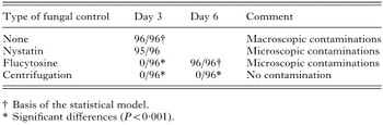
Table 2. Proportion of contaminated wells after fungi control applied on stabilate C

Table 3. Non-significant effect of fungal control on the in vitro Theileria parva development (Stabilate A) – stratified logistic regression on dose
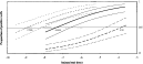
Fig. 1. In vitro titrations using non-centrifuged Theileria parva sporozoite stabilate A (——), the pellet (---) or the supernatant (–––) of 3200 g centrifuged stabilate A: average proportions of positive wells as function of the dose of stabilate (bold lines) and the 95% confidence intervals (fine lines).
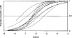
Fig. 2. Average proportions of positive wells as function of the dose of Theileria parva stabilate used for in vitro titration (stabilate A): curves of 7 individual tests (——), their average (–––) and 95% confidence interval of the average (---).
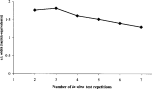
Fig. 3. Effect of the number of test repetitions on the average width of the 95% confidence interval of the ED50 (c.i. width) in Theileria parva sporozoite in vitro titrations.
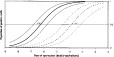
Fig. 4. In vitro comparison of stabilates A (——) and B (---): averages (bold lines) and 95% confidence intervals (fine lines).

Table 4. In vivo titration parameters of the stabilates A and B
 You have
Access
- 6
- Cited by