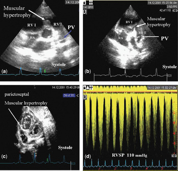
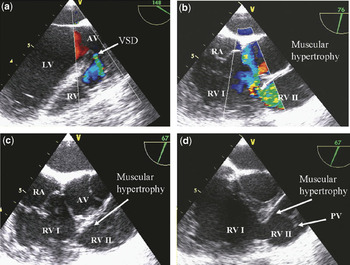
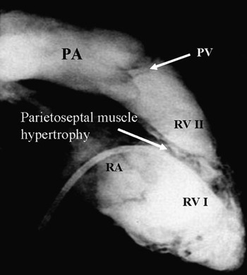
Double-chambered right ventricle is a rare congenital heart disease, characterized by division of the right ventricle by hypertrophy of the septoparietal trabeculations into two parts, the proximal chamber with higher pressure, and the distal chamber with low pressure (Figs 1–3). The term has been defined previously as a congenital anomaly characterized by a low infundibular stenosis, frequently associated with one or several “closing” ventricular septal defects.Reference Lacour-Gayet1 Peacock was the first to describe this anomaly, as a constriction of the proximal portion of the right ventricular infundibulum. There have been several subsequent definitions.Reference Forster and Humphries2–Reference Pongiglione, Freedom, Cook and Rowe5
The short axis view shows prominent hypertrophy of the right ventricular septoparietal musculature in a child aged 2 years, as seen in systole (a) and diastole (b). The subcostal view shows the division of the right ventricle into 2 chambers (c). The regurgitation jet at the tricuspid valve demonstrated the increased right ventricular pressure (d). Abbreviations: RA: right atrium; RV: right ventricle; RV I: proximal right ventricular chamber; RV II: distal right ventricular chamber; PA: pulmonary trunk; PV: pulmonary valve.

Transoesophageal echocardiography shows a small perimembranous VSD with a small left-to-right shunt (a) in 18 years old male patient with history of a small VSD since early childhood. The modified short axis (67°) shows turbulent flow in the middle part of the right ventricle toward the pulmonary valve (b). Viewing the right ventricle in short axis (67°) showed clearly the obstruction and the double chambered right ventricle during systole and diastole (c–d). Abbreviations: RA: right atrium; RV: right ventricle; PA: pulmonary trunk; PV: pulmonary valve; LV: left ventricle; AV: aortic valve; VSD: ventricular septal defect.

Angiography in lateral view shows the two chambers within the right ventricle divided by hypertrophied septoparietal musculature. Abbreviations: RVOT: right ventricular out flow tract; DCRV: double chambered right ventricle; RV: right ventricle; RVI: proximal right ventricular; RVII: distal right ventricular chamber; PA: pulmonary trunk; PV: pulmonary valve.

Amongst the various descriptions, there has been some disagreement regarding that nature of the obstructing anomalous muscular bundles. Some have described a hypertrophied moderator band, taking origin at an anomalous high point on the septal surface of the right ventricle.Reference Lucas, Varco, Lillehei, Adams, Anderson and Edwards4, Reference Pongiglione, Freedom, Cook and Rowe5 Others have described them as hypertrophied accessory septoparietal bands,Reference Gallucci, Scalia, Thiene, Mazzucco and Valfre6 or anomalous muscle bundles arising from the supraventricular crest and extending to the anterior wall of the right ventricle, close to the base of the anterior papillary muscle of the tricuspid valve.Reference Rowland, Rosenthal and Castaneda7 Alva and colleagues,Reference Alva, Ho, Lincoln, Rigby, Wright and Anderson8 however, offered a more precise definition of the site of the obstruction, which was within the apical trabecular component of the right ventricle, distinguishing this variant from the infundibular obstruction seen in entities such as tetralogy of Fallot. In another study, Hindle et al.Reference Hindle, Engle and Hagstrom9 demonstrated histopathologically the nonspecific degenerative changes within the obstructing muscular bundles, along with fibrosis of the endocardium and myocardium. In most of the cases, the obstructing musculature causes progressive obstruction of blood flow, and pressure overload in the right ventricle.Reference Hartmann, Goldring and Ferguson10 Treatment is mainly surgical, albeit that different approaches have been used. Lucas et al.Reference Lucas, Varco, Lillehei, Adams, Anderson and Edwards4 were the first successfully to achieve repair subsequent to right ventriculotomy, but others pointed out the difficulties created by this approach in removing the entire obstructions and correcting associated anomalies.Reference Rowland, Rosenthal and Castaneda7, Reference Barnes, Kwong and Cheung11, Reference Gale, Heimann and Barlow12 Other surgical approaches, such as right atriotomy and pulmonary arteriotomy, have been performed and found to be superior to right ventriculotomy. In this report, we present our experience with 37 patients having double-chambered right ventricles treated at our institution, discussing the clinical and surgical management, and postoperative results over the mid and long term.
Materials and methods
Population studied
We searched the database of the German Heart Institute, Berlin, from January 1988 through September 2005 for all patients admitted or referred for consultation and treatment of double-chambered right ventricle. We included only patients with isolated double-chambered right ventricle, excluding those with other forms of obstruction in the right ventricular outflow tract. We identified 37 patients, 22 being female, with mean age of 21.3 years, all in the first class of the system developed by the New York Heart Association. We evaluated the records obtained in terms of echocardiography, electrocardiography, cardiac catheterization, type of surgery and post-operative course. The diagnosis was made on the basis of the findings at cross-sectional and Doppler echocardiography, along with cardiac catheterisation, of hypertrophied muscle bundles within the outflow from the right ventricle and significant obstruction within the right ventricular cavity.
Clinical history
We analyzed the obstetric and past medical history, taking note of the initial clinical symptoms, the pharmacological therapy, the clinical progress of the disease in terms of symptoms, and the activities of the patients. We recorded the age at the onset of symptoms, and at operation, also noting the interval between diagnosis and surgical intervention. Further note was taken of the clinical situation and complications after operation or in the course of the subsequent treatment, the associated cardiac malformations, and any other syndromes or diagnoses.
We evaluated electrocardiographic findings during the course of the disease, before and after the operation, with regard to cardiac arrhythmia, right ventricle hypertrophy, and any disturbances of atrioventricular conduction.
Echocardiography and angiocardiography
Cross-sectional and Doppler echocardiographic studies were performed according to standard techniques, with commercially available systems. All patients underwent a complete cross sectional echocardiographic examination of the heart and great vessels at rest. M-mode, colour Doppler, and Doppler assessment of the flow of blood, along with the parasternal long and short axis, apical four-chamber, subcostal short axis and our modified subcostal short axisReference Hui, Abd El Rahman and Dsebissowa13 views were particularly useful in detection of muscular obstruction within the right ventricle, having paid careful attention to intracavity and outflow tract obstruction, along with other associated defects such as interventricular communications and other valvar lesions. We performed colour Doppler examination of flow in each plane, measuring acceleration and turbulence first by pulsed wave Doppler imaging, then to record velocities across the right ventricular outflow tract. We calculated the maximal instantaneous gradient in the right ventricle using the modified Bernoulli equation (Fig. 1d). Transoesophageal echocardiography was undertaken in some patients, and provided excellent views of the location of the obstruction in the right ventricle, and any associated ventricular septal defect (Fig. 2). Preoperative cardiac catheterization was performed to confirm the presence of obstruction in doubtful cases, to diagnose other cardiac defects, and to provide further documentation of the haemodynamics (Fig. 3).
Observations
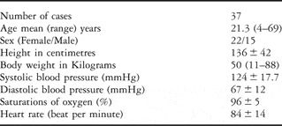
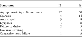
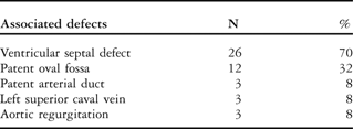
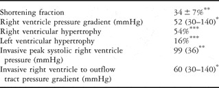
The characteristics of the patients are summarized in Table 1. In 16 (43%), the malformation was diagnosed in infancy. The initial clinical signs were short systolic murmurs of grade 2 to 3 out of 6 heard in the second left intercostals space in 34 (92%) of the cases. The murmurs were associated with thrills in 9 (24%) of the cases. About three-fifths of the patients were asymptomatic, with cyanosis noted in 5 (13%), anoxic spells in 3 (8%), and exertional dyspnoea in 3 (8%). Other symptoms and signs, such as syncope, failure to thrive, increased sweating, and severe congestive heart failure each occurred only in one of the patients (Table 2). In 26 (70%) of patients, there was an associated ventricular septal defect, while 12 (32%) had patency of the oval fossa, and 3 (8%) had mild aortic regurgitation (Table 3). Most of the patients had received no medications prior to surgery, but some had been treated with beta blockers, diuretics, and digitalis. The electrocardiographic examination showed signs of right ventricular hypertrophy in 23 (62%), right axis deviation in 6 (16%), and incomplete right bundle block in 9 (24%) of the cases. Echocardiographic examination revealed good systolic function, with right ventricular hypertrophy in 20 (54%), and left ventricular hypertrophy in 6 (16%). The pressure gradient in the right ventricle ranged from 30 to 140, with a median of 52 mmHg. All patients underwent preoperative diagnostic cardiac catheterization, which showed the right ventricular mean peak systolic pressure to be 99, with standard deviation of 36 mmHg, with a median pressure gradient of 60 mmHg, ranging from 30 to 140 mmHg (Table 4). We performed surgery in 35 of the patients (95%) without mortality. In 2 instances operations were not performed, with one patient refusing operation at that time, and the other awaiting operation at the time of our analysis. The median interval between diagnosis and operation was 9 years, with a range from 0.2 to 41 years. In 25 of 35 (71%) cases, the operation involved enlargement of the right ventricular outflow tract, with resection of fibromuscular tissue in 23 (66%), closure of an associated ventricular septal defect in 22 (63%), closure of the patent oval fossa in 12 (34%), and ligation of a patent arterial duct in 3 (9%). We approached the obstructive lesions through a right ventriculotomy in 19 (54%), right atriotomy in 11 (31%), and the transpulmonary route in 5 (14%) of the patients. The intraoperative findings showed the obstructive lesion to be produced by septoparietal trabeculations in 20 (57%) of the patients, the moderator band in 12 (34%), and the supraventricular crest in 9 (26%).
Characteristics of the patients – with values shown as mean plus or minus standard deviation.

Initial manifestations in terms of symptoms and signs in 37 patients.
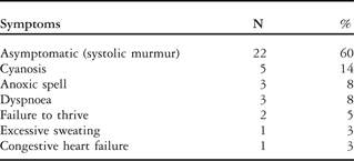
N: number of cases.
Associated cardiac anomalies in 37 patients.
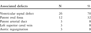
N: number of cases.
Preoperative echocardiographuc and angiographic findings.

*median, **mean, ***percent of cases.
Postoperative complications, such as small pericardial effusions, were encountered in about one-third of the patients, but supraventricular tachycardia, which warranted no special long-term treatment, was seen rarely. The mean follow up period was 7 years, with a range from 0.04 to 11 years. During this time, out of the 35 patients, 33 were asymptomatic, with no special complaints, no significant cardiac arrhythmias, and all were going about their normal activities with good exercise tolerance. Symptoms due to the primary extracardiac diseases were present in 2 patients. In 20 (57%) of the cases, insignificant short systolic murmurs of grade 1 or 2 from 6 were heard at the left second intercostal space. None of the patients have required insertion of a pacemaker for postoperative cardiac arrhythmias.
Electrocardiography revealed development of complete bundle branch block in 16 (46%) of the cases, and supraventricular tachycardia in one patient who was treated with adenosine and a beta blocker in the short term. The follow-up echocardiographic examinations in these patients showed good global systolic function, and no residual pressure gradients measured within the right ventricle.
Discussion
Double-chambered right ventricle is an uncommon congenital heart disease, in some studies accounting for up to 1.5% of the malformations recognised at catheterization.Reference Hartmann, Goldring and Ferguson10 Anderson and colleagues,Reference Alva, Ho, Lincoln, Rigby, Wright and Anderson8, Reference Anderson, Wilkinson, Becker, Rosenquist and Bergsma14, Reference Anderson, Razavi and Taylor15 analysing the right ventricle as tripartite structure, have defined the entity as a congenital cardiac malformation produced by abnormal division of the apical component of the right ventricle by accentuated septoparietal trabeculations. An interventricular communication is the cardiac anomaly most frequently associated with double-chambered right ventricle.Reference Goitein, Neches, Park, Mathews, Lenox and Zuberbuhler16–Reference Pan, Zhang, Ge, Zhao and Shen18 The diagnosis of double-chambered right ventricle may be considered in any patient with a history of deficient ventricular septation, even after surgical or spontaneous closure of the ventricular septal defect. It has been reported in up to one-tenth of patients after spontaneous closure of a pre-existing ventricular septal defect.19 We also found that one-tenth of our patients had previously exhibited ventricular septal defects that had spontaneously closed.
The initial clinical presentation in our patients varied between auscultation of a cardiac murmur, cyanosis, or congestive heart failure. This variation in presentation depends on the presence, site, and size of any associated ventricular septal defect, and the severity of obstruction within the right ventricle. In our study, it was the detection of a heart murmur during routine clinical examination that led to further investigation and subsequent diagnosis of the double-chambered right ventricle in three-fifths of the cases. The initial ventricular septal defect associated with double-chambered right ventricle is typically detected in around seven-tenths of patients studied in early life.19, Reference Vogel, Smallhorn, Freedom, Coles, Williams and Trusler20 The developmental and progressive nature of double-chambered right ventricle leads to relatively early surgical intervention to correct the obstruction and close the ventricular septal defect.
The echocardiographic diagnosis can be challenging, and is often misdiagnosed as obstructed ventricular septal defect, due to the high turbulent jets in the outflow tract of the right ventricle. Without modification of the right ventricular views, and clear imaging of the right ventricular cavity and the muscular obstructions, it is easy to under-diagnosed the lesion.Reference Hui, Abd El Rahman and Dsebissowa13 We have suggested a validated new imaging view of the right ventricle, which may provide more insight into the inflow and outflow portions of the right ventricle, and may be suitable for diagnosis of the obstructed right ventricular outflow tract in these patients.Reference Hui, Abd El Rahman and Dsebissowa13 Pharmacological stress may be indicated to evaluate the increase in pressure gradient during stress and physical exercise. Failure to diagnose the entity is well established as underscoring inadequate surgical treatment.Reference Forster and Humphries2, Reference Barnes, Kwong and Cheung11, Reference Fisher, James, Humphries, Forster and White21 Lucas et al.,Reference Lucas, Varco, Lillehei, Adams, Anderson and Edwards4 for example, reported 2 cases in which the stenotic area within the right ventricle was erroneously sutured, since the lesion had been diagnosed simply as a ventricular septal defect. Simpson et al.Reference Simpson, Sade, Crawford, Taylor and Fyfe22 reported the need for re-operation in 2 patients after surgical closure of a ventricular septal defect who still had turbulent flow postoperatively in the right ventricle.Reference Simpson, Sade, Crawford, Taylor and Fyfe22 Similarly, 2 of our patients developed echocardiographically proven double-chambered right ventricle 6 to 8 years after initial surgical closure of a ventricular septal defect.
The definitive diagnosis of the muscular obstruction is, of course, achieved intraoperatively. In our cohort, in three-fifths of the patients the obstruction involved septoparietal hypertrophy, in one third the moderator band, and in one-quarter the supraventricular crest. Others have reported septoparietal and supraventricular obstructions in three-quarters of cases.Reference Pena, Cabrera and Pastor23 Our own findings support the notion that the morphologically right ventricle is a tripartite structure with confluent inlet, trabecular, and infundibular or outlet components.Reference Anderson, Wilkinson, Becker, Rosenquist and Bergsma14 As shown by Alva and collegues,Reference Alva, Ho, Lincoln, Rigby, Wright and Anderson8 the obstruction occurs mainly due to the hypertrophy of the septoparietal and septomarginal bundles, which can extend to divide the apical trabecular component of the right ventricle. Definition of low and high sites of obstruction relative to the apical trabecular component permits differentiation between the site of obstruction as seen in tetralogy of Fallot, which is typically at the mouth of the subpulmonary infundibulum, and that seen in double-chambered right ventricle, where the obstructing lesion is located within the apical trabecular compartment.
Careful surgical resection of the obstructing muscular bundles is the treatment of choice, with very low intra- and postoperative mortality.Reference Alva, Ortegon and Herrera24, Reference Galal, Al-Halees and Solymar25 Some authorsReference Alva, Ortegon and Herrera24–Reference Tsuchikane, Kobayashi and Kirino26 have reported successful improvement of the pressure gradient in the right ventricle from 84 to 40 mmHg using transcatheter ablation of the septal hypertrophy. These methods, however, may be suitable only for selected adult patients, and may not be practicable in children. Like ourselves, others have achieved surgical correction without mortality,Reference Hachiro, Takagi, Koyanagi, Morikawa and Abe27 with another study recording death of one patient postoperatively due to acute cardiac insufficiency.Reference Pan, Zhang, Ge, Zhao and Shen18 Well-timed surgical repair in patients in good clinical conditions may explain our own zero mortality. Delay of the operation may lead to significant pressure overload and secondary myocardial injury and fibrosis, which may in turn increase the risk of intracardiac repair. In the last documented follow-up after surgery none of our patients was taking regular cardiac medications. This emphasizes the good early and long-term results and the low associated morbidity in patients having undergone corrective surgery of double-chambered right ventricle. Nevertheless, due to the right ventriculotomy in 19 (54%) of the patients, careful follow-up with regard to ventricular arrhythmia was performed by means of serial 24 electrocardiogram. We found no differences with regard to ventricular arrhythmia depending on the surgical approach.
Of our surgical patients, nearly half developed complete right bundle branch block, but there was no need for implantation of a pacemaker in any of our cases. Complete right bundle branch block was found in all independent of the surgical approach. Previous reports have shown the transatrial to be superior to the transventricular approach. It is safer, and produces excellent long-term results.Reference Ford, Bullaboy, Derkac, Hopkins, Jennings and Johnson28, Reference Penkoske, Duncan and Collins-Nakai29 Significant ventricular tachycardia was not detected in any of our patients, and supraventricular tachycardia was found in only one patient, who was successfully treated with a beta blocker. Similarly, Kveselis et al.Reference Kveselis, Rosenthal, Ferguson, Behrendt and Sloan17 reported a relatively low incidence of cardiac complication after surgical repair.
Over the mid and long-term follow-up, nearly all patients who regularly attended our outpatient department were in good cardiac condition, and had no signs of residual defects or arrhythmia. Most of our patients did not need drug treatment. They were free of symptoms, and able to perform normal activities without exercise intolerance or cardiac arrhythmia. The preoperative cardiac symptoms had disappeared. The non-invasive measurement of pressure differences in the right ventricular outflow tract with echocardiography showed no significant residual pressure gradient and normal systolic function. Only trivial haemodynamically insignificant tricuspid and mitral insufficiency was documented.
In conclusion, the rarity of double-chambered right ventricle, and its association with other cardiac anomalies, mainly ventricular septal defect, necessitates careful preoperative assessment by echocardiography, and in some cases by cardiac catheterization. Surgical resection of the hypertrophied muscle bands should carry no risk of death. Mid and long-term follow-up shows excellent surgical results. The possibility of residual obstruction within the right ventricular outflow tract mandates careful long term follow-up. Patients may need long-term follow-up of right and left ventricular function during adulthood should cardiac arrhythmias or failure develop.
Aknowledgment
We are grateful to Anne M. Gale of the Deutsches Herzzentrum Berlin for editorial assistance.