Introduction
The morphology of the taste buds of fungiform papillae can only be examined after biopsy of a single papilla. Changes in morphology with ageing and after denervation depend markedly on which papilla is removed for examination. More than 50 per cent of human fungiform papillae bear no taste buds.Reference Kullaa-Mikkonen, Koponen and Seilonen1In vivo video microscopy has been used to count fungiform papillae and their taste pores.Reference Kullaa-Mikkonen, Koponen and Seilonen1–Reference Miller and Reedy3 The taste pore is part of the taste bud and opens onto the surface of the tongue. Therefore, the number of taste pores has been used as an indication of the number of taste buds. Kullaa-Mikkonen et al. examined cases of filiform atrophy, fissured tongue and hairy tongue, and identified no taste buds in 68, 90 and 80 per cent of fungiform papillae, respectively.Reference Kullaa-Mikkonen, Koponen and Seilonen1 These authors argued that desquamation of epithelial cells onto the fungiform papillae leads to covering of the taste pores. Their findings raise the question of whether video microscopy is the best method for studying taste bud quantity with ageing, and for demonstrating the effects of denervation on fungiform papillae and their taste buds. Video microscopy fails to visualise the taste pores in about 20–30 per cent of fungiform papillae. The taste bud itself cannot be detected using video microscopy.
Confocal microscopy is an in vivo method which provides quantitative data both non-invasively and rapidly.Reference Guthoff, Baudouin and Stave4In vivo laser scanning microscopy is mainly used in the field of ophthalmology for assessment of corneal epithelium.
Confocal microscopy allows the clinician to visualise the tongue epithelium. This technology provides cellular information within the papillae, and thus is ideal for examining taste buds. Early studies demonstrated that the taste bud structure was easy to identify.Reference Just, Zeisner, Stave and Pau5, Reference Just, Stave, Pau and Guthoff6 Several features were described which characterised the peripheral taste organ (i.e. the number of cells of the taste bud at a defined depth, the density of cells of the taste bud, and the area of the taste pore at a defined depth), and these were assessed for varying ages and genders. These data were used as the basis for further in vivo studies measuring taste bud morphology and volume.
The specific aim of this study was to demonstrate the technique and clinical application of in vivo assessment of the taste bud structure in humans, using volumetry.
Materials and methods
Optical instrumentation
Confocal laser scanning microscopy used a diode laser at 670 nm for illumination (Heidelberg Engineering, Heidelberg, Germany). Digital laser scanning confocal microscopy equipment consisted of a basic HRT II device (Heidelberg Retina Tomograph HRT II) and a lens system attachment known as Rostock Cornea Module (Heidelberg Engineering, Heidelberg, Germany). This module was combined with a manual z-axis drive to move the focal plane inside the tongue. This allowed selection of the starting plane for the automated internal z-scan. During the examination, a pressure-free contact with the tongue could be monitored by means of a camera. The distance from the tongue to the microscope was kept stable using a contact element with a 0.5-mm polymethyl methacrylate surface (TomoCap®; Heidelberg Engineering). The latter and the use of contact gel (Vidisic®; Dr Mann Pharma, Berlin, Germany) avoided strong reflections. With the aid of a ×63 water immersion objective (Zeiss ×63/0.95 W, 670 nm, ∞/0; Jena, Germany) and carbomer contact gel, a resolution of 0.430 µm was achieved.Reference Guthoff, Baudouin and Stave4 In laser scanning confocal microscopy, depth resolution is determined as a function of the numerical aperture, the size of the detector diaphragm and the amount of light available. The depth resolution of the system was about 1–2 µm. The size of the field of view in the contact mode was 400 × 400 µm using the ×63 immersion objective.
Imaging
Both axial scans and z-scans of the fungiform papillae could be stored as image files and video sequences. The starting plane could be set manually before using the automatic internal z-scan. Multifaceted Amira® software (Visage Imaging, San Diego, California, USA) was used for three-dimensional reconstruction of the taste bud. A three-dimensional image consisted of 40 image planes with a depth range of about 80 µm. The acquisition time for a volume scan was 6 seconds. Each single section was recorded in 0.024 seconds.
Procedure
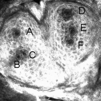
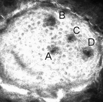
For tongue stabilisation, subjects were instructed to press their tongues through the hole in a parabolic Plexiglas™ disc.Reference Just, Pau, Bombor, Guthoff, Fietkau and Hummel7 Axial scans of the tongue surface enable mapping of the form of the fungiform papillae. In order to facilitate investigation of the fungiform papillae form and of the arrangement of surface taste pores, certain fungiform papillae on each side of the tongue were defined as reference papillae (Figures 1 and 2). Based on these reference fungiform papillae, adjacent papillae could be easily located.
Confocal laser scanning photomicrograph of a single butterfly-shaped fungiform papilla with six taste pores (labelled A to F) visible on the surface (400 × 400 µm).

Confocal laser scanning photomicrograph of a single fungiform papilla with a T-shaped pattern of four taste pores (labelled A to D) visible on the surface (400 × 400 µm).

Analysis
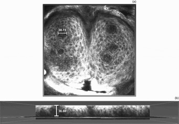
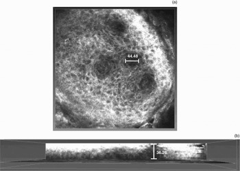
In order to demonstrate age-related changes in the morphology and volume of taste buds, and to assess changes after denervation, each taste bud of each fungiform papilla should be measured separately. Assessment of the following parameters is recommended: (1) the diameter of the fungiform papilla; (2) the diameter of the taste pore at the surface (at a depth of 2–10 µm); (3) the largest diameter across the middle of the taste bud (Figures 3a and 4a); and (4) the height of the taste bud (Figures 3b and 4b).
Volumetric calculations for taste bud A in the butterfly-shaped fungiform papilla used (a) the diameter (38.73 µm) and (b) the height (36.60 µm).

Volumetric calculations for taste bud C in the T-pattern fungiform papilla used (a) the diameter (44.48 µm) and (b) the height (36.26 µm).

We measured the volume of the taste bud as the volume of a cylinder given by V = πr2h (Figures 5 and 6).
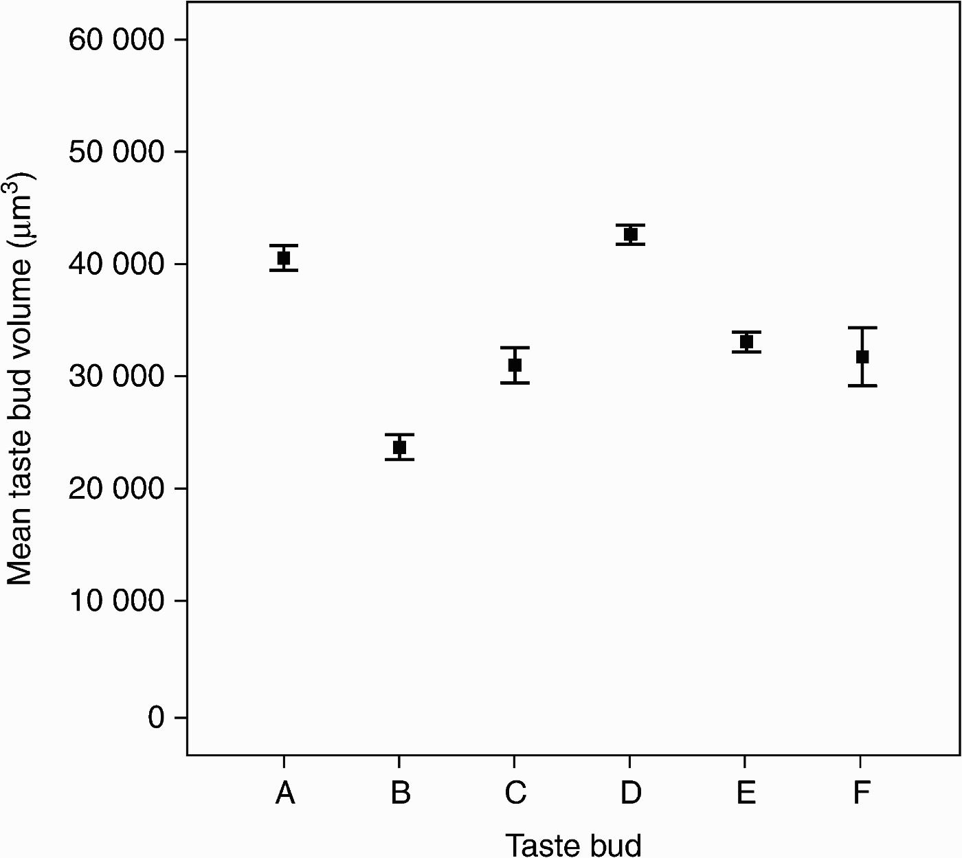
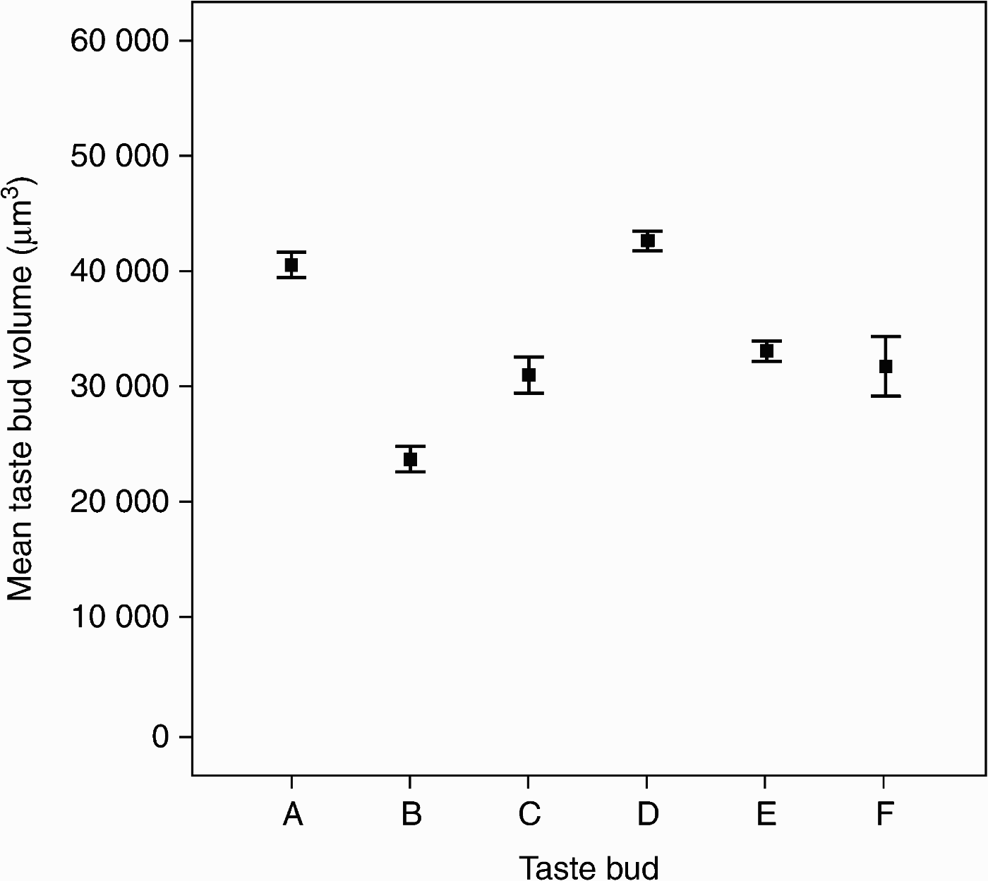
Mean volumes of the six taste buds (A to F) of the butterfly-shaped fungiform papilla, calculated from measurements taken over 9 days. Outliers indicate ±1 standard deviation of the sampling distribution of the mean.
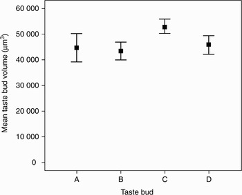
Mean volumes of the four taste buds (A to D) of the fungiform papilla with T-shaped taste pore pattern, calculated from measurements taken over 9 days. Outliers indicate ±1 standard deviation of the sampling distribution of the mean.

Measurements were performed on nine consecutive days, at least twice a day. The mean value for each day was used for the calculation.
Results
Two reference fungiform papillae of a 23-year-old man were examined (Figures 1 and 2). One, a butterfly-shaped fungiform papilla, bore six taste buds (Figure 3). The average diameters of this fungiform papilla and of its taste pores (labelled ‘A’ to ‘F’) were 463, 14.4, 13.7, 14.2, 14.8, 14.3 and 14.4 µm, respectively. Figure 5 demonstrates a small deviation in taste bud volume over nine days, for each taste bud of the butterfly-shaped fungiform papilla.
Figure 4 shows a single fungiform papilla with a T-shaped arrangement of four taste buds, in the same 23-year-old man (see also Figure 2). The average diameters of this fungiform papilla and of its taste pores (labelled ‘A’ to ‘D’) were 434, 14.4, 16.4, 14.2 and 13.7 µm, respectively. The error bar chart shown as Figure 6 demonstrates a slightly higher standard deviation for all four taste buds, compared with taste buds A to F of the butterfly-shaped fungiform papilla.
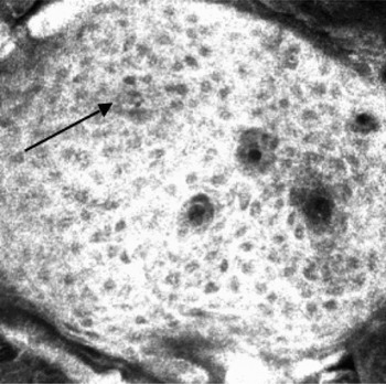
The same fungiform papilla (with T-shaped taste bud pattern) was re-investigated three weeks later. Figure 7 reveals an increase in the diameter of taste bud B and the appearance of a new taste bud. One week after this, taste bud B had disappeared, while all the other taste buds' diameters had deviated only slightly (Figure 8).
Confocal laser scanning photomicrograph of the T-pattern fungiform papilla, taken three weeks after Figure 2. The diameter of taste pore B has increased (arrow) and a new taste pore has appeared (the dark spot between taste pores C and D) (400 × 400 µm).


Discussion
The aim of this study was to use in vivo confocal microscopy and the basic principles of volumetry to quantify taste bud volumes within fungiform papillae. In vivo measurement of fungiform papillae seems to be appropriate to indicate potential changes in taste bud volume. Our measurements were based on calculation of the volume of a cylinder rather than an elliptical cone. Volumetric measurements using this technique gave data for a healthy human, with acceptable deviation of values over nine days. Therefore, we accept volumetric evaluation of taste buds based on a cylinder defined by the radius and height of the taste bud.
The following clinical applications are conceivable.
Firstly, this assessment method may be useful for the re-evaluation of human taste bud volume with ageing. Previous research had indicated that continuous cell differentiation and cell death maintain the shape of taste buds.Reference Mistretta8 However, recent studies have revealed that taste bud cells die due to apoptosis.Reference Zeng and Oakley9 This may lead to persistent changes in taste bud volume. In vivo confocal microscopy may be a useful tool with which to detect changes of taste bud volume.
Secondly, the above volumetric method may be used to study the effects of chorda tympani nerve or lingual nerve transection on the volume of the taste bud. In animal models of chorda tympani nerve transection, taste buds were seen to be smaller and fewer in number on the ipsilateral tongue side, five days after transection.Reference Guagliardo and Hill10 Degenerated taste buds showed a decrease in volume due to a loss of taste bud cells rather than changes in taste bud morphology. In order to reveal similar results in humans, a single fungiform papilla needs to be resected. This raises the question of which fungiform papilla to biopsy. Denervation-related atrophy of a single fungiform papilla cannot be differentiated from selective taste bud atrophy.
• This paper demonstrates the application of in vivo confocal laser scanning microscopy for visualisation of taste bud structure and calculation of taste bud volume
• Possible clinical applications of this technique include study of the effects of lingual and chorda tympani nerve injury, and of the effect of ageing
• Results are presented for a single individual; further study is needed to determine the clinical usefulness of this technique
Thirdly, the above technique could be used to demonstrate recovery of taste buds in patients with severed chorda tympani nerve or severed lingual nerve who show partial or complete recovery of gustatory sensibility after surgery. Proving the regeneration of taste buds is a very challenging task. One method of achieving this would be to monitor and measure the volumes of all taste buds from several reference fungiform papillae pre-operatively, for a period ranging from several days to several weeks. Natural volume changes should be compared with those occurring after bilateral chorda tympani nerve transection. A consistent and significant decrease in the taste bud volume after nerve transaction, combined with an increase following surgical repair, would furnish clinical evidence of taste bud regeneration. At present, there is no such evidence of fungiform taste bud regeneration after chorda tympani nerve transection in humans. Volumetry of fungiform taste buds in patients with recovered taste sensibility would supply objective data on taste bud regeneration.
Conclusions
This paper demonstrates the application of in vivo confocal laser scanning microscopy for visualisation of taste bud structure and calculation of taste bud volume. As this technique is non-invasive and simple to use, confocal microscopy may in future provide a means to visualise taste buds and to measure their volume. This paper presents data from only one healthy, 23-year-old man. Further investigations of healthy subjects are required in order to evaluate the feasibility of using confocal laser scanning microscopy to evaluate the degeneration and regeneration of taste buds.
Acknowledgement
We thank Joachim Stave for his invaluable help with the laser scanning microscopy and for critical reading of the manuscript.