Introduction
Ovarian tissue cryopreservation and transplantation emerges as a promising option for preserving future endocrine function and fertility in various animals (Gosden et al., Reference Gosden, Baird, Wade and Webb1994a; Almodin et al., Reference Almodin, Minguetti-Câmara, Meister, Ferreira, Franco, Cavalcante, Radaelli, Bahls, Moron and Murta2004) and in human beings (Kiran et al., Reference Kiran, Kiran, Coban, Guven and Yuksel2004; Kim, Reference Kim2006), especially in young women or girls who are about to start chemo- and radiotherapy. In 2004, the first live birth after orthotropic transplantation of human cryopreserved ovarian tissue in a woman previously exposed to conventional dose of chemotherapy treatment was reported (Donnez et al., Reference Donnez, Dolmans, Demylle, Jadoul, Pirard, Squifflet, Martinez-Madrid and Van Langendonckt2004).
Most of the present researches have focused on how to produce fully grown and developmentally competent oocytes from cryopreserved tissues after autotransplantation in animals and human. The main problems include which site is most effective and practical for autotransplantation and which fertilization technique is favorable for the fertilization and further developmental potency of oocytes retrieved from grafts. Our previous study had shown that mesometrium was an available transplantation site for follicle survival and development in rabbit (Deng et al., Reference Deng, Xu, Chao, Yu, Zhen, Hashimoto and Morimoto2007). However, further investigation is needed to determine whether the site is suitable for the developmental potency of oocytes. Liu et al. (Reference Liu, Rybouchkin, Van der Elat and Dhont2002) had demonstrated that fertilization of mouse oocytes from in vitro-matured preantral follicle by intracytoplasmic sperm injection (ICSI) improved the percentage of 2-cell formation compared with in vitro fertilization (IVF). However, it remains unknown whether fertilization by ICSI may improve the developmental competence of oocyte retrieved from grafts. It had been reported that the blastocyst formation of in vitro-matured oocytes was lower (Aubard et al., Reference Aubard, Piver, Cognie, Fermeaux, Poulin and Driancourt1999). It still unknown whether decreased blastocyst formation is reflected in the pregnancy rate and birth rate.
The aim of this study was to evaluate mesometrial transplantation of frozen-thawed ovarian tissue in rabbit and compare different fertilization techniques (ICSI and IVF) by investigating the capability of oocyte fertilization, blastocyte formation and pup birth.
Material and methods
Animals and experiment protocols
All experiment protocols and animal use were approved by the Animal Research Ethical Committee, Qilu Hospital of Shandong University. All experiments were carried out on New Zealand white rabbits, aged between 12 and 24 months, weighing 2.0 to 2.5 kg and purchased from Shandong Agriculture Science Academy. Forty female rabbits with normal estrous cycles according to vaginal smear were included in this test. The rabbits were randomly divided into three groups. Group 1 (n = 10) was control, both ovaries were present in these rabbits and included total volume of ovarian tissue. Rabbits in Group 2 (n = 15) were autologous transplanted with small pieces of fresh ovarian tissues into mesometrium as soon as the pieces of ovarian tissues were prepared after bilateral oophorectomy. In Group 3 (n = 15), ovarian tissues were cryopreserved for 7 days after bilateral oophorectomy, and then similarly volume with Group 2 of frozen-thawed ovarian tissues were autologous transplanted into mesometrium. Sperm taken from a 14-month-old male rabbit was used for IVF or ICSI.
Obtaining rabbit ovaries
Each rabbit was anesthetized by 1 ml/kg pentobarbital sodium (intravenous (i.v.)). With its abdominal fur sheared and its skin sterilized with iodine, medical laparotomy was then performed to isolate and remove the ovary. The ovary was washed several times at room temperature in Dulbecco’s phosphate-buffered saline (DPBS, Sigma, St Louis, MO, USA) solution to remove excess blood before being cut into small pieces (1 mm3 each). Two pieces of ovarian tissue from each ovary were randomly selected for histological analysis.
Freezing and thawing
The ovarian tissues were frozen and thawed according to the protocol of Almodin et al. (Reference Almodin, Minguetti-Câmara, Meister, Ferreira, Franco, Cavalcante, Radaelli, Bahls, Moron and Murta2004) with some modification. The pieces of ovarian tissue were equilibrated with DPBS medium containing 1.5 M dimethyl sulphoxide (DMSO, Sigma, D5879, USA), 5.6 mM D-glucose (Gibco, Langley, OK, USA), 0.3 mM pyruvate (Gibco, USA) and 10% (v/v) fetal bovine serum (FBS, Sigma, USA) and gently shaken for 30 min at 4°C. Then, each piece was loaded into a 0.5-ml plastic freezing straw (Pacific vet, No 00-061). The straws were placed in a programmable cryopreservation system (Freeze Control, Cryologic, Mulgrave, Victoria, Australia) that had been pre-cooled to 0°C. The system then started cooling down at the speed of 2°C/min till −9°C, followed by manual seeding at −9°C, being held for a further 5 min, cooling down at 0.3°C/min till −40°C, then 10°C/min till −120°C before finally being plunged into liquid nitrogen (LN2) −196°C for storage.
Seven days after cryopreservation, the straws were taken out from LN2, held in air for 20 s, transferred into a water bath set at 37°C and stood for 10 to 20 s. The tissues were washed thoroughly with progressively lower concentrations of cryoprotectant media, which contained 1.0 M, 0.5 M and 0 M DMSO, respectively, for 5 min each time, and then kept in DPBS with 10% FBS until transplantation. Two pieces of freezing-thawing ovarian tissues of each ovary were randomly chosen for histological analysis.
Transplantation
After the rabbits in Group 2 and Group 3 had been anesthetized as described above, the fresh ovarian tissues of Group 2 and the frozen-thawed ovarian tissues of Group 3 were transplanted into the mesometrium and the transplantation sites were sutured with silk thread for subsequent detection. Twelve pieces of ovarian tissues were transplanted in each rabbit.
Ovary stimulation and oocytes retrieval
Three months after ovarian transplantation, all rabbits were stimulated to superovulate by FSH (purified follicle stimulating hormone, Livzon Group, China) (15 IU/dose, twice/day, 3 days), and subsequent 100 IU HCG (human chorionic gonadotropins, Livzon Group, Zhuhai, Guangdong Province, China) given 12 h later. Approximately 13 h after HCG administration, the ovarian tissues were aseptically removed. The grafts were kept in tissue culture medium-199 (TCM-199, Sigma) with 10% FBS before the oocyte–cumulus complexes (OCC) were mechanically isolated from antral follicles greater than 1.0 mm in diameter using 25G needles under a dissecting microscope. The oocytes that had expanded cumulus and a polar body were classified as mature, and those that had compact cumulus with or without germinal vesicle (GV) were immature. Immature oocytes were cultured individually in 50 μl IVG medium droplets covered with mineral oil (Sigma) in 35 × 10 mm tissue culture dishes (Falcon, Belgium) for 16 h at 37°C with 5% CO2. The culture system used was modified from the method of Yoshimura et al. (Reference Yoshimura, Ando, Nagamatsu, Iwashita, Adachi, Sueoka, Miyazaki, Kuji and Tanaka1996) and Lorenzo et al. (Reference Lorenzo, Rebollar, Illera, Illera, Illera and Alvarino1996). The IVG medium was TCM199 supplemented with 2 mIU/ml follicle stimulating hormone (FSH), 1 mIU/ml luteinizing hormone (LH), 100 mIU/ml prolactin (PRL, Sigma), 1 μg/ml estradiol (E2, Sigma), 0.1 μg/ml recombinant human insulin-like growth factor I (rhIGF-1, Jingmei Biotech Co., Ltd, Baihuan DaDao Avenue, Shenzhen, China), 0.05 μg/ml recombinant human epidermal growth factor (rhEGF, Jingmei Biotech Co., Ltd), 10 000 mIU/ml penicillin (Sigma), 10 μg/ml streptomycin (Sigma) and 10% FBS.
In vitro fertilization
In vivo-matured oocytes in each group were randomly inseminated by IVF or ICSI and inseminated at 1 h post-collection. Immature oocytes were inseminated after IVM, and also randomly assigned to IVF or ICSI.
Sperm preparation
The caudae epididymis was removed from mature male rabbit, and the contents were carefully squeezed out. Semen were treated in a manner similar to those reported previously for in vitro capacitation. Capacitation medium containing mDM (isotonic defined medium, DM + 10 mM NaHCO3) and mHIS (high-ionic-strength Brackett’s defined medium, HIS + 10 mM NaHCO3) (Zeng et al., Reference Zeng, Zhu, Wang, Chen, Zhang and Chen1999). Spermatozoa were introduced in the fertilization drops at a final concentration of 1 × 10⁶. For ICSI, supernatants were mixed with 10% polyvinyl pyrrolidone (PVP; Sigma) to slow down the sperm’s vigorous movement before ICSI (Deng and Yang, Reference Deng and Yang2001).
IVF
Oocytes were incubated with sperm for 5 h, then were moved to embryo culture medium containing TCM199 medium suspended with 1.25 mM pyruvate, 0.1 mM EDTA, 10% FBS and cultured at 37°C, 5% CO2.
ICSI
The micromanipulation was performed in fertilization medium on a warm microscope stage at 37°C. Cumulus cells of mature oocytes were removed by hyaluronidase treatment (200 IU/ml, Sigma) and gentle pipetting. The injection needle used for rabbit sperm was of 5.0 μm inner diameter. After injection, the oocytes were transferred to embryo culture medium.
Embryo culture and transfer
Half of the medium was changed every other day. Pronuclear formation was examined with an inverted microscope 9 h after IVF or ICSI. The oocytes with two distinct pronuclei and a second polar body were considered as fertilization. Fertilized oocytes were cultured in the same medium until the blastocyst stage at day 5 to 6 after fertilization. Pseudopregnancy was obtained by witnessed copulation with vasectomied male rabbits. Hyperstimulation was accomplished with the same FSH and HCG as described above. To perform the transfers, rabbits were anesthetized by 1 ml/kg pentobarbital sodium (i.v.). Two blastocyst embryos were transferred, through the infundibulum, into each oviduct of the pseudopregnant rabbits (Watanabe et al., Reference Watanabe, Nakagawa, Kitajima, Liu, Morimoto, Watanabe and Fan2005).
Statistical analysis
Percentages were compared between groups by means of the χ 2-test. One-way ANOVA was used for analysis of the number of follicles per high-power field (HPF) of ovarian tissue. A value of P < 0.05 was considered significant.
Results
Histology of the ovarian grafts
New blood vessels were seen on the surface of surviving ovarian tissues. Uterus congested obviously after superovulation. The frozen-thawed and transplanted ovarian tissues had no obvious structural differences compared with fresh tissues, with a well-preserved structure of germinative epithelium, albuginea and cortical tissue granule-albican bodies. Follicles at different growth stages could be seen in the cortex, and most of them showed normal morphology. Large antral follicle, which protrudes to the surface of the ovarian tissues, could be seen. The number of follicles per HPF of ovarian tissue was markedly reduced in transplanted ovarian tissues (27.4 ± 12.6, 12.3 ± 8.7 and 11.8 ± 7.5 in Groups 1, 2 and 3, respectively; P < 0.05).
Growth and maturation of oocytes
Isolation from antral follicles by mechanical dissection of ovarian tissues yielded 415, 327 and 304 OCCs in Groups 1, 2 and 3, respectively. There were no significant differences in the percentages of mature oocytes and immature oocytes among the three groups (Table 1). After being cultured for 16 h, some immature oocytes underwent GV breakdown, extruded the second polar body and the cumulus became expanded. No significant difference was observed in the maturation rate after IVM among the three groups (73.1%, 67.2% and 69.8% in Groups 1, 2 and 3, respectively) (Table 1).
Proportion of retrieved oocytes matured in vivo and in vitro in three groups (%)

Fertilization and blastocyst formation after IVF or ICSI
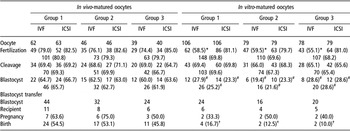
No significant differences were found in the percentage of oocytes undergoing fertilization, cleavage or blastocyst formation among the three groups.
With regard to in vivo-matured oocytes, the fertilization rate, cleavage rate and blastocyst formation rate did not differ significantly among the three groups, irrespective of whether they were fertilized by IVF or ICSI.
With regard to in vitro-matured oocytes, significantly higher fertilization rates were observed when ICSI was used compared with IVF in each groups (Group 1: 81.1% v. 58.5%, Group 2: 79.7% v. 59.5%, Group 3: 81.0% v. 55.1%, respectively, P < 0.05). No significant differences were observed in the cleavage rate and blastocyst formation rate among the three groups, regardless of whether IVF or ICSI was performed.
The fertilization rate of in vitro-matured oocytes after ICSI was similar to that of in vivo-matured oocytes. However, a significantly lower fertilization rate was observed using IVF for the in vitro-matured oocytes in each group compared to the in vivo-matured oocytes (Group 1: 58.5% v. 79.0%, Group 2: 59.5% v. 76.1%, Group 3: 55.1% v. 74.4%) (P < 0.05). The blastocyst formation rate of in vitro-matured oocytes was significantly lower compared with that of in vivo-matured oocytes (Group 1: 25.2% v. 65.7%, Group 2: 21.6% v. 62.7%, Group 3: 28.6% v. 61.9%) (P < 0.05).
Embryo transfer
A total of 160 embryos at blastocyst stage were transferred into the oviducts of 40 pseudopregnancy recipients, of which 22 carried the pregnancy to term (Table 2). There were no significant differences in the pregnancy rate and birth rate of in vivo-matured oocytes or in vitro-matured oocytes among the three groups. However, a significantly lower birth rate was observed for the in vitro-matured oocytes compared to that of in vivo-matured oocytes (Group 1: 16.7% v. 54.5%, Group 2: 12.5% v. 53.1%, Group 3: 10.0% v. 45.8%, P < 0.05).
Fertilization and development competence between oocytes matured in vivo and in vitro among three groups (%)

IVF = in vitro fertilization; ICSI = intracytoplasmic sperm injection.
Pregnancy rate = pregnant recipients/transferred recipients; birth rate = pups live born/transferred embryos.
*Compared to ICSI in each group, difference significant, P < 0.05.
#Compared to in vivo-matured oocytes, difference significant, P < 0.05.
†Compared to in vivo-matured oocytes, difference significant, P < 0.05.
Discussion
We chose New Zealand White rabbits as experimental models because rabbit oocyte has similarity to human oocyte, considering morphology, hardness and tenacity of zona pellucida (Al-Hasani et al., Reference Al-Hasani, Trotnow, Sadtler and Hahn1986).
The highly vascularized renal capsule was the most common transplant site (Waterhouse et al., Reference Waterhouse, Cox, Snow, Jenkin and Shaw2004). There has been report on live offspring by IVF after transplantation of cryopreserved mouse ovarian tissue into the renal capsule (Liu et al., Reference Liu, Van der Elst, Van den Broecke and Dhont2001). However, the renal capsule may not accommodate the full development of follicles of bigger species such as human and sheep. Gosden et al. (Reference Gosden, Boulton, Grant and Webb1994b) found that larger follicles were never found in ovine xenografts and no human antral follicle had been reported to grow larger than 5 mm in the renal capsule of SCID (Oktay et al., Reference Oktay, Newton, Mullan and Gosden1998; Abir et al., Reference Abir, Orvieto, Raanani, Fisch, Schoenfeld, Ginton, Nitke and Ben Rafael2000). In the present study, ovarian tissues were transplanted into the mesometrium owing to the spongier texture, abundant vessels and large surface area at this site. No significant morphological change of ovarian grafts was found in the present study and the follicles developed well after grafting, which was consistent with our previous study (Deng et al., Reference Deng, Xu, Chao, Yu, Zhen, Hashimoto and Morimoto2007).
In this study, the number of rabbit follicles per high-power field was found markedly reduced after grafting. Several possible factors could account for the delete period: (i) mechanical injury: when the ovaries were cut into small pieces before freezing, which caused the loss of large follicles and (ii) ischemic injury: which result from low revascularization. It has been demonstrated that revascularization of the grafts seemed to be a crucial factor for the survival of follicles because much more follicles were lost during grafting than during freezing and thawing (Nisolle et al., Reference Nisolle, Casanas-Roux, Qu, Motta and Donnez2000; Baird et al., Reference Baird, Webb, Campbell, Harkness and Gosden1999). Theoretically, ischemic injury after transplantation can be minimized by alleviation of hypoxic tissue damage by the use of antioxidants or anti-apoptotic agents and facilitation of angiogenesis by manipulating the expression of angiogenic factors (Jemal et al., Reference Jemal, Clegg, Ward, Ries, Wu, Jamison, Wingo, Howe, Anderson and Edwards2004; Greenlee et al., Reference Greenlee, Hill-Harmon, Murray and Thun2001). Nugent et al. (Reference Nugent, Newton, Gallivan and Gosden1998) found that antioxidant treatment using vitamin E improved the survival of follicles in ovarian grafts by reducing ischemic injury.
In the present study, we used a typical dose of gonadotropin to induce superovulation and obtained a similar percentage of mature oocytes in the two transplantation groups. Previous studies found that exogenous PMSG was administered to stimulate mouse ovarian tissue grafted under the kidney capsule failed to increase the yield of oocytes from ovarian grafts (Lee et al., Reference Lee, Ho, Yu and Lu2005). The reason for this is not known, but it may be possible that the vascular remodeling that occurs in the ovarian graft does not allow adequate delivery of the gonadotropin to the grafts. It has been reported that gonadotropin treatment of graft recipients improved the meiotic competence of the oocytes and the embryo development of fertilized oocytes (Kaneko et al., Reference Kaneko, Kikuchi, Noguchi, Ozawa, Ohnuma, Maedomari and Kashiwazaki2006; Yang et al., Reference Yang, Cox, Jenkin, Findlay, Trounson and Shaw2006). Angiogenesis is also stimulated directly or indirectly by gonadotropic hormones (Zygmunt et al., Reference Zygmunt, Herr, Keller-Schoenwetter, Kunzi-Rapp, Munstedt, Rao, Lang and Preissner2002). Although exogenous heat treatments have positive effects on the developmental ability of primordial oocytes xenografted to host recipients, the optimal dose, initiation and duration of gonadotropin stimulation needed further investigation.
Aubard et al. (Reference Aubard, Piver, Cognie, Fermeaux, Poulin and Driancourt1999) reported that sheep oocytes could be obtained from heterotopic and orthotopic grafts, matured by IVM and some of them fertilized by IVF, but none developed to the blastocyst stage. Lee et al. (Reference Lee, Ho, Yu and Lu2005) performed subcutaneous transplantation of frozen ovarian tissue in mouse. Among 14 MII oocytes recovered, six were fertilized by IVF and two developed to the blastocyst stage. Oktay et al. (Reference Oktay, Buyuk, Veeck, Zaninovic, Xu, Takeuchi, Opsahl and Rosenwaks2004) obtained four normal human 2- to 3-day-old embryos following IVM and ICSI after heterotopic ovarian transplant and no pregnancy ensued. Only three live births from autotransplanted ovarian tissues have been reported in humans (Donnez et al., Reference Donnez, Dolmans, Demylle, Jadoul, Pirard, Squifflet, Martinez-Madrid and Van Langendonckt2004; Meirow et al., Reference Meirow, Levron, Eldar-Geva, Hardan, Fridman, Yemini and Dor2007). This unsatisfactory result might be related to the techniques used for fertilization and the quality of oocytes.
It has been reported that ICSI could be applied to increase the fertilization rates of in vitro-matured oocytes (Dell’Aquila et al., Reference Dell’Aquila, Cho, Minoia, Traina, Fusco, Lacalandra and Maritato1997). However, there is no agreement on which fertilization technique should be applied to oocytes recovered from grafts. In the current study, we compared the fertilization rate and developmental potency of oocytes fertilized by IVF or ICSI. Our results showed that the fertilization rate of in vivo-matured oocytes did not show any difference among the three groups fertilized by either IVF or ICSI. Moreover, significantly higher fertilization rates were observed in oocytes matured in vitro when submitted to the ICSI procedure as compared with IVF. These results also indicated that ICSI could be applied successfully to in vitro-matured rabbit oocytes to increase fertilization rate.
In the present study, the blastocyst formation rate of in vitro-matured oocytes was significantly lower than in vivo-matured oocytes. O’Brien et al. (Reference O’Brien, Pendola and Eppig2003) reported that embryos derived from prepubertal lamb have the same capacity to develop to term as embryos derived from adult sheep once the embryo reaches the blastocyst stage. However, Revel et al. (Reference Revel, Mermillod, Peynot, Renard and Heyman1995) found that transfer of blastocysts obtained from in vitro-matured oocytes resulted in a lower pregnancy rate. In the present study, no significant difference was found in the pregnancy rate between in vivo and in vitro oocytes. However, the birth rate of in vivo-matured oocytes was significantly higher than that in vitro-matured oocytes. Long-term in vitro culture of immature oocytes might cause spontaneous zona pellucida hardening and a poorer capacity for fertilization and further development (Baird et al., Reference Baird, Webb, Campbell, Harkness and Gosden1999; Aubard et al., Reference Aubard, Piver, Cognie, Fermeaux, Poulin and Driancourt1999; Fukuda et al., Reference Fukuda, Roudebush and Thatcher1992). Abnormal cytokinesis or genetic abnormalities may be the most important factor. Nogueira et al. (Reference Nogueira, Staessen, Van de Velde and Van Steirteghem2000) reported a high incidence of multinuclear blastomeres and aneuploidy of embryos from in vitro-matured human oocytes. Lechniak et al. (Reference Lechniak, Warzych, Pers-Kamczyc, Sosnowski, Antosik and Rubes2007) also found that the rate of aneuploidy among in vitro-matured pig oocytes was higher than that of in vivo-matured oocytes. Recently, observations suggested that both gonadotropins and growth factors were important for IVM (Carneiro et al., Reference Carneiro, Lorenzo, Pimentel, Pegoraro, Bertolini, Ball, Anderson and Liu2001; Mao et al., Reference Mao, Smith, Rucker, Wu, McCauley, Cantley, Prather, Didion and Day2004). Further research focusing on the culture system should increase the capacity of fertilization and embryonic development of in vitro-matured oocytes. Despite the quality of in vitro-matured oocytes being poorer, systems for in vitro oocyte development have provided an opportunity to maximize the yield of oocytes having full developmental competence from grafts, which, in turn, should increase the number of embryos and raise the chances of success.
Conclusion
In conclusion, mesometrial transplantation of frozen-thawed ovarian tissue provides favorable conditions for follicle development. Oocytes retrieved from mesometrial grafts can develop to the blastocyst stage and produce live offspring; ICSI may optimize their fertilization rate. Thus, cryopreservation and mesometrial transplantation of ovarian tissue shown in the current study and subsequent assisted reproductive techniques provide possible means for fertility preservation in other mammalian species.