Introduction
Mammalian spermatozoa represent a growing list of cell types that exhibits a capacity to generate highly reactive oxygen species (ROS), such as hydrogen peroxide (H2O2), superoxide anion (•O2−), hydroxyl radical (OH•) and the hypochlorite (•OHCl) when incubated under aerobic conditions (Sharma and Agarwal, Reference Sharma and Agarwal1996). ROS are free radicals and peroxides that are derived from the metabolism of oxygen and are continuously produced by various metabolic and physiological processes (Agarwal and Prabakaran, Reference Agarwal and Prabakaran2005).
Lipids are the macromolecules which are most susceptible to ROS attack. They are present in the sperm plasma membranes, in the form of polyunsaturated fatty acids (PUFAs; fatty acids containing more than two carbon–carbon double bonds) (Agarwal and Prabakaran, Reference Agarwal and Prabakaran2005). ROS attack the PUFAs in the cell membranes leading to a chain of chemical reactions called lipid peroxidation (LPO) (Agarwal and Prabakaran, Reference Agarwal and Prabakaran2005). The combination of Fe2+ and ascorbate has been used to induce oxidative stress/LPO via hydroxyl radical (OH•) formation and by increasing the levels of thiobarbituric acid-reactive substances (TBARS) such as malondialdehyde (MDA) that originate from the breakdown of lipid peroxides (Aitken et al., Reference Aitken, Harkiss and Buckingham1993). In sperm cells, spontaneous LPO occurs, but at a lower level than that induced by ferrous sulphate and ascorbate (Storey, Reference Storey1997).

Lipid peroxides formed during this process are generally associated with decreased sperm functions and viability, but have significant enhancing effects on the ability of spermatozoa to bind both homologous and heterologous zonae pellucidae (De Lamirande et al., Reference De Lamirande, Jiang, Zini, Kodama and Gagnon1997). In the mouse, a low concentration of ferrous ascorbate (FeAA) causes an enhancement in LPO that is associated with a 50% increase in the fertilising ability of spermatozoa coupled with improvement in sperm-binding properties to homologous zona pellucida (Aitken et al., Reference Aitken, Clarkson and Fishel1989; Kodama et al., Reference Kodama, Kribayashi and Gagnon1996; De Lamirande et al., Reference De Lamirande, Jiang, Zini, Kodama and Gagnon1997). This increased formation of two-cell embryo is not concerned with altered sperm parameters such as motility or hyperactivity, but appears to be related to a marked increase in binding of spermatozoa to mouse zona pellucida (Kodama et al., Reference Kodama, Kribayashi and Gagnon1996; De Lamirande et al., Reference De Lamirande, Jiang, Zini, Kodama and Gagnon1997). It was hypothesised that the increased binding which results from changes in membrane fluidity and rearrangement of membrane components is caused by LPO (induced by FeAA) (Kodama et al., Reference Kodama, Kribayashi and Gagnon1996). It has been suggested that mild and low oxidative stress for extensive periods could alter the condensation state of sperm DNA, which may enhance the fertilising potential (De Lamirande et al., Reference De Lamirande, Jiang, Zini, Kodama and Gagnon1997). Spermatozoa, after ejaculation, use a strategy involving oxidation and breakdown of membrane lipids to improve their chance to fertilise oocytes (Kodama et al., Reference Kodama, Kribayashi and Gagnon1996). Data obtained provide evidence that Fe2+ and ascorbate at proper concentrations increase sperm-binding capacity to homologous zona pellucida and fertilising potential (Aitken et al., Reference Aitken, Clarkson and Fishel1989; Kodama et al., Reference Kodama, Kribayashi and Gagnon1996).
Thus, the present in vitro studies were aimed at finding the optimum/best dose of FeAA (FeSO4 + ascorbic acid), which would induce oxidative stress/LPO in bull sperm and simultaneously maintain its motility and viability (required for sperm capacitation and acrosome reaction). In assisted reproduction, poor sperm motility rather than low sperm count in the semen is most often the cause of male infertility. It is suggested that maintained motility, viability and mild oxidative stress may enhance the fertilising potential of crossbred cattle bull spermatozoa.
Material and methods
Sperm samples
Semen samples (n = 5) with more than 80% motility and 1200 × 106 to 1400 × 106 per ml sperm count were obtained from healthy local crossbred cattle bulls (HHS, Holstein Friesen × Sahiwal: FC, Friesian crosses: 1F and 4F first and fourth generation of inter-breeding) maintained at the Dairy Farm, Punjab Agricultural University, Ludhiana. Three ejaculates of each bull were analysed and in all five bulls were studied. Each parameter was repeated thrice using five animals. A known volume of semen sample was taken in a centrifuge tube (pre-warmed at 37°C), which was centrifuged (800 × g, 5 min). Seminal plasma was removed, the sperm pellet was washed two or three times with 2.9% sodium citrate (pH 7.4), followed by suspension of pellet in this solution for the preparation of a known volume. This suspension was divided into four equal parts (in four test tubes). In one tube, which served as a control, 2.9% sodium citrate was added, whereas the other three tubes were supplemented with three different concentrations of FeAA (100 : 500 μmol/l (I), 150 : 750 μmol/l (II), 200 : 1000 μmol/l (III); FeSO4 : ascorbic acid) and served as experimental fractions.
Evaluation of sperm motility
The numbers of motile and immotile spermatozoa from control as well as experimental fractions were counted at 37°C, after an interval of 30 min each up to 2 h, by direct light microscopy.
Evaluation of sperm viability
The viability of bull spermatozoa (control as well as experimental fractions) was observed up to 2 h of incubation by using eosin-nigrosin stain prepared by mixing 5% eosin-Y (made in 2.9% sodium citrate) and 10% nigrosin (made in 2.9% sodium citrate) in the ratio of 1 : 4 (Blom, Reference Blom1977). The viability was assessed by taking one drop of FeAA-treated/untreated samples and mixed with four drops of stain, maintained at 37°C. After 2 min, a thin permanent smear was prepared. About 100 spermatozoa were counted in different areas of the smear at 100× magnification. Three slides per ejaculate and per treatment were analysed. The % viability was calculated as the number of viable (pink) sperm/total number of (pink + transparent) sperm multiplied by 100.
Determination of lipid peroxidation
Samples were incubated to induce LPO at 37°C in the presence of different concentrations of FeAA for 2 h. The levels of LPO induced in bull spermatozoa were assessed by the determination of TBARS (Buege and Aust, Reference Buege and Aust1978). For this assay, a known volume of sperm suspension was incubated with 0.1 ml of 150 mmol/l Tris–HCl (pH 7.1) for 20 min at 37°C. Subsequently, 1 ml of 10% trichloroacetic acid (TCA) and 2 ml of 0.375% thiobarbituric acid (TBA) were added followed by keeping it in a boiling water bath for 30 min. Thereafter, it was centrifuged for 15 min at 3000 r.p.m. In the blank tube, the sample was replaced by 2.9% sodium citrate (pH 7.4). The absorbance was read at 532 nm. The molar extinction coefficient for MDA is 1.56 × 10⁵ mol/l per cm. The results were expressed as n moles MDA per μg protein.
Determination of total proteins
Total proteins in the sperm samples were determined spectrophotometrically (Lees and Paxman, Reference Lees and Paxman1972); 20 to 100 μg/ml bovine serum albumen standard was also run simultaneously.
Statistical analysis
‘Analysis of factorial experiment in CRD’ (software programme made by Department of Mathematics, Statistics and Physics, College of Basic Sciences and Humanities, Punjab Agricultural University, Ludhiana, India) or ‘one-way analysis of variance’ was used to evaluate the significance levels between the parameters studied. The critical difference of three factors A (incubation period), B (control and FeAA treatments) and AB (interaction between incubation periods and treatments) obtained were used to find the level of significance. A P value of 0.05 was selected as the criterion for statistically significant differences.
Results
Motility
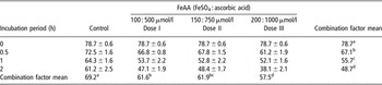
Corresponding to the interaction between the incubation periods and treatments, motility of the control as well as of FeAA-treated samples decreased with time (Table 1). At 0 h, motility of all the samples was similar. It decreased significantly (P ⩽ 0.05) after 0.5 h with supplementation of all doses of FeAA as compared with the control. Among three doses of FeAA, the decrease in motility was non-significant (P ⩾ 0.05) between doses I and II, but significant (P ⩽ 0.05) between doses I and III, as well as, doses II and III (Table 1). After 1 h, the motility decreased significantly (P ⩽ 0.05) with all doses of FeAA as compared with the control, but on comparing all the doses of FeAA, the decrease was non-significant (P ⩽ 0.05) between them. After 2 h, motility decreased non-significantly (P ⩾ 0.05) as compared with the control. Among three doses of FeAA, the decrease was significant (P ⩽ 0.05) between doses I and III as well as II and III, but non-significant (P ⩾ 0.05) between doses I and II (Table 1).
Effects of various concentrations of ferrous ascorbate (FeAA) (ferrous sulphate + ascorbic acid) on % motility of bull spermatozoaFootnote †

† Each value represents mean ± s.e. of 15 observations from five animals. Critical difference at 5% level of significance: for incubation periods is 2.475; for promoter concentrations is 2.475; and for interaction between incubation periods and promoter concentrations is 4.951.
a,b,c,d Mean values in a row or column with different superscript letters are significantly different at 5% level of significance.
Viability
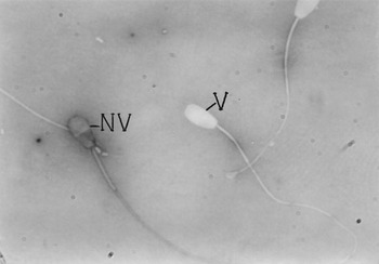
On staining with the eosine-nigrosine stain, the viable (live) sperm cells remained unstained (transparent), whereas non-viable (dead) cells were stained (pink) (Figure 1). Morphology of viable and non-viable spermatozoa remained unaffected with various doses of FeAA.
Cattle bull spermatozoa incubated for 2 h in optimum dose of FeAA (150 μmol/l FeSO4 : 750 μmol/l ascorbic acid) at 37°C, stained with eosine-nigrosine for viable (V) and non-viable (NV) cells.

A gradual and significant (P ⩽ 0.05) decrease in sperm viability was observed from 0 to 2 h in both FeAA treated as well as untreated/control samples (Table 2). Supplementation of sperm samples with various doses of FeAA decreased the % viability significantly (P ⩽ 0.05). Among the three doses of FeAA, % viability decreased non-significantly (P ⩾ 0.05), but it was maximum with the dose II, i.e. 150 μmol/l FeSO4 : 750 μmol/l ascorbic acid. Statistical analysis showed non-significant (P ⩾ 0.05) interaction between treatments and incubation periods. Thus, increase or decrease in % viability with FeAA treatments is not affected by incubation periods or vice versa (Table 2).
Effects of various concentrations of ferrous ascorbate (FeAA) (ferrous sulphate + ascorbic acid) on % viability of bull spermatozoaFootnote †

† Each value represents mean ± s.e. of 15 observations from five animals. Critical difference at 5% level of significance: for incubation periods is 4.92; for promoter concentrations is 6.96; and for interaction between incubation periods and promoter concentrations is non significant.
a,b Mean values in a row or column with different superscript letters are significantly different at 5% level of significance.
Lipid peroxidation
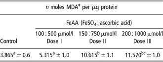
A gradual increase in MDA was observed with all the doses of FeAA. This increase was significant (P ⩽ 0.05) between the control and doses II and III, but non-significant (P ⩾ 0.05) between the control and dose I (Table 3). Among the three doses, LPO level increased significantly (P ⩽ 0.05) between doses I and II as well as doses I and III, but non-significantly (P ⩾ 0.05) between doses II and III. Dose II of FeAA enhanced the level of LPO suddenly and significantly as compared with the other two doses (Table 3).
Effects of various concentrations of ferrous ascorbate (FeAA) (ferrous sulphate + ascorbic acid) on lipid peroxidation (LPO) of bull spermatozoa†
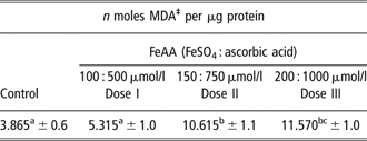
†Each value represents mean ± s.e. of 15 observations from five animals; critical difference at 5% level of significance for promoter concentrations is 3.591.
‡MDA = malondialdehyde.
a,b,cMean values in a row with different superscript letters are significantly different at 5% level of significance.
Discussion
In this study, if the incubation period of bull sperm is prolonged from 0 to 2 h, % motility and % viability decrease by 30% and 20%, respectively. Similar observations have been made in humans (Rao et al., Reference Rao, Soufir, Martin and David1989; Verma and Kanwar, Reference Verma and Kanwar1999), buffalo (Singh et al., Reference Singh, Chand and Georgie1989) and mice (Kodama et al., Reference Kodama, Kribayashi and Gagnon1996). It is known that ferrous ions act as promoters to initiate the chain of LPO, thereby inducing the oxidative stress in sperm membranes (Sharma and Agarwal, Reference Sharma and Agarwal1996). Therefore, in this study, FeAA is used as a promoter system to induce oxidative stress in cattle sperm. Many researchers have suggested that suitable promoter systems such as ferrous and ascorbate ions are required to induce oxidative stress in sperm cells (Jones and Mann, Reference Jones and Mann1977; Jones et al., Reference Jones, Mann and Sherin1979; Aitken et al., Reference Aitken, Clarkson and Fishel1989).
Doses I, II and III of FeAA used in this study decrease motility from initial 69.21% to 61.64%, 61.95% and 57.57%, respectively. Similarly, these doses decreased the viability from initial 83.06% to 71.72%, 71.85% and 67.70%, respectively. Incubation of buffalo spermatozoa with FeAA (0.025 mmol/l FeSO4 : 0.125 mmol/l ascorbic acid) reduces % motility from initial 76% to 42% and viability from 84% to 56% (Singh et al., Reference Singh, Chand and Georgie1989). Hence, it is suggested that reduction in % motility and % viability may be associated with ROS production by FeAA. ROS produced affect the fluidity and integrity of sperm membranes negatively, thereby affecting the motility and viability.
ATP and ATPase play an important role in the signal transduction process coupled with the sperm motility (Mann and Mann, Reference Mann and Mann1981). Any interference in production of ATP, and decline of ATPase activity inhibits the motility of spermatozoa (Kalla and Vasudev, Reference Kalla and Vasudev1981). It is suggested that on supplementation of FeAA to bull spermatozoa, ROS or oxygen free radicals are produced, which result in decrease in the intracellular ATP levels. This decrease in ATP may adversely affect the sperm motility, and also initiate LPO in PUFAs of the sperm membranes, hence culminating in enzyme inactivation and production of spermicidal end products, thereby decreasing the % viability.
In our study, on bull spermatozoa, FeAA (FeSO4 + ascorbic acid) was used as a pro-oxidant to induce LPO in sperm. Present results indicate a gradual increase in MDA production (n moles MDA per μg protein) on supplementation of various doses (I, II and III) of FeAA (5.31, 10.65 and 11.75), as compared with the control (3.86). Thus, TBARS (LPO indicator) increases three to four times with doses II and III of FeAA. Similarly, four- to sixfold increases in TBARS have been observed with 0.4/2.0 mmol/l FeAA supplementation in mouse sperm (Kodama et al., Reference Kodama, Kribayashi and Gagnon1996). It indicates that a combination of ferrous sulphate and ascorbic acid proves a good promoter system for inducing oxidative stress in crossbred cattle bull spermatozoa.
Our study suggests that rate of peroxidation can be considerably enhanced/speeded up if a suitable promoter system such as FeAA supplements the incubation medium (De Lamirande et al., Reference De Lamirande, Jiang, Zini, Kodama and Gagnon1997). The metal cations (Fe2+) are believed to act either by initiating the formation of free radicals or by increasing the rate of breakdown of pre-existing lipid hydroperoxides to peroxyl and alkoxyl radicals and propagate LPO through the generation of short-chain carbonyl fragments, MDA, a substance that can conveniently be measured owing to the formation of a coloured reaction product with TBA (Aitken et al., Reference Aitken, Harkiss and Buckingham1993; De Lamirande et al., Reference De Lamirande, Jiang, Zini, Kodama and Gagnon1997).

It is suggested that in the absence of FeAA, high levels of ROS generated are not associated with significantly enhanced rate of MDA production (Aitken et al., Reference Aitken, Harkiss and Buckingham1993). Therefore, in the present study, FeAA was used as a promoter system of LPO where it enhanced this process at all doses.
In conclusion, out of three doses of FeAA (100 : 500, 150 : 750, 200 : 1000; μmol/l FeSO4 : μmol/l ascorbic acid), dose II, i.e. 150 : 750 μmol/l has been suggested to be the optimum/best dose of FeAA as it induced the oxidative stress to a significant extent, but maintained better motility and viability (required for sperm capacitation and acrosome reaction) as compared with the other two doses. Thus, it is suggested that maintained motility, viability and mild oxidative stress may enhance the fertilising potential of crossbred cattle bull spermatozoa.