




No CrossRef data available.
Article contents
The effect of non-immune stresses on the development of Strongyloides ratti
Published online by Cambridge University Press: 04 May 2005
Abstract
Strongyloides ratti is a parasitic nematode of rats. The host immune response against S. ratti affects the development of its free-living generation, favouring the development of free-living adult males and females at the expense of directly developing, infective 3rd-stage larvae. However, how the host immune response brings about these developmental effects is not clear. To begin to investigate this, we have determined the effect of non-immune stresses on the development of S. ratti. These non-immune stresses were subcurative doses of the anthelmintic drugs Ivermectin, Dithiazanine iodide and Thiabendazole, and infection of a non-natural host, the mouse. These treatments produced the opposite developmental outcome to that of the host immune response. Thus, in infections treated with subcurative doses of Ivermectin, Dithiazanine iodide and in infections of a non-natural host, the sex ratio of developing larvae became more female-biased and the proportion of female larvae that developed into free-living adult females decreased. This suggests that the mechanism by which the host immune response and these non-immune stresses affect S. ratti development differs.
Information
- Type
- Research Article
- Information
- Copyright
- © 2005 Cambridge University Press
INTRODUCTION
Strongyloides ratti is a parasitic nematode of rats. The parasitic stage is female only, which produces genetically male or female eggs, the sex-ratio of which is genetically controlled by an XX/XO, female/male system (Harvey & Viney, 2001). Male eggs develop through 4 larval stages into free-living adult males. Female eggs either develop directly, via 2 larval stages, into infective 3rd-stage larvae (iL3) or via 4 larval stages into free-living adult females (Harvey et al. 2000). Free-living adult males and females mate and their progeny develop into iL3. These infect a new host by skin penetration, then migrate to the intestine and develop into parasitic females (Fig. 1).

Fig. 1. Life-cycle of Strongyloides ratti with two discrete developmental switches shown as grey boxes: 1, a sex-ratio switch; 2, a female-only developmental switch. Larval stages are omitted for clarity. Pre-patency and the duration of infection in an immunocompetent host are approximately 5 and 28 days, respectively. After Harvey et al. (2000).
The development of the free-living generation is controlled by 2 discrete switches (Fig. 1). The sex-ratio switch is affected by the host immune response, with a more male-biased sex-ratio occurring with an increasing anti-S. ratti host immune response (Harvey et al. 2000). This is analogous to the phenomenon in Caenorhabditis elegans where old hermaphrodites or those subject to stress produce progeny with a more male-biased sex-ratio (Goldstein & Curis, 1987).
The female-only developmental choice can be made up until the end of the L2 stage (Viney, 1996) and is affected by both within and without host factors. Thus, a greater proportion of female larvae develop into free-living adult females when they are (i) passed from hosts mounting an anti-S. ratti immune response and (ii) maintained, external to the host, at elevated temperatures (Harvey et al. 2000). These factors also interact, such that female larvae become more temperature sensitive with respect to development (measured as the difference in development between two temperatures) when passed from hosts mounting an anti-S. ratti immune response (Harvey et al. 2000). Thus, the combined effect of the host immune response is to increase the proportion of larvae that develop into free-living adult males and females.
The effect of the host immune response on the development of the free-living generation of S. ratti implies that it can directly or indirectly sense this cue. The developmental effect of the host immune response requires a specific anti-S. ratti response (West et al. 2001). However, it is not known how S. ratti, or any other parasitic nematodes, sense their hosts' immune responses. It is possible that particular components of the anti-S ratti immune response are directly sensed, perhaps by amphids or other sensory structures. Alternatively, parasitic adults and, or developing larvae may sense the stress and damage to which they are subject by an anti-S. ratti immune response (Gemmill, Viney and Read, 1997). Ultrastructural observations of S. ratti have found that as the host immune response develops, there is increasing damage to the worms' intestines which may result in functional starvation (Moqbel & McLaren, 1980). This starvation is likely to be a stress and could be the cue for altered development. A third scenario is that an anti-S. ratti immune response changes the physiology of the host (Hoste, 2001), especially that of the gut mucosa, to the extent that these changes could be sensed by parasitic females and, or developing larvae and used to direct their development. A notable feature of the developmental consequence of the host immune response is that eggs and larvae can enact the female-only developmental decision outside of the host. This implies that developing larvae have a ‘memory’ of the immune status of the host from which they were passed or that this information in some way persists in the environment.
To further understand how S. ratti senses the host immune response we have investigated the effect of stresses, independent of the host immune response, on the development of the free-living generation. We hypothesized that if parasitic females and developing larvae sense ‘stress’ per se, then non-immune stresses will result in the same effect on the development of the free-living generation as the host immune response. We used subcurative doses of anthelmintics (Ivermectin, Dithiazanine iodide and Thiabendazole) and infection of a non-natural host, as non-immune stresses. The rationale for this was that these treatments reduce the reproductive output of the infection without removing the infection, which is phenomenologically similar to the effect of the host immune response on S. ratti (Paterson & Viney, 2002).
MATERIALS AND METHODS
Parasites and infections
Isofemale line ED321 Heterogonic (Harvey et al. 2000) was maintained by serial passage in female Wistar rats (Bantin & Kingman Universal Ltd, Hull, UK) and used throughout. Female rats were infected subcutaneously with 500 iL3; male Wistar rats and C57BL/6J mice were infected with 1000 iL3. Food and water were provided ad libitum unless otherwise stated. Anthelmintic treatment was administered daily from day 1 p.i. Faeces were collected and cultured as previously described (Viney, Matthews & Walliker, 1993) and maintained at 19 °C for 3 days or 25 °C for 2 days, unless otherwise stated, at which times the proportion of directly developing iL3, free-living adult males, free-living adult females and the daily reproductive output were determined (Viney, 1996). The daily reproductive output is the total number of viable larvae that develop in the faeces collected from 1 rat or mouse overnight and thus represents the daily fecundity of an infection (Paterson & Viney, 2002). The positioning of faecal-collection cages in cage racks and the positioning of faecal cultures in incubators was randomized.
Ivermectin
Twelve animals were divided into 3 equal groups. Two experimental groups were administered with 0·206 or 0·051 mg.kg−1.day−1 Ivermectin (Sigma, UK) in 1% (v/v) dimethyl sulphoxide (DMSO) in tap water; the control group received 1% (v/v) DMSO only, as drinking water. The recommended Ivermectin dose for human S. stercoralis infection is 0·2 mg.kg−1.day−1 (Datry et al. 1994) and therefore the doses used are approximately equal to and one quarter of this dose. Faeces were collected on days 5, 7, 10 and 12 p.i. and free-living development determined at 19 and 25 °C.
The host immune response affects parasitic females by reducing the number of parasitic females in the gut, their per capita fecundity, their length and the number of in utero eggs (Kimura et al. 1999; Paterson and Viney, 2002; Wilkes et al. 2004). To determine how Ivermectin affected these infection parameters, 30 animals were divided into 2 equal groups. The experimental group was administered with 0·206 mg.kg−1.day−1 Ivermectin; the control group received 1% (v/v) DMSO, as above. The daily reproductive output was determined for 4 animals from each group on days 5, 9 and 12 p.i., and the number of parasitic females in these same animals was determined on days 6, 10 and 13 p.i., as described by Paterson and Viney (2002). Per capita fecundity was calculated for each rat by dividing the daily reproductive output on day x p.i. by the number of parasitic females in the gut on day x+1 p.i. The length of parasitic females and the number of in utero eggs were determined for parasitic females recovered from 1 animal per treatment group per day, as previously described (Wilkes et al. 2004).
Dithiazanine iodide
Twelve animals were divided into 3 equal groups. Two experimental groups were administered per os with 0·075 or 0·0375 mg Dithiazanine iodide (Sigma, UK) dissolved in 100 μl of 1% (v/v) DMSO with 25% (w/v) sucrose; the control group received 100 μl of 1% (v/v) DMSO with 25% (w/v) sucrose only. Dithiazanine iodide is a cyanine dye and has been used for treatment of human strongyloidiasis (Swartzwelder et al. 1957), but its use subsequently diminished (Stemmermann & Nakasone, 1960). The curative dose for S. ratti in rats is 20 mg.kg−1.day−1 (Swartzwelder et al. 1958) and therefore these treatments are approximately one twenty fifth and one fiftieth of this dose. The effect of these treatments on free-living development at both 19 and 25 °C and of 0·075 mg Dithiazanine iodide on the infection parameters was determined as for Ivermectin, above.
Thiabendazole
Twelve animals were divided into 3 equal groups. Two experimental groups were administered per os with 22 or 11 mg Thiabendazole (Sigma, UK) dissolved in 70 μl of sunflower oil; the control group received 70 μl of sunflower oil only. A dose of 176 mg Thiabendazole in sunflower oil clears S. ratti infections (Paterson & Viney, 2002) and therefore these treatments are one eighth and one sixteenth of this curative dose. The effect of these treatments on free-living development at both 19 and 25 °C and of 22 mg Thiabendazole on the infection parameters was determined as for Ivermectin, above.
Infection of a non-natural host
Infection of a non-natural host, the mouse, was chosen as a non-immune stress because the daily reproductive output of a S. ratti infection in mice is severely reduced, compared with rats, in an immune-independent manner (Gemmill, Viney & Read, 2000). Male C57Bl/6J mice were used, which have a greater susceptibility to S. ratti infection than female mice (Dawkins et al. 1980) and those of other strains (Gemmill et al. 2000). In order to remove any effect of host sex on the experiment, male Wistar rats were used for the control infections. Due to the very low reproductive output of S. ratti infections in mice, faeces from pairs of mice were used as one sample. The daily reproductive output is half of this value, i.e. per mouse.
Faeces were collected daily from 8 pairs of mice and from 8 rats from days 5 to 8 p.i. inclusive, and free-living development determined at both 19 and 25 °C. To determine the effect of the treatment on infection parameters, above, daily reproductive output and the number of parasitic females in the gut were determined in 4 pairs of mice and 4 rats on days 5, 6, 7 and 8 p.i each, and used to determine per capita fecundity. Parasitic females were recovered from a pair of mice and 1 rat for each sample day and processed, as above, to determine worm length and the number of in utero eggs.
Statistical analyses
To determine if a treatment acted as a stress we analysed the effect of each treatment with respect to total reproductive output, number of parasitic females in the gut, their per capita fecundity, their length and the number of in utero eggs. The total reproductive output of an infection was calculated by summing the daily reproductive output of the infection with interpolation for non-sample days. This gave a single value per infection, such that each infection appears only once in the analysis and thus pseudoreplication is avoided. To investigate the effect of treatment on the total reproductive output of an infection a generalized linear model (GLM), with a normal error structure, was constructed (GLMStat, Kagi Shareware, Ken Beath, Australia) with total reproductive output of an infection as the response variable and treatment as the explanatory variable.
Because the host immune response reduces reproductive output over time (Gemmill et al. 2000) it was of interest to determine the effect of treatment on the rate of change of daily reproductive output. To do this a regression analysis of daily reproductive output was performed against day p.i. for each infection. These regression values, which represent the rate of change of daily reproductive output, were then used as the response variable in a GLM with a normal error structure, with treatment as the explanatory variable.
To determine the effect of treatment on the number of parasitic females in the gut, per capita fecundity, worm length or the number of in utero eggs, a GLM with a normal error structure was constructed for each of these variables with TREATMENT, TIME p.i. and TREATMENT*TIME as explanatory variables. Means reported (±1 S.E.M.) were calculated per treatment across all sample times.
To determine the effect of each treatment on the development of the free-living generation, the proportion of free-living adult males (Fig. 1, Box 1) and the proportion of free-living adult females (Fig. 1, Box 2) were calculated. The proportion of free-living adult males is the number of free-living adult males/total number of worms. The proportion of free-living adult females is the number of free-living adult females/[number of free-living adult females+number of directly developing iL3s] (Harvey et al. 2000). Mean proportions per infection were calculated from the total number of each morph present throughout the infection, such that each infection appears only once in the analysis and thus pseudoreplication is avoided. The effect of treatment on the mean proportion of free-living adult males and free-living adult females was analysed by constructing a GLM with a binomial error structure, with the total number of free-living adult males and free-living adult females as response variables and the total number of worms and the total number of females (free-living adult females+directly developing iL3s) as the binomial denominator, respectively. The model was constructed with a logit link function and the scale was estimated as the mean deviance, with treatment as the explanatory variable. The effect of treatment on the rate of change of these proportions was determined as for total reproductive output (above) except that the regression analysis was carried out with a binomial error structure.
The developmental response to temperature external to the host is affected by the immune status of the host from which the larvae are passed. This temperature sensitivity of the free-living development can be investigated by calculating the mean difference in the proportion of free-living adult males and free-living adult females that develop at 19 and 25 °C as [mean proportion at 25 °C – mean proportion at 19 °C] per infection (Harvey et al. 2000). A difference in proportion of zero indicates no temperature sensitivity, with positive and negative values indicating an increase or decrease in temperature sensitivity, respectively. To determine the effect of treatment on these measures of temperature sensitivity, these mean differences in proportion per infection were arcsine transformed and used as the response variable in a GLM with a normal error structure with treatment as the explanatory variable. P values of <0·05 were taken as significant.
RESULTS
No treatments affected the rate of change in daily reproductive output or the rate of change of the proportion of free-living adult males or free-living adult females and therefore these results are not reported here.
Ivermectin
Treatment with 0·206 mg.kg−1.day−1 Ivermectin significantly reduced the total reproductive output, compared with the control (F1,6=36·7, P=0·0009) (Fig. 2A); treatment with 0·051 mg.kg−1.day−1 Ivermectin had no effect (F1,6=0·49, P=0·51) and is therefore not considered further. Treatment with 0·206 mg.kg−1.day−1 Ivermectin significantly reduced the per capita fecundity compared with the control (5·01±1·5; 18·3±5·1, respectively), possibly via the significant reduction in the length of parasitic females (2·01±0·03 mm; 2·15±0·03 mm, respectively) (Table 1). Therefore, by these criteria, treatment with 0·206 mg.kg−1.day−1 Ivermectin can be considered to be a non-immune stress.

Fig. 2. The effect of Ivermectin on (A) daily reproductive output and the proportion of (B) free-living adult males and (C) free-living adult females. Plotted values are the means (±1 S.E.M.) per time-point and best fit regression lines for 0·206 mg.kg−1.day−1 ([squf ]––), 0·051 mg.kg−1.day−1 ([utrif ]--) Ivermectin and controls ([lozf ]––). Regression lines were calculated for arcsine square root transformed data that were back-transformed before plotting.

We hypothesized that a non-immune stress would have the same effect on the development of the free-living generation as the host immune response, namely an increase in the proportion of free-living adult males and free-living adult females. Treatment with 0·206 mg.kg−1.day−1 Ivermectin resulted in a significantly lower mean proportion of free-living adult males (Fig. 2B) and free-living adult females (Fig. 2C), compared with the control (F1,6=8·64, P=0·026; F1,6=7·46, P=0·034, respectively). There was no effect of treatment on the difference in the proportion (i.e. temperature sensitivity of development) of free-living adult males and free-living adult females, compared with the control (F1,6=0·0067, P=0·94; F1,6=2·18, P=0·19, respectively). Therefore, contrary to our hypothesis, treatment with 0·206 mg.kg−1.day−1 Ivermectin, which appears to act as a non-immune stress, results in a decrease in the proportion of free-living adult males and females.
Dithiazanine iodide
Treatment with both 0·075 and 0·0375 mg Dithiazanine iodide significantly reduced the total reproductive output, compared with the control (F1,6=27·14, P=0·002; F1,6=19·81, P=0·0043, respectively) (Fig. 3A), although there was no significant difference between the two drug concentrations (F1,6=0·047, P=0·84). The 0·075 mg treatment significantly reduced the per capita fecundity (3·01±0·65; 18·3±5·1, respectively), possibly via the significant reduction in the number of in utero eggs (6·14±0·4; 7·34±0·46, respectively), compared with the control (Table 1). Therefore, by these criteria, treatment with 0·075 and 0·0375 mg Dithiazanine iodide can be considered to be non-immune stresses.

Fig. 3. The effect of Dithiazanine iodide on (A) daily reproductive output and the proportion of (B) free-living adult males and (C) free-living adult females. Plotted values are the means (±1 S.E.M.) per time-point and best fit regression lines for 0·075 mg ([squf ]––), 0·0375 mg ([utrif ]---) Dithiazanine iodide and controls ([lozf ]––). Regression lines were calculated for arcsine square root transformed data that were back-transformed before plotting.
Treatment with Dithiazanine iodide slowed the rate of development of the free-living stages such that determination of the numbers of free-living adult males, free-living adult females and directly developing iL3s occurred 1 day later in treatment groups, compared with controls. Treatment with 0·075 and 0·0375 mg Dithiazanine iodide significantly decreased the mean proportion of free-living adult males (Fig. 3B) and free-living adult females (Fig. 3C), compared with the control (Males: F1,6=31·49, P=0·0014; F1,6=23·4, P=0·003; Females: F1,6=16·7, P=0·0065; F1,6=10·17, P=0·019, respectively). There was no effect of either treatment on the difference in the proportion of free-living adult males or the difference in the proportion of free-living adult females, compared with the control (Males: F1,6=5·95, P=0·051; F1,6=0·12, P=0·74; Females: F1,6=0·59, P=0·47; F1,6=0·19, P=0·68, respectively). Therefore, contrary to our hypothesis, these treatments with Dithiazanine iodide, which appear to act as non-immune stresses, result in a decrease in the proportion of free-living adult males and females.
Thiabendazole
Treatment with 22 mg Thiabendazole significantly reduced the total reproductive output, compared with the control (F1,6=8·03, P=0·03) (Fig. 4A); treatment with 11 mg Thiabendazole had no effect (F1,6=3·69, P=0·076) and is therefore not considered further. Treatment with 22 mg Thiabendazole significantly reduced the number of parasitic females in the gut (28·8±12·8; 179·4±17·3, respectively), their length (1·89±0·028 mm; 2·18±0·029 mm, respectively) and their number of in utero eggs (1·14±0·3; 5·6±0·4, respectively), compared with the control (Table 1). Therefore, by these criteria, treatment with 22 mg Thiabendazole can be considered to be a non-immune stress.

Fig. 4. The effect of Thiabendazole on (A) daily reproductive output and the proportion of (B) free-living adult males and (C) free-living adult females. Plotted values are the means (±1 S.E.M.) per time-point and best fit regression lines for 22 mg ([squf ]––), 11 mg ([utrif ]---) Thiabendazole and controls ([lozf ]––). Regression lines were calculated for arcsine square root transformed data that were back-transformed before plotting.
Treatment with 22 mg Thiabendazole significantly increased the mean proportion of free-living adult males (F1,6=7·62, P=0·033) (Fig. 4B), but had no effect on the mean proportion of free-living adult females, compared with the control (F1,6=0·22, P=0·66) (Fig. 4C). There was no effect of treatment on the difference in the proportion of free-living adult males or free-living adult females, compared with the control (F1,6=0·81, P=0·4; F1,6=0·77, P=0·41, respectively). Therefore, in at least partial accordance with our hypothesis, treatment with 22 mg Thiabendazole, which appears to act as a non-immune stress, results in an increase in the mean proportion of free-living adult males but has no effect on the mean proportion of free-living adult females.
Infection of a non-natural host
Infection of a non-natural host significantly reduced the total reproductive output, compared with the control (F1,14=72·55, P<0·0001) (Fig. 5A). This treatment significantly reduced the number of parasitic females in the gut (24±6·2; 332·3±35·6, respectively), their length (1·74±0·006 mm; 2·08±0·003 mm, respectively) and their number of in utero eggs (3·04±0·053; 5·42±0·038, respectively) (Table 1), compared with the control. Therefore, by these criteria, infection of a non-natural host can be considered to be a non-immune stress.

Fig. 5. The effect of infection of a non-natural host on (A) daily reproductive output and the proportion of (B) free-living adult males and (C) free-living adult females. Plotted values are the means (±1 S.E.M.) per time point and best fit regression lines for infection in mice ([squf ]––) and rats ([lozf ]––). Regression lines were calculated for arcsine square root transformed data that were back-transformed before plotting.
Infection of a non-natural host significantly reduced the mean proportion of free-living adult males (Fig. 5B) and free-living adult females (Fig. 5C), compared with the control (F1,14=5·671, P=0·032; F1,14=95·56, P<0·0001, respectively). There was no effect of treatment on the difference in proportion of free-living adult females, compared with the control (F1,14=3·64, P=0·077). However, there was a significant effect of treatment on the difference in the proportion of free-living adult males, compared with the control (F1,14=15·74, P=0·0014). Therefore, contrary to our hypothesis, infection of a non-natural host, which appears to act as a non-immune stress, results in a decrease in the proportion of free-living adult males and females.
DISCUSSION
The work presented here has investigated the effect of putative non-immune stresses on the development of the free-living generation of S. ratti. This was done to investigate the hypothesis that the known effects of the host immune response on the development of the free-living generation (i.e. greater proportion of larvae developing into free-living adult males and females) are due to the stress that the immune response places on parasitic females and developing larvae and, therefore, that inducing stress by other means would have the same developmental consequence. The hypothesis was not supported, because treatment with subcurative doses of Ivermectin and Dithiazanine iodide and infection in mice resulted in the opposite developmental outcome (i.e. a lower proportion of larvae develop into free-living males and free-living adult females). Treatment with a third anthelmintic, Thiabendazole, resulted in a significant increase in the proportion of free-living males (consistent with our hypothesis), but no difference in the proportion of free-living females (inconsistent with our hypothesis). Thus, overall these drug and host-induced stresses resulted in a decrease in the development of the sexual adult forms of S. ratti. The effect of these non-immune stresses is in contrast to Plasmodium chabaudi (malaria) infections in mice where the rate of gametogenesis (the development of sexual forms), a process analogous to the development of free-living adults of S. ratti, increases under adverse conditions (Sinden et al. 1996) and the putative stress of subcurative doses of the anti-malarial chloroquine (Buckling, Crooks & Read, 1999).
The effect of the host immune response on an S. ratti infection is to reduce the intestinal worm burden, the length of parasitic females, the number of in utero eggs and to alter the intestinal position of worms (Moqbel & McLaren, 1980; Kimura et al. 1999; Paterson & Viney, 2002; Wilkes et al. 2004). The combined effect of this immune pressure is to reduce the total reproductive output of the infection. The criterion for considering the treatments used here as stresses is that they reduced the total reproductive output of the infection, without eliminating the infection. All of the treatments used here reduced the total reproductive output of the infection and, therefore, can be considered to be stresses. Other criteria, such as measurement of the expression of heat shock proteins (Walker et al. 2003), could be used as an alternative means to assess stress in this species, both with respect to the effect of the host immune response and the treatments used here. The reduction in total reproductive output was brought about by a reduction in the per capita fecundity of parasitic females (Ivermectin and Dithiazanine iodide) or by a reduction in the number of parasitic females (Thiabendazole and infection of mice). The reduction in the per capita fecundity of parasitic females is likely to occur via the significant reductions in worm length (Ivermectin) and the number of in utero eggs (Dithiazanine iodide), though Thiabendazole and infection of mice also significantly reduced these parameters.
Our hypothesis may not have been supported because of the direct effect of the drugs on the control of the development of the free-living generation of S. ratti. However, these drugs are chemically unrelated and have different modes of anthelmintic activity (Saz & Bueding, 1966; Prichard, 1970; Paiement et al. 1999), yet have similar effects on parasite development. For this reason, we consider it extremely unlikely that the developmental effects seen are caused by the direct action of the drugs on the developmental pathways in S. ratti. It is also possible that the effects on the development of the free-living generation that we observe are due to the differential survival of the different free-living morphs. However, again, given the different nature of these drugs, we consider it extremely unlikely that this has occurred. Furthermore, the effects of these drug treatments and infection of mice, on the development of the free-living generation were broadly similar. We therefore consider that the effects of these drug treatments and infection of mice affects the development of the free-living generation of S. ratti via a common mechanism, but one that is different to that of the host immune response (Harvey & Viney, 2001).
How these treatments bring about these developmental effects is unknown. The nematocidal effect of Ivermectin is due to the inhibition of pharyngeal pumping, which may result in starvation (Paiement et al. 1999); Dithiazanine iodide inhibits glucose uptake (Saz & Bueding, 1966) and Thiabendazole damages intestinal epithelial cells (Beugnet et al. 1996). Therefore, these treatments are likely to functionally starve parasitic females and developing larvae. This may mean that there are resultant energetic constraints on parasitic female egg production and subsequent larval development, which may result in changes in development of the free-living generation that maximizes fitness under these energy-constraining regimes. Thus, one can envisage that the energy required for a first-stage larva to develop into a free-living adult (4 moults, significant body growth, especially of the gonad) is significantly greater than to develop into an iL3 (2 moults, limited body growth and no gonadal development). Comparison of the fecundity of lines of S. ratti that differed in their degree of direct and indirect development are consistent with the production of eggs that develop into free-living adults being 3·76 times more ‘expensive’ to produce than eggs that develop directly into iL3 (Fenton et al. 2004). Therefore, under these drug treatments, which may have limited the energy available to the worms, more direct development into iL3s may be favoured with this development being brought about by a reduction in development into free-living adult males and females, as we observed (Harvey et al. 2000). Understanding the energetic consequences of infection in a mouse is more difficult. However, one can envisage that the niche presented by a mouse gut is sufficiently different to that of a rat to be nutritionally suboptimal and present the same energetic constraints on parasite development. However, this, in turn, must suggest that the host immune response (that favours the development of ‘expensive’ free-living adults rather than iL3s) has no such energetic effect on S. ratti.
These developmental effects, whether by this hypothesized energetic constraint or some other mechanism may occur via the parasitic females which alter the further development of their eggs and, or by direct effects on very early developing larvae. We note that 76% of Ivermectin is passed in faeces (Perez et al. 2001) and that Dithiazanine iodide is visible in faeces (Swartzwelder et al. 1958) which allows the possibility that the developmental effects of these drugs may continue to occur outside of the hosts. This possibility could be investigated by the addition of these drugs to faecal cultures of S. ratti to determine their developmental effect, if any.
The host immune response has deleterious effects on a range of infection parameters (Kimura et al. 1999; Paterson & Viney, 2002; Wilkes et al. 2004) and on the subsequent development of the free-living generation (Harvey et al. 2000). This correlation suggested causality. However, the results presented here show that the deleterious effects of these treatments on parasitic females affect the development of the free-living generation in an opposite way to the host immune response. The results therefore suggest that the mechanisms by which these apparent stresses on parasitic females bring about the developmental consequences, differ.
In human infection with S. stercoralis, disseminated infections can occur, usually in immunosuppressed individuals (Grove, 1989). These disseminated infections appear to require the precocious development of iL3s within the gut and subsequent auto-infection of the host. Ivermectin is used in the therapy of human strongyloidiasis (Torres et al. 1993) in which there is the possibility that treatment is not always curative. In this study with S. ratti, subcurative doses of Ivermectin resulted in greater direct development of iL3s. Therefore, if these findings apply to S. stercoralis infections in humans, subcurative doses of Ivermectin may favour auto-infection and thus, perhaps, disseminated strongyloidiasis.
We thank Elizabeth Dilkes, Emma Fisher, Alison Shorto and Clare Wilkes for technical support, Mike Gardner for statistical advice and Mike Gardner, Simon Harvey and Fiona Thompson for their comments on this manuscript. This work was supported by a grant from the Medical Research Council. M.C. was supported by a University of Bristol Scholarship.
References
REFERENCES
Beugnet, F.,
Kerboeuf, D.,
Nicolle, J. C. and
Soubieux, D. (
1996).
Use of free living stages to study the effects of thiabendazole, levamisole, pyrantel and ivermectin on the fine structure of Haemonchus contortus and Heligmosomoides polygyrus.
Veterinary Parasitology
63,
83–94.CrossRefGoogle Scholar
Buckling, A.,
Crooks, L. and
Read, A. F. (
1999).
Plasmodium chabaudi: effect of antimalarial drugs on gametocytogenesis.
Experimental Parasitology
93,
45–54.CrossRefGoogle Scholar
Datry, A.,
Hilmarsdottir, I.,
Mayorgasagastume, R.,
Lyagoubi, M.,
Gaxotte, P.,
Biligui, S.,
Chodakewitz, J.,
Neu, D.,
Danis, M. and
Gentilini, M. (
1994).
Treatment of Strongyloides stercoralis infection with ivermectin compared with albendazole – results of an open study of 60 cases.
Transactions of the Royal Society of Tropical Medicine and Hygiene
88,
344–345.CrossRefGoogle Scholar
Dawkins, H. J.,
Grove, D. I.,
Dunsmore, J. D. and
Mitchell, G. F. (
1980).
Strongyloides ratti: susceptibility to infection and resistance to reinfection in inbred strains of mice as assessed by excretion of larvae.
International Journal for Parasitology
10,
125–129.CrossRefGoogle Scholar
Fenton, A.,
Paterson, S.,
Viney, M. E. and
Gardner, M. P. (
2004).
Determining the optimal developmental route of Strongyloides ratti: an evolutionarily stable strategy approach.
Evolution
58,
989–1000.CrossRefGoogle Scholar
Gemmill, A. W.,
Viney, M. E. and
Read, A. F. (
1997).
Host immune status determines sexuality in a parasitic nematode.
Evolution
51,
393–401.CrossRefGoogle Scholar
Gemmill, A. W.,
Viney, M. E. and
Read, A. F. (
2000).
The evolutionary ecology of host-specificity: experimental studies with Strongyloides ratti.
Parasitology
120,
429–437.CrossRefGoogle Scholar
Goldstein, P. and
Curis, M. (
1987).
Age related changes in the meiotic chromosomes of the nematode Caenorhabditis elegans.
Mechanisms of Ageing and Development
40,
115–130.CrossRefGoogle Scholar
Grove, D. I. (
1989).
Clinical manifestations. In
Strongyloidiasis a Major Roundworm Infection of Man. (
ed. Grove, D. I.), pp.
155–173.
Taylor & Francis,
London.
Harvey, S. C.,
Gemmill, A. W.,
Read, A. F. and
Viney, M. E. (
2000).
The control of morph development in the parasitic nematode Strongyloides ratti.
Proceedings of the Royal Society of London, B
267,
2057–2063.CrossRefGoogle Scholar
Harvey, S. C. and
Viney, M. E. (
2001).
Sex determination in the parasitic nematode Strongyloides ratti.
Genetics
158,
1527–1533.Google Scholar
Hoste, H. (
2001).
Adaptive physiological processes in the host during gastrointestinal parasitism.
International Journal for Parasitology
31,
231–244.CrossRefGoogle Scholar
Kimura, E.,
Shintoku, Y.,
Kadosaka, T.,
Fujiwara, M.,
Kondo, S. and
Itoh, M. (
1999).
A second peak of egg excretion in Strongyloides ratti infected rats: its origin and biological meaning.
Parasitology
119,
221–226.CrossRefGoogle Scholar
Moqbel, R. and
McLaren, D. J. (
1980).
Strongyloides ratti: structural and functional characteristics of normal and immune-damaged worms.
Experimental Parasitology
49,
139–152.CrossRefGoogle Scholar
Paiement, J. P.,
Leger, C.,
Ribeiro, P. and
Prichard, R. K. (
1999).
Haemonchus contortus: effects of glutamate, ivermectin, and moxidectin on insulin uptake activity in unselected and ivermectin selected adults.
Experimental Parasitology
92,
193–198.CrossRefGoogle Scholar
Paterson, S. and
Viney, M. E. (
2002).
Host immune responses are necessary for density-dependence in nematode infections.
Parasitology
125,
1–8.CrossRefGoogle Scholar
Perez, R.,
Cabezas, I.,
Sutra, J. F.,
Galtier, P. and
Alvinerie, M. (
2001).
Faecal excretion profile of moxidectin and ivermectin after oral administration in horses.
Veterinary Journal
161,
85–92.CrossRefGoogle Scholar
Prichard, R. K. (
1970).
Mode of action of the anthelmintic thiabendazole in Haemonchus contortus.
Nature, London
228,
684–685.CrossRefGoogle Scholar
Saz, H. J. and
Bueding, E. (
1966).
Relationships between anthelmintic effects and biochemical and physiological mechanisms.
Pharmacological Reviews
18,
871–894.Google Scholar
Sinden, R. E.,
Butcher, G. A.,
Billker, O. and
Fleck, S. L. (
1996).
Regulation of infectivity of Plasmodium to the mosquito vector.
Advances in Parasitology
38,
53–117.CrossRefGoogle Scholar
Stemmermann, G. N. and
Nakasone, N. (
1960).
Strongyloides stercoralis infestation – malabsorption defect with reaction to dithiazanine iodide.
Journal of the American Medical Association
174,
104–107.Google Scholar
Swartzwelder, J. C.,
Frye, W. W.,
Muhleisen, J. P.,
Miller, J. H.,
Lampert, R.,
Chaverria, A. P.,
Abadie, S. H.,
Anthony, S. O. and
Sappenfield, R. W. (
1957).
Dithiazanine, an effective broad-spectrum anthelmintic.
Journal of the American Medical Association
165,
2063–2067.CrossRefGoogle Scholar
Swartzwelder, J. C.,
Muhleisen, J. P.,
Abadie, S. H.,
Frye, W. W.,
Jones, J. T.,
Robertson, B. D. and
Herbert, D. R. (
1958).
Therapy of strongyloidiasis with dithiazanine.
American Medical Association Archives of Internal Medicine
101,
658–661.CrossRefGoogle Scholar
Torres, J. R.,
Isturiz, R.,
Murillo, J.,
Guzman, M. and
Contreras, R. (
1993).
Efficacy of ivermectin in the treatment of Strongyloidiasis complicating AIDS.
Clinical Infectious Diseases
17,
900–902.CrossRefGoogle Scholar
Viney, M. E. (
1996).
Developmental switching in the parasitic nematode Strongyloides ratti.
Proceedings of the Royal Society of London, B
263,
201–208.CrossRefGoogle Scholar
Viney, M. E.,
Matthews, B. E. and
Walliker, D. (
1993).
Mating in the nematode parasite Strongyloides ratti: proof of genetic exchange.
Proceedings of the Royal Society of London, B
254,
213–219.CrossRefGoogle Scholar
Walker, G. A.,
Thompson, F. J.,
Brawley, A.,
Scanlon, T. and
Devaney, E. (
2003).
Heat shock factor functions at the convergence of the stress response and developmental pathways in Caenorhabditis elegans.
FASEB Journal
17,
508–526.CrossRefGoogle Scholar
West, S. A.,
Gemmill, A. W.,
Graham, A.,
Viney, M. E. and
Read, A. F. (
2001).
Immune stress and facultative sex in a parasitic nematode.
Journal of Evolutionary Biology
14,
333–337.CrossRefGoogle Scholar
Wilkes, C. P.,
Thompson, F. J.,
Gardner, M. P.,
Paterson, S. and
Viney, M. E. (
2004).
The effect of the host immune response on the parasitic nematode Strongyloides ratti.
Parasitology
128,
661–669.CrossRefGoogle Scholar

Fig. 1. Life-cycle of Strongyloides ratti with two discrete developmental switches shown as grey boxes: 1, a sex-ratio switch; 2, a female-only developmental switch. Larval stages are omitted for clarity. Pre-patency and the duration of infection in an immunocompetent host are approximately 5 and 28 days, respectively. After Harvey et al. (2000).
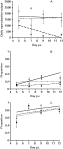
Fig. 2. The effect of Ivermectin on (A) daily reproductive output and the proportion of (B) free-living adult males and (C) free-living adult females. Plotted values are the means (±1 S.E.M.) per time-point and best fit regression lines for 0·206 mg.kg−1.day−1 ([squf ]––), 0·051 mg.kg−1.day−1 ([utrif ]--) Ivermectin and controls ([lozf ]––). Regression lines were calculated for arcsine square root transformed data that were back-transformed before plotting.
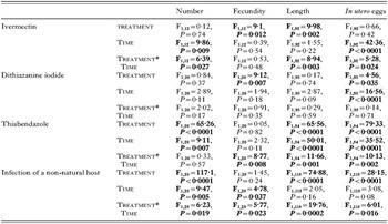
Table 1. The effect of Ivermectin, Dithiazanine iodide, Thiabendazole and infection of a non-natural host on the number of parasitic females in the gut, their per capita fecundity, their length and the number of in utero eggs compared to the respective controls (Significant effects are shown in bold.)
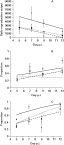
Fig. 3. The effect of Dithiazanine iodide on (A) daily reproductive output and the proportion of (B) free-living adult males and (C) free-living adult females. Plotted values are the means (±1 S.E.M.) per time-point and best fit regression lines for 0·075 mg ([squf ]––), 0·0375 mg ([utrif ]---) Dithiazanine iodide and controls ([lozf ]––). Regression lines were calculated for arcsine square root transformed data that were back-transformed before plotting.
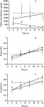
Fig. 4. The effect of Thiabendazole on (A) daily reproductive output and the proportion of (B) free-living adult males and (C) free-living adult females. Plotted values are the means (±1 S.E.M.) per time-point and best fit regression lines for 22 mg ([squf ]––), 11 mg ([utrif ]---) Thiabendazole and controls ([lozf ]––). Regression lines were calculated for arcsine square root transformed data that were back-transformed before plotting.
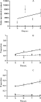
Fig. 5. The effect of infection of a non-natural host on (A) daily reproductive output and the proportion of (B) free-living adult males and (C) free-living adult females. Plotted values are the means (±1 S.E.M.) per time point and best fit regression lines for infection in mice ([squf ]––) and rats ([lozf ]––). Regression lines were calculated for arcsine square root transformed data that were back-transformed before plotting.
 You have
Access
- 13
- Cited by