INTRODUCTION
Impairments after traumatic brain injury (TBI) include deficits in motor skills, processing speed, attention, word finding, memory, social skills, and executive functioning. These impairments interfere with successful return to work, school, family, and community—often in relation to the level of injury severity (e.g., Millis et al., Reference Millis, Rosenthal, Novack, Sherer, Nick, Kreutzer, High and Ricker2001; Rohling et al., Reference Rohling, Meyers and Millis2003; Schretlen & Shapiro, Reference Schretlen and Shapiro2003; Serino et al., Reference Serino, Ciaramelli, Di Santantonio, Malagu, Servadei and Ladavas2006).
TBI results in both gray matter and white matter (WM) tissue loss. Magnetic resonance imaging (MRI) studies help to clarify relationships between cognitive deficits and underlying neuropathology (Bigler, Reference Bigler2003; Gale et al., Reference Gale, Baxter, Roundy and Johnson2005; Johnson et al., Reference Johnson, Bigler, Burr and Blatter1994), even in cases of mild TBI (Kurca et al., Reference Kurca, Sivak and Kucera2006). Diffuse axonal injury (DAI) to WM occurs during rapid acceleration/deceleration and rotation of variously dense brain structures (Adams et al., Reference Adams, Graham, Murray and Scott1982; Strich, Reference Strich1961). Conventional MRI is insensitive to DAI except when accompanied by visible bleeds (e.g., Rugg-Gunn et al., Reference Rugg-Gunn, Symms, Barker, Greenwood and Duncan2001) and has limited ability to detect subtle WM injury.
Diffusion Tensor Imaging
Diffusion of water molecules in healthy WM is highly directional or anisotropic, occurring more readily along axons because of natural barriers (cell membranes, myelin, and adjacent axons) (Le Bihan, Reference Le Bihan1995). Damaged WM bundles have increased diffusion perpendicular to axons and lower anisotropy relative to healthy WM. Two commonly used diffusion measures are fractional anisotropy (FA) and mean diffusivity (MD). FA is a relative measure of diffusion directionality along the axon relative to the perpendicular axis (Pierpaoli & Basser, Reference Pierpaoli and Basser1996). Lower FA is observed in TBI, especially in regions where DAI occurs, whereas MD is the average diffusion in all directions and should be higher in TBI.
There is now general consensus that diffusion tensor imaging (DTI) is more sensitive than conventional MRI for detecting DAI in acute and chronic stages of TBI (e.g., Rugg-Gunn et al., Reference Rugg-Gunn, Symms, Barker, Greenwood and Duncan2001). For example, DTI detects changes in WM within hours or days of injury. In humans, anisotropy changes have been detected within 24 (Arfanakis et al., Reference Arfanakis, Haughton, Carew, Rogers, Dempsey and Meyerand2002) and 32 (Newcombe et al., Reference Newcombe, Williams, Nortje, Bradley, Harding, Smielewski, Coles, Malya, Gillard, Hutchinson, Pickard, Carpenter and Menon2007) hours and within 6 days in mildly injured adolescents (Wilde et al., Reference Wilde, McCauley, Hunter, Bigler, Chu, Wang, Hanten, Troyanskaya, Yallampalli, Li, Chia and Levin2008). Huisman et al. (Reference Huisman, Schwamm, Schaefer, Koroshetz, Shetty-Alva, Ozsunar, Wu and Sorensen2004) found lower FA after TBI within 7 days in the internal capsule and corpus callosum, and these were correlated with injury severity and early outcomes.
Because Wallerian degeneration continues after initial recovery, it is important to describe long-term WM changes in TBI. Within the past year, a few studies have used DTI to document changes in WM in adults with TBI in chronic recovery (e.g., Mathias et al., Reference Mathias, Bigler, Jones, Bowden, Barrett-Woodbridge, Brown and Taylor2004; Xu et al., Reference Xu, Rasmussen, Lagopoulos and Haberg2007; Yuan et al., Reference Yuan, Holland, Schmithorst, Walz, Cecil, Jones, Karunanayaka, Michaud and Wade2007). Salmond et al. (Reference Salmond, Menon, Chatfield, Williams, Pena, Sahakian and Pickard2006) found widespread decreases in cerebral WM FA in adults with TBI who were at least 6 months postinjury. A small sample of severely injured adults who were 4–6 years postinjury had significant decreased FA in the major WM tracts (Xu et al., Reference Xu, Rasmussen, Lagopoulos and Haberg2007). Benson et al. (Reference Benson, Meda, Vasudevan, Kou, Koushik, Hanks, Millis, Makke, Latif, Coplin, Maythaler and Haacke2007) found that changes in WM FA in a chronically injured group predicted severity of injury, in particular posttraumatic amnesia better than Glasgow Coma Scale scores (GSC). Kraus et al. (Reference Kraus, Susmaras, Caughlin, Walker, Sweeney and Little2007) examined WM and cognitive changes in adults with mild, moderate, or severe TBI who averaged 10 years postinjury. Compared to healthy adults, FA was decreased in all 13 regions of interest (ROIs) in adults with moderate-to-severe TBI. A general WM load measure was correlated with executive functions, attention, and memory domains.
In the current study, we use an ROI approach to compare WM FA and MD of severely injured adults with TBI who averaged 7 years postinjury to healthy controls. We expected lower WM FA and higher WM MD in the group with TBI. We were particularly interested in the centrum semiovale (CS), superior frontal (SPF), and inferior frontal (INF) ROIs due to the diffuse nature of TBI and the long-lasting executive function difficulties. Additionally, we explored associations between time since injury and WM integrity.
METHODS
Participants
Nine adults with TBI were recruited from rehabilitation programs, newsletters, and support groups. Ten control participants were recruited through advertisements. All were native English speakers without a history of neurological disease, substance abuse, learning difficulties, or psychiatric disorder. One TBI participant was excluded because of an unavailable medical record. One control was dropped for partial completion and another for being an outlier on vocabulary—a verbal IQ estimate. Thus, data from eight adults with TBI and eight controls who were close in age and education were included (Table 1). Groups did not approach significant differences for age and education (p < .05). No participants had aphasia (i.e., aphasia quotient <93.8 on the Western Aphasia Battery; Kertesz, Reference Kertesz1982), and all successfully read sentences and paragraphs (reading subtest). Verbal IQ was estimated with the National Adult Reading Test (NART; Nelson & Willison Reference Nelson and Willison1991), a stable estimate when administered at least 1 year postinjury (Riley & Simmonds, Reference Riley and Simmonds2003). Controls averaged higher verbal IQ than adults with TBI, whereas years of education did not statistically differ (p = .10) (Table 1).
Means, SDs, and statistical comparisons of demographics and neurocognitive performance for adults with TBI (n = 8) and healthy controls (n = 8)

Note.
Group comparisons of neurocognitive performance were analyzed with MANCOVAs in which estimates of verbal intelligence quotient (IQ) were covaried.  ηp2 effect size values are interpreted as follows: large is >.2, medium is >.1, and small is >.05. d effect size values are interpreted as follows: large is >.8, medium is >.5, and small is >.2 (Cohen, Reference Cohen1988). CVLT-II, California Verbal Learning Test—II; D-KEFS, Delis-Kaplan Executive Function System.
* p < .05.
** p < .01.
All participants with TBI had sustained severe injuries in vehicular accidents that averaged 7.0 years (standard deviation [SD] = 8.6) prior to the study. Early clinical MRIs or CT reports were available and with the exception of one participant, all had substantial cortical neuropathology. Length of coma ranged from 5 to 90 days (mean = 21.75, SD = 28.69). Injury severity was based on coma length, GCS scores when available, and initial neuropathology (Teasdale & Jennett, Reference Teasdale and Jennett1974).
Neurocognitive Measures
All experimental procedures were approved by the Institutional Review Board of the university. The study included two sessions: neurocognitive assessment and MRI. The average time between sessions was 14.6 days (SD = 15.6). Neurocognitive tests were selected based on their known sensitivity to memory and executive functions associated with TBI.
Verbal fluency and trail subtests were administered from the Delis-Kaplan Executive Function System (D-KEFS) (Delis et al., Reference Delis, Kramer, Kaplan and Ober2001), including letter fluency, category fluency, category switching, and trail making. The go/no-go task, a measure of motor response execution and inhibition, was based on a task described by Braver et al. (Reference Braver, Barch, Gray, Molfese and Snyder2001); individuals with TBI have been shown to be impulsive on this task (Dockree et al., Reference Dockree, Kelly, Roche, Hogan, Reilly and Robertson2004). The California Verbal Learning Test—II (Delis et al., Reference Delis, Kramer, Kaplan and Ober2000) measures verbal learning during immediate and delayed recall and recognition memory and assesses learning strategies.
Imaging Procedures
MRI data acquisition
MRI was performed on a 3Tesla Trio scanner using an eight-channel array head coil (Siemens, Erlangen, Germany). T1 images were acquired coronally using a three-dimensional magnetization prepared rapid gradient echo (MP-RAGE) sequence [repetition time (TR) = 2530 ms, echo time (TE) = 3.65 ms, inversion time (TI) = 1100 ms, 240 slices, 1 × 1 × 1–mm voxel, flip angle = 7°, field of view (FOV) = 256 × 256 mm]. Proton density (PD)–weighted images were acquired axially using a hyperecho turbo spin echo (TSE) sequence (TR) = 8550 ms, (TE) = 14 ms, 80 slices, 1 × 1 × 2–mm voxel, flip angle = 120°, FOV = 256 mm). DTI data were acquired axially, aligned with the TSE images, using a dual–spin echo, single-shot, pulsed-gradient, echo planar imaging sequence (TR = 8300 ms, TE = 86 ms, 64 contiguous slices, 2 × 2 × 2–mm voxel, FOV = 256 mm, two averages, b value = 1000 s/mm2). Thirteen unique volumes were collected to compute the tensor: a b = 0 s/mm2 image and 12 images with diffusion gradients applied in noncollinear directions. A dual–echo flash field map sequence with voxel parameters common to the DTI was used to correct the DTI data for geometric distortion caused by magnetic field inhomogeneity (TR = 700 ms, TE = 4.62 ms/7.08 ms, flip angle = 90°, magnitude, and phase difference contrasts).
Anatomical image processing
Imaging data were processed using tools from the FMRIB software library (FSL, http://www.fmrib.ox.ac.uk/). Nonbrain tissue was removed using brain extraction tool (BET). The PD brain was aligned to the T1 brain using the FMRIB’s linear registration tool (FLIRT). Dual-channel segmentation was performed on the T1 and aligned PD brains using FMRIB’s automated segmentation tool (FAST), producing four tissue classes (cerebral spinal fluid, white, gray, and blood). The T1 brain was registered to the FMRIB software library (FSL) template brain (MNI-152 brain, Montreal Neurological Institute) using a 12-parameter affine transformation.
DTI processing
FMRIB’s diffusion toolbox (FDT) was used to correct the diffusion-weighted images for motion and eddy current distortion and then compute the diffusion tensor. Maps of MD and FA were derived. FA is the anisotropic component of the tensor (Basser, Reference Basser1995), ranging between 0 (perfectly isotropic) and 1 (diffusion occurring in only one direction). The geometric distortion caused by the magnetic field inhomogeneity was determined from the field map image, and FMRIB’s utility for Geometrically Unwarping EPIs (FUGUE) was used to dewarp the b = 0 diffusion image and the scalar maps (FA, MD).
ROI definition
The same semiautomated procedures reported in Wozniak et al. (Reference Wozniak, Krach, Ward, Mueller, Muetzel, Schnoebelen, Kriel, Kiragu and Lim2007) were used in this work to define ROIs. Subject-specific WM masks for the dewarped DTI scalar maps were determined by transforming the partial volume estimate (PVE) WM map from the anatomic segmentation onto the distortion-corrected DTI image. Voxels in the dewarped DTI images were classified as WM if the DTI-aligned PVE value exceeded 50% in that voxel.
Three ROIs were selected for evaluation because of the diffuse nature of injury and incidence of executive dysfunction in TBI: the CS, which is all WM voxels superior to corpus callosum; the INF region, which is all WM voxels rostral to the genu of the callosum and inferior (ventral) to the anterior and posterior commisure (AC–PC) plane; and the SPF region, which is all WM voxels rostral to the genu of the callosum and superior to the AC–PC plane (Figure 1). A trained operator determined the boundary of the ROIs on each MNI-aligned T1 image; these planes were aligned on the DTI and convolved with the WM mask to define the WM ROI masks. Mean FA and MD values were computed for the WM voxels within each of the three ROIs.
(a) The CS region is superior to the corpus callosum. (b) The INF region is rostral to the genu of the corpus callosum and inferior (ventral) to the AC–PC plane. The SPF region is rostral to the genu of the corpus callosum and superior to the AC–PC plane.

RESULTS
Multivariate analysis of covariance (MANCOVA) was used to compare groups’ neurocognitive performance and DTI measures. NART verbal scores served as the covariate to statistically account for any variance that could be attributed to differences in verbal IQ. Tables 1 and 2 contain univariate F tests and  effect sizes, whereas the omnibus results are reported below. Associations between FA, MD, and time since injury were explored using Pearson partial correlations in which verbal IQ was partitioned out.
Neurocognitive Findings
Three neurocognitive domains were examined to describe the population samples: executive functions (alternating trails, inhibition, and verbal fluency), delayed recall, and speed (Table 1).
Executive functions
The MANCOVA revealed that adults with TBI were slower than controls at alternating trails (Wilks’ Lambda = 8.21, p = .004 ). Specifically, they were significantly slower when combining number and letter sequences. Adults with TBI tended to perform worse than controls on verbal fluency tasks revealed by a statistical trend (Wilks’ Lambda = 2.80, p = .09
 ). Univariate tests revealed that they had fewer correct responses when switching categories than controls. Groups did not differ significantly on letter fluency, category fluency, or repetition errors. On the go/no-go task, a test of inhibition, adults with TBI made more errors (Wilks’ Lambda = 4.44, p = .04
 ). Thus, as groups, adults with TBI were within average, whereas controls were just above average, resulting in significant differences overall in two of the three executive domains and in several specific tasks.
Delayed recall
Overall, adults with TBI recalled significantly less than controls under delayed conditions (Wilks’ Lambda = 4.18, p = .04 ). As expected, adults with TBI recalled less than controls without cues, whereas recall by adults with TBI improved when provided with cues.
Speed: Simple trails
Adults with TBI were significantly slower than controls overall (Wilks’ Lambda = 9.39, p = .002 ). Specifically, they were slower when sequencing letters and tracing lines.
DTI Findings
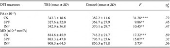
FA and MD differed between groups in the MANCOVA omnibus comparisons when verbal IQ was controlled for (FA: Wilks’ Lambda = 8.83, p = .004 ; MD: Wilks’ Lambda = 8.93, p = .004
 ).Footnote a FA was significantly lower and MD was significantly higher in adults with TBI compared to controls in all three ROIs in the univariate comparisons at varying levels of probability ranging from p < .05 to p < .0001 (Table 2). Thus, even when accounting for any differences in estimates of verbal IQ, differences in WM in adults with TBI were significantly apparent in the expected direction.
Means, SDs, and statistical comparisons of FA and MD for adults with TBI (n = 8) and healthy controls (n = 8)

Note.
Group comparisons were analyzed with MANCOVAs in which estimates of verbal IQ were covaried.  ηp2 effect size values are interpreted as follows: >.2 is large, >.1 is medium, and >.05 is small.
* p < .05.
** p < .01.
*** p < .001.
**** p < .0001.
Association Findings for Adults With TBI
Negative associations between FA and MD across ROIs are reported here to validate the integrity of these measures. Pearson partial correlation coefficients in which verbal IQ was controlled revealed that CS FA was negatively correlated with CS MD (r = −.94, p = .002), SPF FA was negatively correlated with SPF MD (r = −.88, p = .02), and INF FA was negatively correlated with INF MD (r = −.89, p = .02). There were no significant correlations of FA or MD between ROIs.
Using partial correlations, time since the injury was strongly and negatively associated with SPF FA (r = −.96, p = .002) and positively associated with SPF MD (r = .83, p = .04) but was not significantly associated with FA or MD of the CS or INF. Thus, those who had lived with chronic TBI longer had lower FA and higher MD in the SPF ROI.
DISCUSSION
This study sought to identify changes in cerebral WM in adults with severe TBI in the chronic recovery stage. While DTI may be a biomarker for DAI in acute recovery (Arfanakis et al., Reference Arfanakis, Haughton, Carew, Rogers, Dempsey and Meyerand2002; Huisman et al., Reference Huisman, Schwamm, Schaefer, Koroshetz, Shetty-Alva, Ozsunar, Wu and Sorensen2004; Newcombe et al., Reference Newcombe, Williams, Nortje, Bradley, Harding, Smielewski, Coles, Malya, Gillard, Hutchinson, Pickard, Carpenter and Menon2007; Wilde et al., Reference Wilde, McCauley, Hunter, Bigler, Chu, Wang, Hanten, Troyanskaya, Yallampalli, Li, Chia and Levin2008), few others have identified WM changes years after injury (e.g., Kraus et al., Reference Kraus, Susmaras, Caughlin, Walker, Sweeney and Little2007). Our results provide additional evidence that DTI is a useful technique for describing WM integrity, and compared to controls, FA and MD differ as hypothesized when statistically controlling for any difference due to verbal IQ (Kraus et al., Reference Kraus, Susmaras, Caughlin, Walker, Sweeney and Little2007; Xu et al., Reference Xu, Rasmussen, Lagopoulos and Haberg2007). That is, adults who averaged 7 years after the injury demonstrated decreased FA and increased MD in the three ROIs.
WM changes observed in this chronic sample likely represent multiple injury processes over time including direct, primary mechanisms (e.g., DAI/shearing, hemorrhages, contusions/contra-coup injury); secondary mechanisms (e.g., swelling); and tissue atrophy from Wallerian degeneration. Salmond et al. (Reference Salmond, Menon, Chatfield, Williams, Pena, Sahakian and Pickard2006) showed changes in FA and MD of adults beyond 6 months postinjury in 10 ROIs, and Kraus et al. (Reference Kraus, Susmaras, Caughlin, Walker, Sweeney and Little2007) found similar changes in adults who averaged 10 years postinjury. The current study showed robust group differences in FA and MD within the CS, similar to findings from other studies. In our study, the CS region was dorsal to corpus callosum (e.g., motor and sensory tracts, frontobasal ganglia tracts, arcuate fasciculus, longitudinal fasciculus) as was the superior parietal/SPF regions in the study of Salmond et al. (Reference Salmond, Menon, Chatfield, Williams, Pena, Sahakian and Pickard2006). In the study of Kraus et al. (Reference Kraus, Susmaras, Caughlin, Walker, Sweeney and Little2007), the CS included the corona radiata, superior longitudinal fasciculus, and corticospinal tracts. Similarly, group differences were also found in FA and MD in the SPF and INF regions, although our frontal ROIs were not comparable to the frontal regions of Salmond et al. (Reference Salmond, Menon, Chatfield, Williams, Pena, Sahakian and Pickard2006) or Kraus et al. (Reference Kraus, Susmaras, Caughlin, Walker, Sweeney and Little2007).
Associations of FA and MD within ROIs were included to verify the integrity of our DTI measures. FA and MD should be negatively associated, and indeed, our results support this. However, unlike the findings of other studies (e.g., Kraus et al., Reference Kraus, Susmaras, Caughlin, Walker, Sweeney and Little2007), no associations were found between ROIs. Possible reasons for this are the small sample and the limited range of severity. Our study was limited to those with severe injury, whereas Kraus et al. (Reference Kraus, Susmaras, Caughlin, Walker, Sweeney and Little2007) included those with mild, moderate, and severe injuries.
A noteworthy finding in the current study was the relationship between time since injury and WM integrity in the SPF ROI. Given that typical aging is associated with changes in WM in frontal lobes and CS, one might suspect that this was due to aging (e.g., Pfefferbaum et al., Reference Pfefferbaum, Sullivan, Hedehus, Lim, Adalsteinsson and Moseley2000). Post hoc Pearson partial correlation that accounted for verbal IQ revealed that age was not associated with time since injury (r = .03, p = .95), the SPF FA or MD (r = −.15, p = .77; r = −.12, p = .82), or the CS FA or MD (r = .48, p = .34; r = −.35, p = .50). Age tended to be negatively associated with the INF FA (r = −.77, p = .07) and was positively associated with the INF MD (r = .90, p = .01). Thus, associations between FA and MD in the SPF ROI and time since injury were not due to aging, though aging did have its effect on the INF region. Relationships between time since injury and WM integrity in the SPF region could represent compounding effects of multiple injury processes over time that interact with age. We do not know whether WM in the SPF region is particularly susceptible to injury-related changes over time, yet this finding certainly warrants further consideration given the propensity of long-lasting executive dysfunction in the population with TBI.
The performance of those with TBI was significantly lower than the performance of healthy adults on several neurocognitive measures even when controlling for verbal IQ; however, their average performance was within the normal range and would not necessarily mean that they were impaired. Indeed, these adults with TBI were all employed, either full time or part time, and living in the community. Thus, in the presence of significant reductions in WM integrity, those who sustain severe injuries may still perform quite well over time. We could find only one published case study that found similar results (Skoglund et al., Reference Skoglund, Nilsson, Ljunberg, Jonsson and Rydenhag2008). Additional studies with larger sample sizes of severely injured adults who display more severe neurocognitive impairments are needed to clarify whether WM changes are predictive of eventual neurocognitive and long-term functional outcomes. Future studies should include larger sample sizes that allow one to model relationships between demographics, FA and MD, and neurocognitive performance reliably. Until then, we cannot be certain that WM integrity and eventual, long-term neurocognitive performance are related.
ACKNOWLEDGMENTS
The authors thank the Center for Magnetic Resonance Research at the University of Minnesota, Minneapolis, MN, for the use of time, space, equipment, and personnel to operate the 3Tesla magnet during this study. Acknowledgments are also extended to Pradeep Ramanathan and Casey Tuck for their assistance in scanning participants. Importantly, we want to thank the Brain Injury Association—Minnesota (www.braininjurymn.org) for assisting us in recruiting volunteers for the study. Edward Carney assisted us with statistics and for this we are grateful. A special “thank you” is extended to all of those with TBI, who so willingly gave us their time and energy to participate in this study.