Implications
Dietary supplementation with an immunomodulatory herb, such as pale purple coneflower (Echinacea pallida: EPAL), could provide benefits to growing rabbits. Phagocytic activity, which is a defense mechanism against pathogens, was increased in the fattening rabbits fed the supplemented diets. However, dietary supplementation with EPAL had no impact on the growth performances, bacterial community, blood parameters or humoral immune responses in the growing rabbits.
Introduction
The addition of medicinal plants with immune-stimulating properties, such as Echinacea species, is a valuable strategy as pointed out in the previous part of this study (Dabbou et al., Reference Dabbou, Rotolo, Kovitvadhi, Bergagna, Dezzutto, Barbero, Rubiolo, Schiavone, De Marco, Helal, Zoccarato and Gasco2016). An improvement in growth performances and a reduction in the mortality rate were observed in growing rabbits fed diets with Echinacea purpurea (EPUR) supplementation (Ahmed et al., Reference Ahmed, Kamel, El-Sabeiy and Zeitouny2008; Arafa et al., Reference Arafa, Salem and Farid2010). The higher number of white blood cells has been reported in groups of rabbits, laying hens and fattening pigs treated with EPUR (Böhmer et al., Reference Böhmer, Salisch, Paulicks and Roth2009; Arafa et al., Reference Arafa, Salem and Farid2010). EPUR dietary addition has been shown to enhance phagocytic activity in fattening pigs (Böhmer et al., Reference Böhmer, Salisch, Paulicks and Roth2009) and the level of specific antibody response against vaccination in poultry, quail, swine and rabbit (Maass et al., Reference Maass, Bauer, Paulicks, Böhmer and Roth-Maier2005; Böhmer et al., Reference Böhmer, Salisch, Paulicks and Roth2009; Ma et al., Reference Ma, Shi, Niu, Wang and Zhong2009). To date, the research on animal studies has tended to focus on EPUR rather than Echinacea angustifolia (EANG) or Echinacea pallida (EPAL; Hermann et al., Reference Hermann, Honeyman, Zimmerman, Thacker, Holden and Chang2003; Maass et al., Reference Maass, Bauer, Paulicks, Böhmer and Roth-Maier2005; Böhmer et al., Reference Böhmer, Salisch, Paulicks and Roth2009; Ma et al., Reference Ma, Shi, Niu, Wang and Zhong2009; Arafa et al., Reference Arafa, Salem and Farid2010; Sahin et al., Reference Sahin, Ozlem and Mehmet2012) and it has been shown that there is a broad variation of the active compounds in the Echinacea species (Dabbou et al., Reference Dabbou, Rotolo, Kovitvadhi, Bergagna, Dezzutto, Barbero, Rubiolo, Schiavone, De Marco, Helal, Zoccarato and Gasco2016). Therefore, the objective of this present study has been to evaluate pre- and postnatal effects on the performances, bacterial community, blood parameters and immunity of rabbits after the introduction of EPAL (pale purple coneflower) as a dietary addition. The consequences of EPAL dietary supplementation on rabbit does were presented in the previous part of this study (Dabbou et al., Reference Dabbou, Rotolo, Kovitvadhi, Bergagna, Dezzutto, Barbero, Rubiolo, Schiavone, De Marco, Helal, Zoccarato and Gasco2016).
Material and methods
Animals, housing, diets, experimental design and growth performances
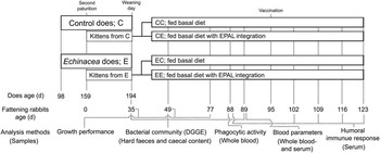
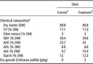
The trial was conducted at the experimental rabbit facility of the Department of Agricultural, Forest and Food Sciences in Carmagnola (Turin, Italy). Eighty kittens were taken from the second parturition of control group (C) and Echinacea group (E) of the previous study (Dabbou et al., Reference Dabbou, Rotolo, Kovitvadhi, Bergagna, Dezzutto, Barbero, Rubiolo, Schiavone, De Marco, Helal, Zoccarato and Gasco2016). The kittens were weaned at 35-day old, were randomly separated into four groups of 20 and were housed in individual wire cages (0.41 m long×0.30 m wide×0.28 m high) when their mothers were 194-day old. The fattening rabbits were fed a growing commercial basal diet with or without the supplementation (3 g/kg of EPAL): the CC group (rabbits from the C does fed the control diet), CE group (rabbits from the C does fed the supplemented diet), EC (rabbits from the E does fed the control diet) and EE group (rabbits from the E does fed the supplemented diet). Feeds and clean water were provided ad libitum. The preparation and chemical analysis of the feeds and supplement (EPAL powder), which were obtained from the same provider, were performed as outlined in the previous part of this study (Dabbou et al., Reference Dabbou, Rotolo, Kovitvadhi, Bergagna, Dezzutto, Barbero, Rubiolo, Schiavone, De Marco, Helal, Zoccarato and Gasco2016). The diet composition for the growing rabbits is illustrated in Table 1. Throughout the whole trial, the facility was climate and light controlled in order to maintain a temperature of 22±2°C and a 16L : 8D photoperiod. Live weight and feed intake were measured at 35, 49 and 77 days old to calculate the average daily weight gain, average daily feed intake and feed conversion ratio, whereas morbidity and mortality rate were recorded according to Gidenne et al. (Reference Gidenne, Combes, Feugier, Jehl, Arveux, Boisot, Briens, Corrent, Fortune, Montessuy and Verdelhan2009). Ten fattening rabbits were selected from each group to evaluate the bacterial community and blood parameters, while the remaining ten rabbits from each group were kept until 123 days old to study the phagocytic activity and specific antibody response. The timeline of the experimentation is illustrated in Figure 1.
The timeline of the experimental plan.

Composition of the growing rabbit diets

1 Minerals and vitamins in the growing diets: calcium 1% of DM, phosphorus 0.4% of DM, sodium 0.3% of DM, lysine 0.7% of DM, methionine 0.4% of DM, vitamin A 12 500 IU/kg, vitamin D3 1250 IU/kg, vitamin E 100 mg/kg, ferrous carbonate 704 mg/kg, copper sulphate 98 mg/kg, manganese oxide 209 mg/kg, zinc oxide 186 mg/kg, sodium selenite 0.57 mg/kg and potassium iodide 2.5 mg/kg. These data were provided by Ferrero Mangimi S.p.A, (Farigliano CN, Italy), who formulated and prepared the experimental diets.
2 The experimental diets were analysed in the laboratory of the Department of Agricultural, Forest and Food Sciences, Turin, Italy.
Bacterial community
The hard faeces of the growing rabbits were collected at 35, 49 and 89 days from five animals per group, whereas the caecal content samples (n=5 per group) were taken immediately after slaughtering at 89 days. The samples were kept in sterile polyethylene bags and immediately stored at −20°C until analysis. The samples (10 g) were homogenized in 90 ml of Ringer’s solution (Oxoid, Milan, Italy) for 2 min in a stomacher (LAB Blender 400 and Sto-circul-bag stomacher bags, PBI, Milan, Italy) at room temperature; a deposit was allowed to set for 1 min, and 1 ml of the supernatant was used for the DNA extraction. A Powersoil DNA kit (MO-BIO, Carlsbad, CA, USA) was used, according to the manufacturer’s instructions. A total of 5 µl of RNAse (Promega, Milan, Italy) was added to the DNA and incubated at 37°C for 30 min before storage at −20°C. DNA was quantified using a NanoDrop 1000 spectrophotometer (Thermo Scientific, MA, USA) and was standardized at 50 ng/μl. PCR was performed with the universal primers 338F-GC and 518R (Muyzer et al., Reference Muyzer, Waal and Uitterlinden1993; Invitrogen, Carlsbad, CA, USA) amplifying the variable V3 region of the 16S rRNA gene. The amplicons obtained (about 250 bp) were analysed by means of DGGE, with a Dcode universal mutation detection system (BioRad, Milan, Italy). The gels were run for 4 h at 200 V, stained with SYBR® Gold Nucleic Acid Gel Stain (Life technologies, Milan, Italy) for 30 min, and analysed under UV using UVIpro Platinum 1.1 Gel Software (Eppendorf s.r.l., Milan, Italy).
Blood parameters
Blood samples were taken from 10 rabbits per group at 89 days old during the slaughter process. Sterile tubes containing ethylenediaminetetraacetic acid were used to collect the samples for haematological analysis, which was conducted using an automated laser cell counter calibrated for rabbits (MS4-S Hematology Analyzer, Melet Schloesing, Osny, France). A plain serum tube was instead used as a container to obtain serum for biochemistry analysis and protein electrophoresis, which was conducted using an automated photometer system (Screen Master Touch, Hospitex Diagnostics, Sesto Fiorentino, Italy) and a semi-automated agarose gel electrophoresis system (Sebia Hydrasys, Evry, France), respectively.
Changes in the number and species of white blood cells after infection and blood production disorders can be revealed by means of haematological parameters (Varga, Reference Varga2014). The liver function (albumin), liver damage (aspartate aminotransferase and alanine aminotransferase), renal function (blood urea nitrogen and creatinine) and lipid metabolism (cholesterol and triglyceride) of the rabbits were established through an evaluation of the serum biochemistry (Varga, Reference Varga2014). A serum protein fraction was used to introduce the an inflammatory response (α 1 globulin), metabolic enzymes (α 2 globulin), immunoglobulin G (γ globulin), iron-binding glycoprotein (β 1 globulin) and immunoglobulin A and M (β 2 globulin; Tizard, Reference Tizard2013). Therefore, these parameters were evaluated in this study to illustrate the effects of EPAL supplementation.
Phagocytic activity
At 88 days, blood samples were taken from the lateral saphenous vein of 10 rabbits per group. The samples were placed in sterile tubes containing lithium-heparin, kept at 4°C and immediately transported to the laboratory to perform a phagocytosis test, which involved a direct counting procedure using bacterial cells (Ragap et al., Reference Ragap, Khalil and Mutawie2012). The heparinized blood samples (100 µl) were left until they reached room temperature (22°C), and then they were mixed with 10 µl of diluted bacterial solution (100 µl of broth with 1×10¹⁰ cells/ml of Streptococcus canis and 900 µl of PBS, pH 7.2) in 14 ml sterile glass tubes (15×125 mm). These mixtures were incubated at 37°C for 30 min. Blood samples, without the addition of bacterial cells, were used as a negative control. Immediately after incubation, all the samples were placed at 4°C to stop the reaction, and blood smearing was performed. After drying, the samples were stained with gram stain, according to the manufacturer’s instructions (Sigma-aldrich®, Buchs, Switzerland). The results were evaluated under an oil immersion light microscope. The neutrophils and monocytes that engulfed the gram positive cocci were counted as phagocytic cells. Phagocytic activity was calculated on the basis of the percentage of phagocytic cells observed (Ragap et al., Reference Ragap, Khalil and Mutawie2012). Triplicate counting was performed for each sample.
The phagocytosis test is able to confirm the response of sentinel cells (Macrophage and dendritic cells) and proinflammatory cytokine production against the pathogen invasion. After a bacterial pathogen passes the physical barrier and invades the host, sentinel cells can detect pathogen-associated molecular patterns on bacteria antigen through toll-like receptors. The sentinel cells then secrete vasoactive molecules and proinflammatory cytokines (interleukin 1, interleukin 6 and tumor necrotic factor α), which activate a systemic response and phagocytic activities (Tizard, Reference Tizard2013).
Specific humoral immune response
The rabbits were vaccinated against rabbit haemorrhagic disease virus (RHDV; IZOVAC MEVAX®, IZO S.p.A., Brescia, Italy) at 95 days old, and serum was collected at 88, 102, 109, 116 and 123 days old through centrifugation (1600×g for 15 min) of the blood samples (collected from the lateral saphenous vein) after incubation at room temperature (22°C) for 4 h. The serum was stored at −20°C to evaluate the percentage of inhibition (PI) against the RHDV antibody, which was conducted by competitive ELISA (Istituto Zooprofilattico Sperimentale della Lombardia e dell’Emilia, Brescia, Italy). The wells were coated with the antibody against RHDV. The serum samples (5 µl) and RHDV antigen (25 µl) were added to each well (1/10 serum dilution). Duplicates of the positive and negative controls were tested for each plate. The protocol recommendation by the Istituto Zooprofilattico Sperimentale della Lombardia e dell’Emilia (Brescia, Italy) was followed. The colour was developed by adding 50 µl of 3,3',5,5'-tetramethylbenzidine substrate, and the reaction was then stopped by blocking solution. An automated ELISA microplate reader was used to determine an optical density at 492 nm (Titertek Multiskan MCC/340, Labsystems, Helsinki, Finland). The results were expressed as PI on the basis of the optical density values according to the following formula: PI=[1−(optical density of the sample)/(average negative control optical density)]×100.
In this study, the RHDV vaccine contained dead virus. Therefore, this vaccination only stimulated the humoral immune response (Tizard, Reference Tizard2013). Dead virus is engulfed, then is represented by major histocompatibility complex (MHC) class II and is presented to helper T cell by antigen presenting cells which activates B cell differentiation to plasma cells for specific antibody production (Tizard, Reference Tizard2013). Therefore, the stimulation of the humoral immune response through vaccination can be used to the confirm phagocytosis process, antigen presenting activity and antibody network.
Statistical analysis
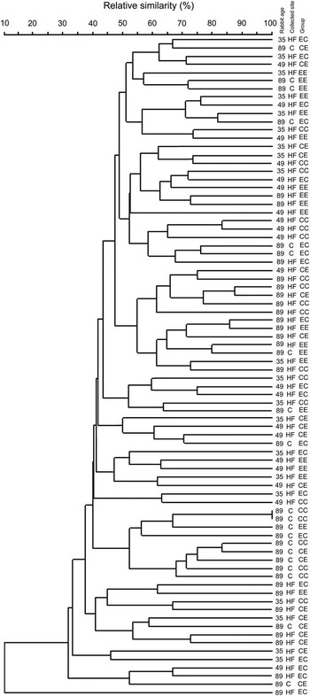
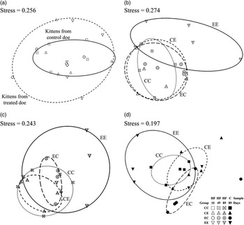
All the statistical analyses were performed using the SPSS software package (IBM SPSS, 2012). The growth performances, blood parameters, phagocytic activity and PI were processed with one-way ANOVA (groups as the fixed factors) whose normal distribution was observed in the parametric data by Kolmogorov–Smirnov test. Significant differences in treatment means were established by Duncan’s multiple range tests at P<0.05. The entire fingerprint database of the bacterial community was created using Bionumerics version 5.1 software (Applied Maths, Sint Martens Latem, Belgium). A dendrogram of similarity was retrieved using the dice coefficient and unweighted pair group method for the arithmetic average clustering algorithm (Figure 2). The symmetrical matrix of the all pairwise distances among the proximities of the DGGE profiles of the rabbit’s bacterial community was calculated using the Euclidean distance to establish the distance matrix. Two dimensions were chosen for the ordination. The distance matrix was used for nonmetric multidimensional scaling (nMDS) plots, which were performed in IBM SPSS (2012) using the PROXSCAL scaling algorithm. The nMDS plots of the bacterial communities from hard faeces at 35, 49 and 89 days and the caecal contents at 89 days are presented in Figure 3a–d, respectively. Kruskal’s S-Stress was calculated. On nMDS plots, the near point shows higher similarity than the far point (Combes et al., Reference Combes, Fortun-Lamothe, Cauquil and Gidenne2011).
Dendrogram of the PCR-DGGE profiles of the rabbit bacterial community from hard faeces samples (HF) at 35, 49 and 89 days, and caecal content samples (C) at 89 days between CC (rabbits fed basal diets from does fed the control diet), CE (rabbits fed treated diets from does fed the control diets), EC (rabbits fed basal diets from does fed the supplemented diets) and EE group (rabbits fed treated diets from does fed the supplemented diets).

nMDS plot of the PCR-DGGE profiles of the rabbit bacterial community between CC (rabbits fed basal diets from does fed the control diets), CE (rabbits fed treated diets from does fed the control diets), EC (rabbits fed basal diets from does fed the supplemented diets) and EE group (rabbits fed treated diets from does fed the supplemented diets) from hard faeces samples (HF) at days 35 (a), 49 (b) and 89 (c), and samples of the caecal content at 89 days old (d).

Results
Growth performances and bacterial community
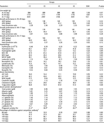
No significant differences were found in growth performance (Table 2). There was no morbidity or mortality during the experimental period. The dendrogram and two-dimensional nMDS of the bacterial community of the rabbits were generated using the digitalizated PCR-DGGE fingerprints obtaining from the DNA extracted directly from the hard faeces and caecal content samples (Figures 2 and 3, respectively). The cluster analysis (Figure 2) showed a shift in the microbial community, due to the an increase in age. Moreover, it was possible to observe a difference between the hard faeces at 89 day old and the caecal content fingerprints (Figure 2). Higher within-group variability was observed in the kittens from the treated groups, compared to the control (Figure 3a). No clear difference was observed between the CC, CE and EC samples, whereas the variability of the bacterial community of the EE group was higher than the others throughout the experimental period (Figure 3b–d).
Effects of pre- and postnatal dietary phytoadditives (Echinacea pallida; EPAL) on growth performances, blood parameters, phagocytic activity and specific antibody response in growing rabbits

CC=rabbits fed the basal diets from does fed the control diet; CE=rabbits fed diets with EPAL from does fed the control diet; EC=rabbits fed the basal diets from does fed diets with EPAL; EE=rabbits fed diets with EPAL from does fed diets with EPAL; ADFI=average daily feed intake; ADG=average daily weight gain; MCV=mean corpuscular volume; MCH=mean corpuscular haemoglobin; MCHC=mean corpuscular haemoglobin concentration; AST=aspartate aminotransferase; ALT=alanine aminotransferase; BUN=blood urea nitrogen.
1 Each globulin fraction was expressed as a percentage of the total protein.
2 The globulin was calculated as the difference between the total protein and albumin.
3 The specific antibody production was performed by the vaccination against rabbit hemorrhagic disease virus at 95-day-old and the data is presented for each collection day.
Blood parameters and immunological test
The blood parameters, phagocytic activities and inhibition percentage of the competitive antibody are illustrated in Table 2. All the evaluated parameters fell into the normality range (Varga, Reference Varga2014). No differences were observed for the haematology, serum biochemistry or serum protein electrophoresis between the groups (P>0.05). An increase in phagocytic activity was observed in the rabbits fed the treated diets, compared to the rabbits fed the control diets (CE and EE v. CC and EC; P<0.001). Because of a specific antibody response against the vaccination, the inhibition percentage was lower than 25% 1 week before the vaccination in all the groups which was considered as negative (Istituto Zooprofilattico Sperimentale della Lombardia e dell’Emilia, Brescia, Italy). A higher level of antibody response was observed 1 week after the vaccination. The inhibition percentage surged to above 80% in the 2nd week after the vaccination, and then became constant after this period. No significant difference was observed for the antibody production against RHDV between the treated groups, which were compared each week.
Discussion
Growth performances
The growth performance of the Echinacea supplemented groups did not show any significant differences. The present results differ from those of Arafa et al. (Reference Arafa, Salem and Farid2010) who found, in a similar study using EPUR at 130 mg/kg BW, a significant decrease in the mortality rate and an increase in the live weight in 6-week-old growing rabbits fed E diets (P<0.05). Dietary herb supplementation usually leads to an improvement in the flavour, which in turn accounts leads to an increase in feed ingestion and better performance (Franz et al., Reference Franz, Baser and Windisch2010; Christaki et al., Reference Christaki, Bonos, Giannenas and Florou-Paneri2012). Ahmed et al. (Reference Ahmed, Kamel, El-Sabeiy and Zeitouny2008) highlighted a significant improvement in the final BW, daily weight gain and feed conversion ratio in growing rabbits, which had orally been given 7.5 mg of EPUR extracts/kg BW/day in liquid form. However, the outcomes of the above reported references are not exactly comparable with the present trial due to some dissimilarities in the experimental designs concerning: the tested Echinacea species, concentration of the supplement, administration route (oral by means of a liquid mixture), supplement preparation (extraction) and the supplemented periods in the doe diets. Positive outcomes on productive performance have been reported in rabbits after EPUR addition (Ahmed et al., Reference Ahmed, Kamel, El-Sabeiy and Zeitouny2008; Arafa et al., Reference Arafa, Salem and Farid2010), whereas studies conducted on other livestock species have not found any improvement (Hermann et al., Reference Hermann, Honeyman, Zimmerman, Thacker, Holden and Chang2003; Maass et al., Reference Maass, Bauer, Paulicks, Böhmer and Roth-Maier2005; Böhmer et al., Reference Böhmer, Salisch, Paulicks and Roth2009; Sahin et al., Reference Sahin, Ozlem and Mehmet2012).
Bacterial community
The bacterial community in rabbits changes continuously from birth to adulthood. This ecosystem involves nutrient utilization, the immune system, development of the digestive tract and pathogen prevention (Combes et al., Reference Combes, Michelland, Monteils, Cauquil, Soulié, Tran, Gidenne and Fortun-Lamothe2013). The period from birth to 49 days (time window of permissiveness) is a critical period for rabbits, because several microorganisms present in an environment can randomly access the rabbit’s digestive tract, depending on the diet formulation, management programme and/or environment conditions (Combes et al., Reference Combes, Michelland, Monteils, Cauquil, Soulié, Tran, Gidenne and Fortun-Lamothe2013). In the aforementioned study, the variability of the bacterial community in caecum at the neonatal stage was remarkable between each rabbit. Thereafter, this variability decreased continuously with age. Finally, the bacterial diversity was nearly similar between each rabbit (Combes et al., Reference Combes, Fortun-Lamothe, Cauquil and Gidenne2011). A similar pattern has been observed in the present study, although hard faeces were used as the samples. The different characteristics between the caecal contents and the hard faeces provided in a dissimilarity of the bacterial community which was also reported by Michelland et al. (Reference Michelland, Combes, Monteils, Cauquil, Gidenne and Fortun-Lamothe2010), as shown in Figure 2.
Nutrition is one of the most important factors that can influence the microbial community (Michelland et al., Reference Michelland, Combes, Monteils, Cauquil, Gidenne and Fortun-Lamothe2011). In the present study, the variability between the kittens from the E does was higher at weaning than the kittens from the C does (Figure 3a). This result may be due to mother and/or diet effects, because the kittens lived in the same cage and were fed the same diet as their mother until weaning. If EPAL contained properties that could modify the bacterial community of does, the initial gut microbial composition of their kittens at weaning would also be changed. The addition of EPAL in the fattening period (CE group) or discontinuous supplementation (EC group) may not be sufficient to provoke effects on the bacterial community. Therefore, in order to modify the bacterial community in fattening rabbits through EPAL supplementation, it should be performed on their mother and the rabbits should be fed the supplemented diet continuously over the fattening period. The variability in the bacterial community between each rabbit of EE is higher than that of the other groups throughout the experimental period (Figure 3). At the moment, it is impossible to conclude whether the modification in EE is a positive or negative outcome. Further analysis on bacterial species should be performed to clarify this aspect.
Blood parameters and immunological test
The physiological and biochemical status of animals can be expressed through haematology and blood chemistry. The collection should be performed more than 6 weeks after the supplementation, because no statistical difference between groups has been observed in samples that were collected at 3 weeks (Arafa et al., Reference Arafa, Salem and Farid2010). In the present study, blood was taken from the rabbits after 54 days of supplementation. Therefore, a difference would have been observed, if the supplement had affected these parameters. A higher proliferation rate of spleen lymphocytes in EPAL supplemented mice has been reported in an in vitro study, but the haematology indices were not influenced (Zhai et al., Reference Zhai, Liu, Wu, Senchina, Wurtele, Murphy, Kohut and Cunnick2007). EPUR supplementation in rabbits, laying hens and fattening pigs has been seen to increase the total amount of white blood cells, while in this study, EPAL in this study did not (Böhmer et al., Reference Böhmer, Salisch, Paulicks and Roth2009; Arafa et al., Reference Arafa, Salem and Farid2010). Therefore, the variation in the chemical compounds between EPUR and EPAL could be a possible explanation.
Echinacea genus herbs are particularly well known as immunostimulant agents, and they mainly affect innate immunity (Barnes et al., Reference Barnes, Anderson, Gibbons and Phillipson2005; Zhai et al., Reference Zhai, Liu, Wu, Senchina, Wurtele, Murphy, Kohut and Cunnick2007). The purified glycoproteins (arabinogalactans, fructofuranosides and heteroxylans), as well as alkylamides, especially isobutylamides and ketones (only in EPAL), in Echinacea plants appear to play important roles on phagocytic stimulation (Classen et al., Reference Classen, Thude, Blaschek, Wack and Bodinet2006; Kraus et al., Reference Kraus, Bae, Wu and Wurtele2007). The supplementation of arabinogalactan-protein from an EPAL extract has been seen to increase the production of IL-6 and nitrite, which are by-products of activated macrophages (Classen et al., Reference Classen, Thude, Blaschek, Wack and Bodinet2006). An increase in phagocytic activity has been reported in fattening pigs after EPUR juice addition (Böhmer et al., Reference Böhmer, Salisch, Paulicks and Roth2009), and in the present study after EPAL dietary integration. Therefore, the active chemical compounds in EPAL could be the cause of the improvement in phagocytosis.
As far as adaptive immunity is concerned, the EPAL extract significantly increased the production of IL-4 and IL-10 from the type 2 T helper cells and immunoglobulin M involved in humoral immune responses. A greater production of IL-2 and IFN-γ from the type 1 T helper cells, which is associated with cell-mediated immunity, was also observed in the Echinacea treated group (Classen et al., Reference Classen, Thude, Blaschek, Wack and Bodinet2006; Zhai et al., Reference Zhai, Liu, Wu, Senchina, Wurtele, Murphy, Kohut and Cunnick2007). β 2 globulin fractions should increase when immunoglobulin M production is higher. However, differences in the plant preparation before usage (extraction v. raw materials) and in the study design (in vitro v. in vivo) may alter the outcomes, as observed in the present study. A higher level of total globulin has been reported in rabbits after EPUR supplementation (Arafa et al., Reference Arafa, Salem and Farid2010). The specific antibody response against inactivated vaccine as a cost effective way of assessing the network functionality of antigen presenting cells (via MHC class II), T helper cell activation and antibody production has been evaluated in this study and has here been shown not to be affected by the chosen treatments. Therefore, the selection of the Echinacea species (EPUR v. EPAL) and of the experimental design (with or without stress environment) can lead to diverse outcomes.
The alkamide, polysaccharide and proteoglycan in Echinacea plants have been suggested as active compounds that can stimulate antibody production (Egert and Beuscher, Reference Egert and Beuscher1992; Hudson et al., Reference Hudson, Vimalanathan, Kang, Amigue, Livesey and Arnason2005). In an in vitro study, it has been shown that EANG extracts contain antiviral properties against the herpes simplex virus (HSV), the influenza virus (FV) and rhinovirus, whereas aqueous extracts of EPUR only show antiviral activities on HSV and FV, but no significant change has been found for EPAL extracts (Hudson et al., Reference Hudson, Vimalanathan, Kang, Amigue, Livesey and Arnason2005). Instead, the antibody response against the red blood cells of sheep by plaque-forming cell assays has not been affected by the supplementation of Echinacea extracts (Zhai et al., Reference Zhai, Liu, Wu, Senchina, Wurtele, Murphy, Kohut and Cunnick2007). The level of specific antibody response against vaccination in EPUR treated animals has been shown to be greater than in animals fed control diets (Maass et al., Reference Maass, Bauer, Paulicks, Böhmer and Roth-Maier2005; Böhmer et al., Reference Böhmer, Salisch, Paulicks and Roth2009; Ma et al., Reference Ma, Shi, Niu, Wang and Zhong2009), but some studies have not reported positive outcomes (Hermann et al., Reference Hermann, Honeyman, Zimmerman, Thacker, Holden and Chang2003). The diversity in the active chemical compounds between Echinacea species may be the cause of the contrasting results. Further studies on the pharmacological activities of the different active compounds and on the synergistic effects are necessary to clarify these differences.
Conclusion
An increase in phagocytosis has been observed in rabbits fed diets with EPAL supplementation, without any impact on growth performances, blood parameters, bacterial community or humoral immune response.
Acknowledgements
This research was supported by the ‘University of Torino (ex 60%)’ grant (Es. fin. 2013–2015). The authors would like to thank Dr Claudio Malavasi (Biotrade snc®, Modena, Italy) for the dry ground EPAL samples, Dr Paolo Cerrina (Ferrero S.p.a Cuneo, Italy) for his support with the feed formulation, Mrs Vanda Malfatto for her technical support, and Mr Dario Sola, Mr Paolo Montersino and Mr Mario Colombano for the rabbit care.