




No CrossRef data available.
Article contents
- Abstract
- INTRODUCTION
- VENOMOUS AND TOXIC ARTHROPODS
- TICK PARALYSIS
- PARALYSES AND TOXICOSES OF THE ARGASID TICK FAMILY
- PARALYSIS AND TOXICOSES OF THE GENUS ARGAS
- PARALYSIS AND TOXICOSES OF THE GENUS ORNITHODOROS
- PARALYSIS BY O. LAHORENSIS
- SAND TAMPAN TOXICOSIS AND PARALYSIS
- PARALYSIS AND THE GENUS OTOBIUS
- PARALYSES OF THE IXODID FAMILY
- PARALYSES AND THE GENUS IXODES
- PARALYSIS AND THE GENUS HAEMAPHYSALIS
- PARALYSIS AND THE GENUS AMBLYOMMA
- PARALYSIS AND THE GENUS DERMACENTOR
- PARALYSIS AND TOXICOSES OF THE GENUS HYALOMMA
- PARALYSIS AND TOXICOSES OF THE GENUS RHIPICEPHALUS
- OTHER FORMS OF TOXICOSIS CONSIDERED AS NON-PARALYTIC
- TICK EGG TOXINS (IXOVOTOXINS)
- FUNCTIONAL SIGNIFICANCE OF TICK TOXINS
- TOXICOSES FROM AN EVOLUTIONARY PERSPECTIVE
- SOURCES OF TICK TOXINS
- CONCLUSION
- ACKNOWLEDGEMENTS
- References
Biochemical perspectives on paralysis and other forms of toxicoses caused by ticks
Published online by Cambridge University Press: 19 April 2005
Article contents
- Abstract
- INTRODUCTION
- VENOMOUS AND TOXIC ARTHROPODS
- TICK PARALYSIS
- PARALYSES AND TOXICOSES OF THE ARGASID TICK FAMILY
- PARALYSIS AND TOXICOSES OF THE GENUS ARGAS
- PARALYSIS AND TOXICOSES OF THE GENUS ORNITHODOROS
- PARALYSIS BY O. LAHORENSIS
- SAND TAMPAN TOXICOSIS AND PARALYSIS
- PARALYSIS AND THE GENUS OTOBIUS
- PARALYSES OF THE IXODID FAMILY
- PARALYSES AND THE GENUS IXODES
- PARALYSIS AND THE GENUS HAEMAPHYSALIS
- PARALYSIS AND THE GENUS AMBLYOMMA
- PARALYSIS AND THE GENUS DERMACENTOR
- PARALYSIS AND TOXICOSES OF THE GENUS HYALOMMA
- PARALYSIS AND TOXICOSES OF THE GENUS RHIPICEPHALUS
- OTHER FORMS OF TOXICOSIS CONSIDERED AS NON-PARALYTIC
- TICK EGG TOXINS (IXOVOTOXINS)
- FUNCTIONAL SIGNIFICANCE OF TICK TOXINS
- TOXICOSES FROM AN EVOLUTIONARY PERSPECTIVE
- SOURCES OF TICK TOXINS
- CONCLUSION
- ACKNOWLEDGEMENTS
- References
Abstract
Tick toxicoses, of which paralysis is the most widespread and dominant form, are important elements of pathogenesis induced by ticks. Tick paralysis is the most widespread and dominant form of tick toxicoses. Non-paralytic forms of tick toxicoses do occur and evidence suggests that these forms of toxicoses are not evolutionary related. While functional significance has been suggested for tick toxins, the advantages for tick survival in general are not clear. This review considers the molecular nature of tick toxins, the possibility that tick toxins have originated more than once independently and whether these toxins could have unrecognized benign functions.
Information
- Type
- Research Article
- Information
- Copyright
- © 2004 Cambridge University Press
INTRODUCTION
Literature on the nature of tick toxicoses has been particularly scant over the last five years. A seminal work regarding this aspect is the extensive monograph on tick toxicoses by Gothe (1999) and a review on tick paralysis (Masina & Broady, 1999). Most other publications dealing with tick toxicoses are of a clinical or epidemiological nature. On the mechanisms and nature of tick toxicoses only three reports have been published over the last five years. These include investigations into the neuropathogenic effects of Argas walkerae larvae (Maritz et al. 2000, 2001) and an investigation into the cardio-pathogenic effects of Ornithodoros savignyi (Mans et al. 2002). In view of this scarcity in recent literature, tick toxicosis are here reviewed. Tick toxicosis has been a research focus for almost eighty years and during this time, several excellent reviews on this subject have been written that cover the history of toxicosis research as well as its aetiology and pathology (Gregson, 1943, 1973; Stampa, 1959; Neitz, 1962; Murnaghan & O'Rouke, 1978; Gothe, Kunze & Hoogstraal, 1979; Gothe, 1984, 1999; Wikel, 1984; Gothe & Neitz, 1991; Sonenshine, 1993; Masina & Broady, 1999).
VENOMOUS AND TOXIC ARTHROPODS
A distinction should be made between venomous organisms (those that actively secrete toxic components) and those that contain toxins (toxic components present in biological tissues). In ticks both forms of toxins are recognized. Certain tick species can cause pathological changes in their host by inoculation of non-infectious components during feeding. On the basis of this, tick toxins have been identified in whole tick extracts, salivary gland secretions (SGS), salivary gland extracts (SGE) and tick eggs. Tick toxins can, however, only be assigned a venomous status if they are secreted during feeding. The major form of tick toxicosis associated with feeding is tick paralysis, while various other forms of toxicoses induced by individual tick species include sand tampan toxicoses from Ornithodoros savignyi, Hyalomma truncatum toxicoses that include sweating sickness, Mhlosinga and Magudu, necrotic stomatitis nephrosis syndrome and toxicoses from Boophilus microplus, Dermacentor marginatus, Rhipicephalus appendiculatus, Ixodes redikorzevi and O. gurneyi (Gothe, 1999). Hypersensitivity and other immunological reactions can also be included within the scope of general tick toxicoses (Wikel, 1984). It is also clear that many tick saliva components e.g. proteases, protease inhibitors, hyaluronidases, anticoagulants, platelet aggregation inhibitors and haemolytic agents could have toxic effects at the attachment site of the tick on the host (Vermeulen & Neitz, 1987). For practical reasons a distinction is made in this review between tick paralysis and tick toxicosis. The latter is regarded to include all toxicoses except paralysis.
TICK PARALYSIS
Tick paralysis was the earliest form of toxicosis described for ticks and records describing death caused by ticks date back to 1824 (cited in Standbury & Huyck, 1945). For an interesting history on the subject, the reader is referred to the thorough account by Gregson (1973). Paralysis is the most important tick toxicosis for veterinary and human medicine (Gothe & Neitz, 1991). Of the approximately 869 tick species, paralysis has been described for 55 species of hard ticks and 14 soft tick species (Gothe, 1999). However, for many of these species only a few, often insufficient or uncertain records regarding the actual toxicity exist in literature (Table 1, Table 2). It would thus seem as if species with toxins are rather the exception than the norm. The most important paralysis-inducing ticks include the hard ticks I. holocyclus (Australia), D. andersoni, D. variabilis (North America), I. rubicundus and R. evertsi evertsi (South Africa). Soft ticks for which paralysis has been described, demonstrated or suspected include Argas walkerae, A. arboreus, O. capensis, O. lahorensis (Eurasia), O. savignyi (see section on sand tampan toxicoses) and Otobius megnini (South Africa). No logical pattern can be observed for paralysis ticks within the Acari (Fig. 1) and it can only be concluded that more ixodid tick species cause paralysis than argasid species. In fact, paralysis only occurs after prolonged periods of feeding and in soft ticks this type of feeding behaviour is only observed for larvae or nymphae, which feed for several days in contrast to other stages, which complete feeding within minutes.

Fig. 1. The phylogenetic distribution of the major tick paralysis-inducing genera. Indicated are the number of species that are implicated in causing paralysis or toxicoses and the total number of species within the genus. The phylogenetic tree was compiled from data of cited sources (Klompen et al. 2000; Murrel, Campbell & Barker, 2001) and data on toxins from Gothe (1999).


Paralysis is associated with definite feeding phases
In experimentally-induced paralysis, secretion of neurotoxin coincides with a definite repletion phase and in hard ticks is limited to females only. For R. evertsi evertsi paralysis, toxicity is associated with a short period between day 4 and 5 of feeding and a tick body mass of 15–21 mg (Gothe & Lämmler, 1982; Neitz & Gothe, 1986). Paralysis induced by the tick I. holocyclus sets in after 4–5 days of feeding, while paralysis with D. variabilis is only detected after approximately 6–8 days after attachment (Gregson, 1973; Masina & Broady, 1999). In the soft tick A. walkerae, it is only larvae that cause paralysis, which occurs after 5–6 days of feeding (Gothe, 1984; Gothe & Neitz, 1991). In all instances, paralysis coincides with the rapid engorgement phase that is marked by the production and secretion of numerous protein products by the salivary glands. Paralysis is exhibited as an ascending flaccid tetraplegia due to an impaired functioning of the nervous system (Gothe & Neitz, 1991). While these are generally observed symptoms, most neurotoxins have specific characteristics not necessarily shared with toxins from other tick species.
PARALYSES AND TOXICOSES OF THE ARGASID TICK FAMILY
Gregson (1973) postulated that paralysis induced by soft ticks is distinct from paralysis of hard ticks in that paralysis is only caused by the immature stages. The only argasids for which definite paralysis is observed are ticks in the genus Argas and Ornithodoros.
PARALYSIS AND TOXICOSES OF THE GENUS ARGAS
Argas larvae that cause paralysis of fowl under laboratory conditions include A. africolumbae, A. arboreus, A. persicus, A. radiatus, A. sanchezi and A. walkerae (Gothe, 1984). In all cases, paralysis symptoms coincide with the rapid engorgement phase (5–6 days) and persist until all larvae attain a maximal state of engorgement or until termination of the parasitic phase. Symptoms abate as the number of larvae diminishes, with total recovery after all larvae have fallen off the host. A. miniatus has also been implicated in causing paralysis (Hoogstraal, 1985). Although no paralysis is caused in humans by Argas species, severe irritation has been associated with the bites of A. reflexus (Hoogstraal, 1985). In terms of pathology of Argas paralysis, A. walkerae has been studied in most detail.
Paralysis by A. walkerae
Electromyographical studies indicated that the fast conducting nerve fibres of the peripheral nervous system are affected and the paralysis can be classified as a motor polyneuropathy that does not affect the afferent paths (Gothe et al. 1971; Gothe & Kunze, 1971; Gothe & Neitz, 1991). The toxin seems to affect the liberation of acetylcholine as well as its receptor's sensitivity at the myoneural synapse (Gothe & Neitz, 1991). A monoclonal antibody directed against the toxin from R. evertsi evertsi also recognizes protein complexes of molecular mass 60–70 kDa from crude A. walkerae extracts and prevents paresis (partial paralysis) of day-old chicks (Crause et al. 1994). The toxic fraction was purified using a bioassay to detect toxic activity, based on injection of day-old chicks (Viljoen et al. 1990). The purified fraction showed two bands with molecular masses of 32 and 60 kDa using reducing SDS–PAGE while one band (pI~4·5) was obtained by iso-electric focusing. Macromolecular complexes (80–100 kDa) were observed using size exclusion chromatography (Viljoen et al. 1990). Recently, the monoclonal antibody directed against the toxin from R. evertsi evertsi was used in an attempt to purify the neurotoxin from A. walkerae extracts and whilst a 68 kDa toxin was detected using Western blot analysis, an 11 kDa protein was purified that showed cross-reactivity with the mAb using enzyme linked immunosorbent assay (ELISA), although not being detected during Western blot analysis (Maritz et al. 2000). These conflicting results between purification using detection methods of bio-assay and monoclonal antibody have not been resolved yet. Crude A. walkerae larval extracts inhibited potassium-stimulated and veratridine-evoked release of [3H] glycine from rat brain synaptosomes, suggesting that the toxin might be targeting ion channels involved in depolarization (Maritz et al. 2001).
PARALYSIS AND TOXICOSES OF THE GENUS ORNITHODOROS
In the genus Ornithodoros, only O. lahorensis has been truly implicated in causing paralysis. Paralysis of marine birds by O. capensis has been doubted (Hoogstraal, 1985). Other members of this genus can, however, cause severe reactions in the host and symptoms ranging from pain, blisters, local irritation, oedema, fever, pruritus, inflammation and systemic disturbances have been indicated for O. amblus, O. capensis, O. coniceps, O. coriaceus, O. gurneyi, O. muesbecki, O. savignyi and O. rostratus (Hoogstraal, 1985). From these the only form of toxicosis investigated in depth is sand tampan toxicosis caused by Ornithodoros savignyi.
PARALYSIS BY O. LAHORENSIS
Paralysis of sheep and cattle by O. lahorensis has been reported in Jugoslavia, Macedonia, Caucasus, Kazakstan, the Central Asia, Turkmenistan and possibly Turkey (Gothe, 1999). Paralysis is caused by high numbers (100–200) of nymphal ticks of the slow-feeding third-stage. Paralysis progresses rapidly from the rear to the front body accompanied by occasional convulsions. Death occurs on the third or fourth day after the initial onset of paralysis (Gregson, 1973).
SAND TAMPAN TOXICOSIS AND PARALYSIS
One of the first indications of toxicosis due to sand tampans (O. savignyi) was a report by Kone (1948) that described the death of 10 bovines from a herd of 98 cattle within six hours of exposure to the tampans. Their ages varied from 18 months to three years (Kone, 1948). These cattle were en route to Nigeria and were being vaccinated at a vaccination post in N'Guigmi. Before vaccination, they were tethered within a pen that housed numerous sand tampans. Within two hours of confinement the first animal succumbed and others soon followed. The symptoms displayed were indicative of agonizing pain and their rapid development before death suggested anaphylactic shock. Rousselot (1956), who was part of the French delegation at the joint FAO/OIE meeting on the control of tick-borne diseases of lifestock, reported the involvement of several hundred cattle on the borders of Lake Chad in which a high mortality rate from asphyxiation was indicated. Unfortunately, this report was included under the heading of tick paralysis in the meeting report and might have led to subsequent confusion regarding the involvement of O. savignyi as an agent of tick paralysis (Neitz, 1962; Hoogstraal, 1985; Gothe & Neitz, 1991; Gothe, 1999). Exsanguination was another cause of death initially attributed to mortality of cattle, sheep and camels induced by O. savignyi (Hoogstraal, 1956; Du Toit & Theiler, 1964). Howell (1966) stated that feeding of only three ticks kills a guinea pig, suggesting that a toxin is secreted by this tick species.
Purification of sand tampan toxins
It was subsequently shown that salivary gland secretion (SGS) obtained from O. savignyi by pilocarpine stimulation has an LD50 of ~200 μl/kg when injected subcutaneously into 10 g albino mice (Howell, Neitz & Potgieter, 1975). Undiluted SGS (50 μl~5 salivary gland equivalents) kills a mouse within 8 minutes. The toxic activity is temperature stable for at least 15 minutes at 80 °C. An acidic toxin (pI~5) was purified from salivary gland secretion and shown to be a highly abundant protein (~9% of the total salivary gland secretion protein) (Neitz, Howell & Potgieter, 1969). It had a molecular mass of ~15400 Da and in this purified form could kill a mouse within 90 minutes if injected at a concentration of 400 μg/10 g mouse. A non-toxic component (Mr~16000 Da) that showed N-terminal amino acid sequence similarity to the toxin was also described (Neitz, 1976). Recently, the acidic toxin (tick salivary gland protein 2-TSGP2), a non-toxic homologue (TSGP3) and a basic toxin (TSGP4, pI~8) were purified from salivary gland extracts (SGE) (Mans et al. 2001, 2002). The toxins, the non-toxic homologue and another non-toxic protein (TSGP1), are the most highly abundant proteins in the SGE (TSGPs) and comprise individually ~4–5% of the total SGE protein (Mans et al. 2001). Based on this high abundance it was suggested that the TSGPs may be involved in tick salivary gland granule biogenesis. The N-terminal sequences and molecular masses obtained for the acidic toxin and its homologue corresponded very well with each other and the sequences obtained before (Fig. 2). It was also shown that 24 μg of the acidic toxin or 34 μg of the basic toxin in purified form are sufficient to kill a 20 g mouse within 30 minutes (Mans et al. 2002). The difference observed between the toxicities of the original and more recent toxin preparations could be ascribed to the fact that the toxins in purified form seem to be labile and that the current success in obtaining such active toxic fractions can be ascribed to modern high performance liquid chromatography technology that allows purification of the toxins within a few hours, compared to several days for the previous isolation attempts.

Fig. 2. N-terminal sequences obtained for the sand tampan toxins (TSGP2 and TSGP4) and a non-toxic homologue (TSGP3). The sequence for the non-toxic TSGP1 is also indicated, which shows sequence similarity to a highly abundant protein from O. moubata (20A1). The Toxin* and non-toxin* shown derive from sequences previously obtained (Neitz, 1976). Data were taken from Mans et al. (2001).
Clinical pathology of sand tampan toxicosis
In controlled experiments where animals (sheep, rats and mice) were injected subcutaneously with SGS, visible symptoms were minimal (Mans et al. 2002). This was also observed in an experiment where a 300 kg bull was confined to a camp that contained approximately 2000 tampans for two hours on four consecutive days. Most animals show symptoms of shock just prior to death. In large animals the histopathology indicates congestion and oedema in the myocardium, integument, spleen, kidneys, lungs and lymph glands. Haemorrhage within the lungs also occurs in smaller animals such as rats and mice. No other pathological changes can normally be observed and Howell suggested that animals die of heart failure (Mans et al. 2002). Addition of SGE to a rat heart perfusion system causes arrhythmia and bradycardia, followed by cardiac arrest. Monitoring of mouse electrocardiograms after subcutaneous injection of purified toxins showed that ventricular tachycardia (TSGP2) and a Mobitz-type ventricular block (TSGP4) are induced (Mans et al. 2002). This suggests that the pathogenicity of sand tampan toxicosis is due to a targeting of the hosts cardiac system and as such is distinct from tick paralysis.
The origins of sand tampan toxicosis
It was shown that the TSGPs belong to the lipocalin protein family (Mans, Louw & Neitz, 2003). In haematophagous organisms lipocalins perform a variety of functions that include the regulation of inflammation during feeding of the hard tick R. appendiculatus by sequestration of histamine (Paesen et al. 1999, 2000). Lipocalins from the blood-sucking bugs Rhodnius prolixus and Triatoma pallidipenis inhibit various haemostatic processes within the host. The toxic TSGP2 and non-toxic TSGP3 show high sequence identity (46%) with moubatin, a platelet aggregation inhibitor from the closely related non-toxic tick, O. moubata (Waxman & Connolly, 1993; Keller et al. 1993). No anti-platelet activity or anti-blood coagulation capabilities are associated with the TSGPs. Phylogenetic and western blot analysis suggests that the toxins and the non-toxic TSGP3 only evolved after the divergence of O. savignyi and O. moubata by gene duplication from existing lipocalins. This gives convincing evidence of a recent origin for specific tick toxins within a single genus and species. This has important implications as to the origins of all types of tick toxicoses and suggests that various forms of tick toxicoses originated at different times independently. Of interest is the fact that SGE from O. moubata also shows a high abundance of proteins with molecular masses that correspond with the lipocalins from O. savignyi. Their distribution in terms of mass and iso-electric point are, however, slightly different (Fig. 3). This indicates that O. moubata probably has its own set of lipocalin proteins that are non-toxic and which might be more closely related to the original toxin ancestral lipocalins.

Fig. 3. Proteomes obtained for salivary gland extracts from O. savignyi and O. moubata. In each case, the salivary gland extracts are those from 10 ticks. Indicated are TSGP1-4 from O. savignyi. At least three highly abundant proteins in the SGE from O. moubata have similar molecular masses and iso-electric points as TSGP1-3.
PARALYSIS AND THE GENUS OTOBIUS
Paralysis-like symptoms of a 16-month old boy by O. megnini have been reported (Peacock, 1958). Symptoms included neck retraction and respiratory difficulty, although no ankle, knee jerks or sensory loss were observed. Ear infestations of numerous people by O. megnini did not result in paralysis although irritation was common (Chellappa, 1973; Eads & Campos, 1984). In horses, death following infestation by O. megnini has been observed, although symptoms observed are not those commonly associated with paralysis (Rich, 1957). While a neurological pathology that includes muscle tremors and muscle contractions is observed, electromyographic measurements suggest these may be due to increased motor unit activity (Madigan et al. 1995). No conclusive evidence supports the classification of O. megnini as a paralysis tick. The fact that this tick feeds within the ears of its hosts where inflammatory reactions could affect the balance of the host and lead to symptoms that could be interpreted as being neurological in origin should be considered.
PARALYSES OF THE IXODID FAMILY
Paralysis has been described for almost all the genera of the ixodid family (Fig. 1, Table 1). It is generally agreed that paralysis caused by I. holocyclus differs from paralysis caused by Dermacentor and Rhipichephalus species.
PARALYSES AND THE GENUS IXODES
By far the most implicated tick species in paralysis are from the genus Ixodes (Table 1, Table 2; Fig. 1). Paralysis ticks from this genus are distributed worldwide. Definite confirmation of paralysis exists for most Ixodes species. In the case of I. redikorzevi paralysis probably has been misinterpreted with a toxicosis for which symptoms of fever and torticollis have been reported (Kassis et al. 1997).
Ixodes holocyclus
I. holocyclus has been reported to paralyse dogs, cats, cattle, horses and humans. Adult ticks and nymphs have been associated with paralysis, while larvae cause local irritation only (Masina & Broady, 1999). SGE affects dogs and mice, although only suckling mice (4–5 g) are generally affected, while adult mice (20–25 g) do not show paralysis symptoms (Stone & Binnington, 1986). An increase in blood pressure occurs during paralysis, in contrast to paralysis caused by D. andersoni where blood pressure remains normal. Original purification attempts of the neurotoxin from I. holocyclus showed that the paralysis toxin was associated with high molecular mass complexes (40–80 kDa) (Masina & Broady, 1999). A toxic lethal fraction (Mr<20 kDa) without paralysing activity was also identified in I. holocyclus and it was suggested that this might be the agent of cardiovascular failure previously attributed to holocyclotoxin (Stone, Doube & Binnington, 1979). Recently, the paralysis toxin was purified and was shown to have a molecular mass of ~5 kDa that binds to rat synaptosomes in a temperature-dependent manner (Thurn, Gooley & Broady, 1992). This temperature dependence coincides with earlier observations that there is a temperature-dependent inhibition of evoked acetylcholine release during paralysis (Cooper & Spence, 1976). It was also shown that three different isoforms (HT-1, HT-2 and HT-3) of the toxin exist (Thurn, Gooley & Broady, 1992). The cloning of the holocyclotoxin gene for HT-1 was reported recently (Masina & Broady, 1999). The sequence includes a signal peptide (18 residues), start and stop codons, the polyadenylation signal and a polyA tail. The calculated molecular mass (5·9 kDa) corresponds to the mass obtained for the native toxin and a basic iso-electric point (pI~8·86) is predicted. The arrangement of cysteines suggests that the disulphide bond pattern of holocyclotoxin might be similar to scorpion toxins and could indicate structural similarity (Masina & Broady, 1999). Antibodies raised against the recombinant HT-1 protected neonatal mice against the native toxin thereby confirming the sequence identity (Masina & Broady, 1999). The reader is referred to the recent review for a more thorough account of the biology of I. holocyclus (Masina & Broady, 1999) and to Gothe (1999) for a historical overview of the paralysis induced by this tick species.
PARALYSIS AND THE GENUS HAEMAPHYSALIS
Eight out of 150 species of Haemaphysalis have been implicated in paralysis. However, paralysis has only been confirmed for H. kutchensis and H. punctata (Table 1). In most other cases, additional tick species known to cause paralysis were also present on the host. Such mixed infestations are one of the major problems encountered when evaluating clinical reports on tick paralysis.
PARALYSIS AND THE GENUS AMBLYOMMA
Paralysis caused by Amblyomma species has been confirmed by more than one report (Table 1). However, as with most other species responsible for paralysis, not all individuals within a given Amblyomma species will necessarily cause paralysis. All the reports concerning paralysis caused by Amblyomma ticks are of a clinical nature and no experimental investigations have yet been conducted to confirm paralysis-inducing capabilities.
PARALYSIS AND THE GENUS DERMACENTOR
While 10 species from the genus Dermacentor have been implicated in paralysis, extensive records exist for only D. andersoni, D. variabilis and D. occidentalis (Table 1). The pathological mechanisms of only D. andersoni have been extensively studied.
Paralysis caused by Dermacentor andersoni
Infestation by D. andersoni affects motor neurons of the efferent pathway but not the afferent. The neuromuscular junction of peripheral nerves is targeted through inhibition of acetylcholine release from the synapse, suggesting a pre-synaptic target (Gothe & Neitz, 1991). Feeding ticks affect dogs, sheep, cattle, guinea pigs, hamsters and man, but not cats, rabbits, rats and mice. In those animals affected the symptoms could be fairly rapidly reversed on removal of ticks, except for marmots that frequently do not recover (Emmons & McLennan, 1980). Gross symptoms of paralysis in marmots include the loss of the animal's normal piercing cry (paralysis of the vocal cords) followed by an ataxia and weakness of the hind limbs. The condition progressess until the fore limbs are paralysed and the animals are unable to move and lie on their sides; there is retention of urine and faeces (Emmons & McLennan, 1980). No paralysis could be observed when SGE from fast feeding ticks was injected subcutaneously into mice and lambs and intraspinally into puppies (Gregson, 1943). Fractionated extracts also failed to produce any symptoms. However, paralysis was observed when saliva from fed females was continuously injected subcutaneously into marmots and hamsters (Gregson, 1973). Variation in the ability of individual ticks to cause paralysis was also observed. This highlights the problems associated with paralysis by D. andersoni. It also points out why progress on this specific toxicosis has been so slow in recent years. No development of immunity has yet been reported for D. andersoni, while immunity to the holocyclotoxin from I. holocyclus is well established. Hyper-immune serum against holocyclotoxin also fails to relieve D. andersoni paralysis (Gregson, 1973). This indicates that no cross-reactivity exists between these toxins and suggests that D. andersoni and I. holocyclus-derived toxins are evolutionarily distant or not at all related.
PARALYSIS AND TOXICOSES OF THE GENUS HYALOMMA
H. truncatum is the only species from this genus definitely implicated in paralysis (Table 1). In addition, it is also implicated as a vector of sweating sickness, a non-paralytic toxicosis.
Sweating sickness, Mhlosinga and Magudu
Sweating sickness occurs in Central, Eastern and Southern Africa and has been recorded in Sri Lanka and India. In nature it affects only cattle, especially young calves, but can also be transmitted to other artiodactyls such as sheep, goats or pigs (Neitz, 1959, 1962). The latter is particularly useful due to the ease of diagnosis of sweating sickness symptoms such as hyperaemia of the skin and the development of pharyngeal and laryngeal lesions that manifest as changes in the tone and pitch of squeals during handling (Neitz, 1956). H. truncatum has been implicated as being responsible for sweating sickness in the Kwa-Zulu Natal region of Southern Africa (Neitz, 1954, 1956). Sweating sickness positive (SS+) and sweating sickness negative (SS−) tick strains are found. Exposure and recovery from the disease leads to immunity. Two similar, but milder forms of toxicosis named Mhlosinga and Magudu are also induced by H. truncatum. It appears as if Magudu is more closely related to typical sweating sickness as animals recovered from sweating sickness were resistant to Magudu but not to Mhlosinga. In Kenya, sweating sickness and Hyalomma ticks go by the same name of ‘Ol masheri’. Other names used to describe sweating sickness include wet calf disease, vuursiekte (Afrikaans), schwitzkrankheit (German), la hydrose tropicale (French) and Foma (Swazi) (Neitz, 1959).
Sweating sickness symptoms and clinical pathology
The name sweating sickness derives from the profuse moist eczema that is the most noticeable symptom. Symptoms are normally observed after 5–7 days of tick feeding. Other symptoms include fever, pyrexia, anorexia, hyperaemia and hyperaestasia of the skin and mucous membranes, salivation, lachrymation, serous or crupous rhinitis, epitaxis, diarrhoea and diphtheroid stromatitis, pharyngitis, laryngitis, oesophagitis, vaginitis or posthitis. Subepicardial and subendocardial petechia, hyperaemia and oedema of the lungs, liver and kidney congestion, atrophy of the spleen, fatty degeneration of the liver and general inflammation and necrosis of the mucous membranes of the buccal cavity, larynx, pharynx, and oesophagus have been observed (Neitz, 1959). During the symptomatic period the fibrin content in the plasma increases but decreases again with recovery (Neitz, 1959). Addition of a few drops of citrated plasma of sick animals to Hayem's fluid (mercuric chloride, sodium chloride, sodium sulphate) causes haemaglutination. Magudu and Mhlosinga are characterized by pyrexia, anorexia and listlessness. The fibrin content of the plasma also increases over time and haemaglutination of citrated plasma is also observed as seen for sweating sickness (Neitz, 1962). The only difference between Magudu and Mhlosinga is that no immunological cross-reactivity can be observed (Neitz, 1962).
Sweating sickness as a tick-derived toxicosis
Tick-derived toxins were suspected as the causal agents since the disease could not be transferred from sick to healthy animals. Animals were, however, protected by hyperimmune sera. The disease could be transmitted over 15 generations of ticks (Neitz, 1962), after which toxicity was suddenly lost (W. O. Neitz, personal communication). Animals injected with salivary gland secretions from partially or fully engorged SS+ ticks, before challenge with SS+ ticks failed to protect against sweating sickness. Animals injected with partially fed or fully engorged SS+ tick suspension before challenge with SS+ ticks, were protected against sweating sickness, although the tick suspension itself did not induce any sweating sickness symptoms (Bezuidenhout & Malherbe, 1981). This suggests that the salivary glands are not the organs from which the toxins originate and that tick suspensions in themselves probably do not contain the active toxin or sufficient quantities of toxin to induce sweating sickness, although enough of the toxin must be present to allow development of a toxin neutralizing response. Toxicosis could be associated with several novel proteins found in the positive strain which are absent in the negative strain (Burger et al. 1991; Spickett et al. 1991). These include three non-immunogenic proteins with molecular masses of approximately 24, 26 and 42 kDa and three immunogenic proteins with molecular masses of 20, 30 and 32 kDa (Fig. 4). Immune responses against these unique immunogenic proteins were observed in cattle that were treated with hyperimmune sera at an advanced stage of sweating sickness (day 7) and rechallenged on day 27. The immunogenic bands became prominent at day 35 (Spickett et al. 1991). Treatment with hyperimmune sera protected against sweating sickness and the recovered animals showed no sweating sickness symptoms upon re-challenge.

Fig. 4. Novel proteins associated with sweating sickness positive strains of H. truncatum. A. Western blot analysis of salivary gland extracts (SGE) obtained from positive and negative strains of H. truncatum. Lane A is naïve serum on SS+ SGE. Lane B is naïve serum on SS- SGE. Lane C is SS+ anti-sera on SS- SGE. Lane D is SS- anti-sera on SS- SGE. Lane E is SS+ anti-sera on SS+ SGE. B. SDS–PAGE analysis of SS- and SS+ SGE. Indicated are unique protein bands observed in the SS+ SGE.
The presence of rickettsiae in sweating-sickness-inducing strains (SS+) and their absence in negative strains, cast doubt on a tick-derived origin for the toxins and suggested that toxicosis might be associated with this pathogen or symbiont (Bezuidenhout & Malherbe, 1981). As yet, the involvement of the novel proteins identified for sweating sickness positive strains or their association with rickettsial organisms identified in SS positive ticks, have not been ascertained.
PARALYSIS AND TOXICOSES OF THE GENUS RHIPICEPHALUS
Most cases of paralysis caused by Rhipicephalus have been confirmed (Table 1). It is of interest that B. annulatus is the only tick from the genus Boophilus (now part of Rhipicephalus, Murrel, Campbell & Barker, 2001) implicated in paralysis, although this has not yet been confirmed. Within Rhipicephalus, both R. appendiculatus and B. microplus have been implicated in other forms of toxicoses. In terms of the molecular nature of paralysis induced by Rhipicephalus, most studies have been conducted on R. evertsi evertsi.
Brown tick toxicosis
R. appendiculatus causes a leucocytotropic disease in cattle. Symptoms are prolonged fever, oedema of subcutaneous tissues of the ears, eyes, jowls and dewlap, a swelling of the palpable lymphatic glands, anorexia, lachrymation, serous nasal discharge, listlessness and general weakness (Neitz, 1962). This is normally followed by relapses of other tick-borne diseases such as babesiosis, spirochaetosis, anaplasmosis and heartwater. Recovered animals are immune against this toxicosis. Animals resistant to tick-borne diseases from R. appendiculatus-free areas that are introduced into areas where R. appendiculatus is prevalent, succumb to this disease known also as ‘Tzaneen disease’ (De Kock et al. 1937). Treatment with antibiotics known to counter heartwater and anaplasmosis failed to protect animals against this relapse (Thomas & Neitz, 1958). It was suggested that this form of toxicosis weakens the immune system of the affected host to such a degree that no protection against parasitic relapse is present.
Toxicosis induced by B. microplus
Cattle exposed to B. microplus show a weight-loss related to anaemia and loss of appetite (Gothe, 1999). It was considered that this may be due to a disruption in the metabolic processes of the hosts. The possible presence of egg-derived toxins secreted by larvae during feeding via the salivary glands may also have an influence on this phenomenon (see section on tick egg toxins).
Paralysis induced by R. evertsi evertsi
R. evertsi evertsi affects the peripheral nervous system by inducing a motor polyneuropathy in sheep and is appropriately known as spring lamb paralysis (Gothe & Kunze, 1982). Ticks fed on laboratory animals affect mice, rats, hamsters, guinea pigs and rabbits only slightly or not at all (Gothe & Lämmler, 1982). Injection of SGE into sheep, mice and chickens failed to elicit a paralysis response (Viljoen et al. 1986). A very sensitive in vitro assay using a sciatic nerve-gastrocnemius muscle preparation was developed to characterize this toxin (Viljoen et al. 1986). In this assay the dissected nerve was bathed with SGE or purified neurotoxin in a specially constructed nerve bath with a volume of 60 μl. In contrast to the in vivo tests, both SGE and neurotoxin preparations effectively paralysed muscle contraction (Viljoen et al. 1986; Crause et al. 1994). It could thus be argued that the neurotoxin affects the nerve and not the neuromuscular junction. Large quantities of SGE (400–900 μg protein) were used to elicit a response and it could be disputed whether this was a truly specific response. However, total inhibition of nerve impulse propagation was observed with purified preparations (74 μg/ml), which is probably much closer to physiological conditions (Viljoen et al. 1986). The toxin from R. evertsi evertsi that purified as a ~68 kDa protein (Viljoen et al. 1986) was later shown to be the trimeric form of a ~20 kDa protein (Crause et al. 1994). A monoclonal antibody directed against this toxin showed cross-reactivity with both non-paralysis (R. appendiculatus, Hyalomma marginatum rufipes, Boophilus decoloratus and a non-paralysing strain of R. evertsi evertsi) as well as paralysis-inducing ticks (I. rubicundus, A. walkerae and a paralysis-inducing strain of R. evertsi evertsi). Significant was the fact that only paralysis-inducing ticks seem to possess a ~68 kDa reactive antigen (Crause et al. 1994).
OTHER FORMS OF TOXICOSIS CONSIDERED AS NON-PARALYTIC
The fact that some forms of tick toxicosis are considered to be distinct from paralysis indicates that a study into the toxic mechanisms of ticks should take this into account. If toxicoses can be shown to clearly differ in their mechanisms of action, it would provide a specific reference point to catalogue the different toxicoses forms.
TICK EGG TOXINS (IXOVOTOXINS)
Regendanz & Reichenow (1931) postulated that the source of tick paralysis toxin resides in the ovaries of the tick and enters the salivary glands at a late stage of engorgement, only when egg development starts. To corroborate this they produced paralysis-like symptoms in a dog injected with egg extracts from R. sanguineus. This laid the foundation for the investigations into tick egg toxins and their relationship with tick paralysis. The name ‘ixovotoxins’ was proposed for egg-derived toxins and their relationship to paralysis toxins questioned based on the fact that ticks that possess egg toxins do not necessarily cause paralysis during feeding (Oswald, 1938). The name ixovotoxin is particularly apt since egg toxins seem to be limited to hard ticks. Tick egg extracts from 17 ixodid species tested were toxic, while extracts from 5 argasid species (A. persicus, O. coriaceus, O. lahorensis, O. moubata and O. savignyi) were not (Riek, 1957). Cross-reactivity was also observed between hard tick extracts, with anti-sera against hard tick egg extracts that did not cross-react with soft tick extracts (Riek, 1958). Egg toxins have been identified in most hard ticks investigated including A. hebraeum, A. moreliae, A. triguttatum, A. variegatum, Aponomma hydrosauri, B. calcaratus, B. decloratus, B. microplus, D. albipictus, D. reticulatus, D. sinicus, D. variabilis, H. bispinosa, H. leachi, H. dromedarii, H. scupense, H. truncatum, I. hexagonus, I. holocyclus, I. pilosus, I. ricinus, I. rubicundus, R. bursa, R. eversti evertsi and R. sanguineus (Regendanz & Reichenow, 1931; Hoeppli & Feng, 1933; Oswald, 1938; De Meillon, 1942; Gregson, 1941; Steinhaus, 1942; Riek, 1957, 1959; Neitz et al. 1981; Viljoen et al. 1985). Investigations into the relationship between ixovotoxins and paralysis toxins showed that egg toxins differ from paralysis toxins in terms of pathology and molecular properties (Hoeppli & Feng, 1933; Gregson, 1941; Steinhaus, 1942, De Meillon, 1942; Riek, 1957, 1959; Neitz et al. 1981; Viljoen et al. 1985). Research into the ixovotoxins serves the purpose of making the point that deleterious effects caused by tick-derived components within the vertebrate host cannot necessarily be ascribed functional significance.
Clinical symptoms and histopathology of ixovotoxins
The pathological effects of ixovotoxins on guinea-pigs have been studied in various species (Riek, 1957; Neitz et al. 1981; Vermeulen et al. 1984; Vermeulen & Neitz, 1987; Viljoen et al. 1985). Mortality of mice and rabbits injected with ixovotoxins was also demonstrated (Riek, 1957). All toxins cause similar clinical symptoms within guinea-pigs and include hyperaestasia and anorexia, serous nasal and eye discharge accompanied by conjunctivitis and rhinitis, apparent paresis (that differ from ascending paralysis) and a loss of voice over a 15–36 hour period. Histopathology includes necrosis of the liver and kidneys and oedema of the urinary bladder, lungs and skin, at the site of injection. Necrosis is accompanied by elevated calcium levels within the cytoplasm that leads to mineralization within the cytoplasm and mitochondria. This suggests that the ion permeability of these cells is compromised and that lesions are probably all of vascular origin. Lipofuscin also accumulates within necrotic cells within the 36 hour period of induced toxicosis. This is of interest as lipofuscin is normally associated with ageing and an impaired lysosomal function, which is intimately associated with damaged mitochondria (Brunk & Terman, 2002). In the case of tick egg toxins, it probably indicates that disruption of selective membrane permeability to calcium leads to mitochondrial damage, which in turn places an oxidative stress on the system and impairs lysosomal function, thereby allowing rapid accumulation of lipofuscin.
Ixovotoxins are protease inhibitors
All egg toxins investigated so far function as serine protease inhibitors although no causal link has yet been established between protease inhibition and toxicosis (Vermeulen & Neitz, 1987; Vermeulen et al. 1988). Vertebrate proteases are involved in various important physiological functions such as digestion, maturation of hormones, immune responses, inflammation, blood coagulation, fibrinolysis and morphogenic responses (Holzer & Heinrich, 1980). It should therefore come as no surprise that inhibition of proteases within the vertebrate host will lead to pathological states. The concentrations of the tick egg toxins and protease inhibitors within tick eggs are more than a 1000 times higher than their respective Ki values with trypsin and chymotrypsin (Vermeulen & Neitz, 1987; Vermeulen et al. 1988). This indicates that they probably have a physiological function within the eggs. Several functions have been proposed which include involvement in egg development via the regulation of egg proteases during hatching, as anti-haemostatic components and as anti-microbial agents that protect the egg against microbial invasion (Willadsen & Riding, 1979, 1980; Neitz et al. 1981; Viljoen et al. 1985; Vermeulen & Neitz, 1987; Vermeulen et al. 1988). Toxins and anti-proteases within tick eggs could even protect them against microbial, insect and arthropod scavengers and predators.
Anti-protease kinetics of ixovotoxins
The ixovotoxin (Mr~10 kDa) from A. hebraeum shows specific non-competitive fast-binding inhibition of trypsin (Ki~255 nM), but not chymotrypsin (Neitz et al. 1981; Vermeulen & Neitz, 1987; Vermeulen et al. 1988). A non-toxic inhibitor (Mr~8400 Da) from A. hebraeum shows specific fast-binding competitive inhibition for trypsin (Ki~25 nM) (Vermeulen et al. 1984; Vermeulen & Neitz, 1987; Vermeulen et al. 1988). Ixovotoxin from R. evertsi evertsi (Mr~5–6 kDa) is a specific competitive fast tight-binding inhibitor of trypsin (Viljoen et al. 1985; Vermeulen & Neitz, 1987). The purified ixovotoxins from B. microplus (Mr~30–35 kDa), B. decoloratus (Mr~40 kDa) and H. truncatum (Mr~27 kDa) are all competitive slow binding inhibitors of trypsin. The toxins from B. decoloratus and H. truncatum also inhibit chymotrypsin competitively via a fast tight-binding mechanism, while B. microplus toxin does not inhibit chymotrypsin (Viljoen et al. 1985; Vermeulen & Neitz, 1987; Vermeulen et al. 1988). The toxins from B. microplus and B. decoloratus showed absolute identity during Ouchterlony-double-diffusion, while toxins from R. evertsi evertsi and H. truncatum showed no cross-reactivity with any of these toxins (Vermeulen et al. 1988). This is particularly interesting, considering the molecular mass and kinetic differences between B. microplus and B. decoloratus toxins. Immunization against egg extracts from H. bispinosa induced sensitivity that could also be observed with egg extracts from H. dromedarii and A. variegatum, while B. microplus, I. ricinus, I. holocyclus and I. hexagonus did not show as large a sensitivity reaction (Riek, 1958). Immunization did give cross-protection against egg extracts from B. decloratus, B. microplus, I. holocyclus, I. ricinus and H. dromedarii, but not against paralysis induced by I. holocyclus (Riek, 1957).
Ixovotoxins and protease inhibitors from B. microplus
The toxic component from the eggs of B. microplus was shown to occur in larvae, but not in nymphs and adults (Riek, 1957). Presence of egg toxins in larvae was also confirmed when immunization with larval extracts protected against toxicoses by egg extracts (Riek, 1958). Multiple toxic fractions that cross-reacted with larval proteins were subsequently identified in tick extracts (Riek, 1959). Toxic as well as non-toxic fractions from larvae induced hypersensitivity reactions (Riek, 1958). A double-headed protease inhibitor, that inhibits trypsin (Ki (<0·002 μM) and chymotrypsin (Ki~0·2 μM) was purified from larvae of B. microplus and caused an immediate hypersensitivity reaction when injected intradermally into bovines exposed to this tick species (Willadsen & Riding, 1979). It was subsequently shown that this inhibitor occurs in large amounts in tick eggs and in the initial stages of the larvae before disappearing in the later stages (Willadsen & Riding, 1980). The proteins from tick eggs and larvae, while closely related, differ significantly in terms of their kinetic properties (Willadsen & McKenna, 1983). Two different trypsin-chymotrypsin inhibitors (20800 and 15800 Da) were isolated from tick eggs (Willadsen & McKenna, 1983). A toxic component (Mr~30–35 kDa) was also purified from B. microplus eggs that specifically inhibit trypsin (Ki~4·6 nM) but not chymotrypsin (Viljoen et al. 1985; Vermeulen & Neitz, 1987). A larval inhibitor (22500 Da) was given functional significance during feeding as it was shown that this inhibitor can inhibit bovine plasmin (Ki~0·1 μM) and pig pancreatic kallikrein (Ki~0·33 μM), that it inhibited both extrinsic and intrinsic pathways of the coagulation cascade as well as complement-dependent cell-lysis but did not stimulate lymphocyte proliferation (Willadsen & Riding, 1980). The fact that this inhibitor disappears after the initial larval stage suggests that its biological roles are important within the egg and for the larvae during their initial feeding stages. Recently, a double-headed member (BMTI-A) of the basic pancreatic trypsin inhibitor (BPTI) family was described from B. microplus larvae (Tanaka et al. 1999). It inhibited trypsin (Ki~3 nM), chymotrypsin (Ki~33 nM), elastase (Ki~1·4 nM), plasmin (590 nM) and human plasma kallikrein (120 nM) as well as the intrinsic blood coagulation cascade. It did not, however, inhibit porcine pancreatic kallikrein. These studies indicate that tick proteins may have more than one function within the tick and the host. Immunization of cattle with BmTI-A led to a reduction in total tick number, egg weight and female engorged weight although the number of eggs was not affected (Andreotti et al. 2002). Another trypsin inhibitor (BmTI-B-10 kDa) was also purified from larvae (Tanaka et al. 1999).
While specific egg toxins have been isolated from different tick species, it appears that egg extracts are complex mixtures in which more than one toxic principle might reside. Cross-reactivity assays and similar biochemical data suggests that more than on protein family is involved in toxicosis and anti-proteolytic activity. The identification of these components could assist in the study of the origins of all forms of toxicosis, if considered that tick toxins might be evolutionarily related. The study of protein structure, function and evolution has shown that proteins can exist as single domain or multiple domain structures. As such, a protein fold involved in tick toxicoses can occur within a small protein or even as part of a larger protein. It can be concluded that tick egg toxins seem to be distinct from other forms of toxicosis and due to its source cannot be described as being venomous. They thus serve as evidence of toxic molecules derived from ticks that can influence potential hosts in deleterious ways, although toxicity may not be their main property.
FUNCTIONAL SIGNIFICANCE OF TICK TOXINS
While arthropods such as spiders and scorpions are notoriously venomous organisms that utilize their toxins for protection as well as predation, the advantages for ticks being toxic is unclear. It has been suggested that tick paralysis may be a vestigial function conserved in ticks, when ticks evolved a parasitic lifestyle (Stone et al. 1989). Paralysis toxins have been attributed to functional significance during feeding of the tick, in that host mobility and grooming is impaired. This might be relevant, as tick paralysis sets in at the later stages of tick engorgement, when the tick is most liable to be killed by grooming practices. Paralysis would also affect the respiratory system leading to elevated breathing rates and an increase in carbon dioxide expiration. This together with pheromone secretion could attract ticks to the paralysed animal, which accelerates the finding and feeding of ticks. It has been argued that this might be true for most ticks, even though no clinical symptoms can be observed in the majority of feeding events (Gothe, 1984). Toxins might also exert local anaesthesia, prevent blood coagulation or act as a general feeding stimulant (Stone et al. 1989). A role as a regulator of protein synthesis has also been suggested for the paralysis toxin from R. evertsi evertsi based on localization to chromatin in the nuclei (Crause et al. 1993). Problems with attributing functional significance to tick toxins lies in the fact that tick paralysis is not a widespread phenomenon found in all tick species.
TOXICOSES FROM AN EVOLUTIONARY PERSPECTIVE
Another consideration is whether tick toxins have a common ancestor shared with toxins from other toxic arthropods as this would assign biological significance or function to tick toxins. Toxins might also have specific functions related to their toxicity that were specifically acquired during adaptation to a blood-feeding environment. Toxicity could also be a byproduct of proteins occuring in a novel environment and recognition of host targets, a chance event. To investigate these possibilities, it is important to delineate clearly various forms of toxicosis and find their shared properties or differences (mechanism of pathogenesis, homology) as this will give valuable information as to their origins. Such comparative studies are the one way in which a holistic view of tick toxicosis will be attained. There is no consensus yet whether the paralysis toxins from different tick species are homologues. Based on sequence similarity of holocyclotoxin with scorpion toxins (data not yet published) it was speculated that other paralysis toxins from ticks might also be related (Masina & Broady, 1999). On the other hand, neurotoxins from predatory mites, Pyemotes tritici (Superorder: Acariformes) are unique with molecular masses of ~30 kDa (Tomalski & Miller, 1991; Tomalski et al. 1993). Toxins from different arachnid subclasses (spiders and scorpions) also do not all fall into the same protein families (Mênez, 1998; Escoubas, Diochot & Corzo, 2000; Rash & Hodgson, 2002). Ticks are also more closely related to the non-toxic ricinulei (tick-like spiders) than to spiders and scorpions (Lindquist, 1984; Shultz, 1990). Phylogenetic analyses have also shown that the sister group to ticks are the Holothyrida (Dobson & Barker, 1999). Holothyrida is a group of free-living scavenging mites which mainly live on body fluids of dead arthropods. It has been suggested that ticks shared this same trait before adaptation to a blood-feeding environment (Walter & Proctor, 1998). An evolutionary origin for tick toxins shared with other toxic arthropods would thus seem to be a remote possibility. The conclusion derived from this is that evolutionary relationships between toxins from the different arthropod classes cannot yet be inferred with certainty. Another consideration is that ticks originated either ~390 or ~120 million years ago (Klompen et al. 1996; Dobson & Barker, 1999). This was at a time when most modern hosts that are affected by paralysis toxins did not exist. If tick toxins do confer survival advantage, this character probably only emerged within the tick lineage during the adaptation of ticks to a blood-feeding environment or even later when modern hosts were encountered.
SOURCES OF TICK TOXINS
The origins of tick toxicoses should be distinguished from the source of the toxin. The former relates to the evolutionary history of toxins and the latter to its more immediate history and the organisms or tissues it derives from. A number of different possibilities exist as to the source of tick toxins (Fig. 5). They can be a natural tick product that is either in itself toxic or is transformed into a toxic component in the host, or toxins are derived from breakdown of host tissues or a product of a symbiotic organism in the tick (Gregson, 1973). Various other possibilities have also been reviewed (Neitz et al. 1983). An updated version of possible toxin sources is given in Fig. 5: (A) Toxins might derive directly from a pathogen or symbiotic organism living within the tick. This sort of relationship has been suggested for organisms that secrete tetrodotoxin. For example, micro-organisms have been implicated in the origins of tetrodotoxin in frogs and the puffer fish, Fugu rufipes (Yotsu et al. 1987; Daly et al. 1997). In the case of ticks, pathogenic or symbiotic organisms can be ruled out as possible source, due to the fact that toxicosis or paralysis could not be transferred when healthy animals were inoculated with blood from affected animals. Toxicosis in this case would only be transferred if toxicoses were associated with an infectious agent. If tick toxins are synthesized by a symbiotic organism within the tick and transported to the salivary glands where it accumulates and is concentrated to sufficient concentration, then inoculation with affected host-derived material would not produce toxicosis.

Fig. 5. Possible sources of tick toxins. A. Toxins (T) might derive directly from a pathogen or symbiotic organism living within the tick. B. A non-toxic (NT) component from a symbiotic organism is transformed by the tick to a toxic form. C. A non-toxic component from a symbiotic organism is transformed by the host to a toxic form. D. A non-toxic component from a tick is transformed by a symbiotic organism to a toxic form. E. A toxin could derive from a bacteriophage or virus that infects a tick symbiont or pathogen. F. A toxin might derive directly from the tick. G. The tick secretes a non-toxic component which is transformed within the host, to a toxin. H. A non-toxic component from the host is ingested by the tick and transformed to a toxic form before resecretion into the host.
This same rationale follows for non-toxic components from symbiotic organisms being transformed within the tick (B) or the host (C) to toxins, or for non-toxic components from ticks being transformed to toxins by a symbiotic organism (D), or toxins derived from a virus or bacteriophage that infects the tick symbiont or pathogen (E). The case for a bacteriophage that might target a symbiont within the tick (E) is supported by data on the botulinum toxins. Clostridium botulinum produces at least 3 forms of botulinum toxins (neurotoxins C, D and E) that derive from bacteriophages (Caya, 2001). The gene for another toxin (neurotoxin G) is located on an extrachromosomal plasmid.
In some cases ample evidence indicates that some tick toxins are derived from the tick itself (F). These include the sand tampan toxins that are part of a number of highly abundant proteins found within the tick salivary glands and group by phylogenetic analysis within the tick lipocalin family (Mans et al. 2001, 2002, 2003). A further example is tick paralysis which occurs after a slow feeding period that lasts for 5–7 days; it is associated with rapid engorgement indicating that toxins are secreted during this phase. This suggests that paralysis toxins are synthesized by ticks per se. The toxin from R. evertsi evertsi has been purified from salivary glands and has been localized to secretory granules and the nucleus suggesting that this protein is from a tick origin. However, no paralysis could be induced with tick salivary gland extracts obtained from fed ticks, although feeding ticks caused paralysis in sheep. The same phenomenon was observed for D. variabilis where induction of paralysis by salivary gland extracts failed. This suggests that, in these cases, the toxins secreted by these ticks are either inactivated by components not secreted by the tick or that toxins are activated during the secretory process, or else that the toxin is not derived from the tick itself.
Non-toxic components from the tick can also be converted into toxins in the host (G). Non-toxic material from the host could also be ingested by the tick during feeding, transformed into a toxic component and be re-secreted into the host (H). This possibility might not be an impossible scenario, as resecretion of host proteins during the rapid engorgement phase have been indicated for hard ticks (Wang & Nuttall, 1994). It could explain why removal of ticks leads to rapid recovery of affected hosts. If we hold to the principle of Occams razor, the most simple route (F) should also be the most likely route. The other possibilities should also be borne in mind when investigating tick toxicoses as the enigma of toxin origins for most tick toxins still exists.
CONCLUSION
Tick toxicosis and tick paralysis in particular are still one of the most enigmatic of pathogeneses caused by ticks. The difficult nature of the research into tick toxicoses will continue to hamper us in years to come. However, a comprehensive view of tick–host interactions cannot be compiled without a thorough understanding of the mechanisms of tick toxicoses and their origins. Elucidation of the molecular mechanisms by which tick toxins perform their action should expand our understanding of tick evolution.
ACKNOWLEDGEMENTS
Part of this work was funded by the National Research Foundation of South Africa and the University of Pretoria.
References
REFERENCES
ANDREOTTI, R., GOMES, A., MALAVAZI-PIZA, K. C., SASAKI, S. D., SAMPAIO, C. A. M. &
TANAKA, A. S. (
2002).
BmTI antigens induce a bovine protective immune response against Boophilus microplus tick.
International Immunopharmacology
2, 557–563.CrossRefGoogle Scholar
BEZUIDENHOUT, J. D. &
MALHERBE, A. (
1981).
Sweating sickness: A comparative study of virulent and avirulent strains of Hyalomma truncatum. In
Tick Biology and Control (
ed. Whitehead, G. B. & Gibson, J. D.), pp.
7–12.
Grahamstown, South Africa, Tick Research Unit, Rhodes University.
BRUNK, U. T. &
TERMAN, A. (
2002).
The mitochondrial-lysosomal axis theory of aging: accumulation of damaged mitochondria as a result of imperfect autophagocytosis.
European Journal of Biochemistry
269, 1996–2002.CrossRefGoogle Scholar
BURGER, D. B., CRAUSE, J. C., SPICKETT, A. M. &
NEITZ, A. W. H. (
1991).
A comparative study of proteins present in sweating-sickness-inducing and non-inducing strains of Hyalomma truncatum ticks.
Experimental and Applied Acarology
13, 59–63.CrossRefGoogle Scholar
CAYA, J. G. (
2001).
Clostridium botulinum and the ophthalmologist: a review of botulism, including biological warfare ramifications of botulinum toxin.
Survey of Ophthalmology
46, 25–34.CrossRefGoogle Scholar
CHELLAPPA, D. J. (
1973).
Note on spinose ear tick infestation in man and domestic animals in India and its control.
Madras Agricultural Journal
60, 656–658.Google Scholar
COOPER, B. J. &
SPENCE, I. (
1976).
Temperature-dependent inhibition of evoked acetylcholine release in tick paralysis.
Nature
263, 693–695.CrossRefGoogle Scholar
CRAUSE, J. C., VAN WYNGAARDT, S., GOTHE, R. &
NEITZ, A. W. H. (
1994).
A shared epitope found in the major paralysis inducing tick species of Africa.
Experimental and Applied Acarology
18, 51–59.CrossRefGoogle Scholar
CRAUSE, J. C., VERSCHOOR, J. A., COETZEE, J., HOPPE, H. C., TALJAARD, J. N., GOTHE, R. &
NEITZ, A. W. (
1993).
The localization of a paralysis toxin in granules and nuclei of prefed female Rhipicephalus evertsi evertsi tick salivary gland cells.
Experimental and Applied Acarology
17, 357–363.CrossRefGoogle Scholar
DALY, J. W., PADGETT, W. L., SAUNDERS, R. L. &
COVER, J. F. Jr. (
1997).
Absence of tetrodotoxins in a captive-raised riparian frog, Atelopus varius.
Toxicon
35, 705–709.CrossRefGoogle Scholar
DE KOCK, G., VAN HEERDEN, C. J., DU TOIT, R. &
NEITZ, W. O. (
1937).
Bovine theileriosis in South Africa with special reference to Theileria mutans.
Onderstepoort Journal of Veterinary Science and Animal Industry
8, 9–125.Google Scholar
DE MEILLON, B. (
1942).
A toxin from the eggs of South African ticks.
South African Journal of Medical Science
7, 226–235.Google Scholar
DOBSON, S. J. &
BARKER, S. C. (
1999).
Phylogeny of the hard ticks (Ixodidae) inferred from 18S rRNA indicates that the genus Aponomma is paraphyletic.
Molecular Phylogenetics and Evolution
11, 288–295.CrossRefGoogle Scholar
DU TOIT, R. &
THEILER, G. (
1964).
Ticks and tick-borne diseases in South Africa.
Scientific Bulletin of the Department of Agriculture and Technical Services of the Republic of South Africa
Number 364.Google Scholar
EADS, R. B. &
CAMPOS, E. G. (
1984).
Human parasitism by Otobius megnini (Acari: Argasidae) in New Mexico, USA.
Journal of Medical Entomology
21, 244.CrossRefGoogle Scholar
EMMONS, P. &
McLENNAN, H. (
1980).
Some observations on tick paralysis in marmots.
Journal of Experimental Biology
37, 355–362.Google Scholar
ESCOUBAS, P., DIOCHOT, S. &
CORZO, G. (
2000).
Structure and pharmacology of spider venom neurotoxins.
Biochimie
82, 893–907.CrossRefGoogle Scholar
GOTHE, R. (
1984).
Tick paralyses: Reasons for appearing during ixodid and argasid feeding. In
Current Topics in Vector Research (
ed. Harris, K. F.), Vol. 2, pp.
199–223.
New York, Praeger Publishers.
GOTHE, R. (
1999).
Zecken Toxikosen Hieronyms, Munich, JSBN 3-88751-083-17, 377 pp.
GOTHE, R., HAGER, H., JEHN, E., KUNZE, K. &
THOENES, W. (
1971).
Pathological-anatomical studies of peripheral nerves in fowl tick paralysis caused by Argas (Persicargas) persicus larvae.
Zeitschrift für Tropenmedizin und Parasitologie
22, 285–291.Google Scholar
GOTHE, R. &
KUNZE, K. (
1971).
Stimulus conduction of efferent and afferent peripheral nerve fibers in fowl tick paralysis caused by Argas (Persicargas) persicus larvae.
Zeitschrift für Tropenmedizin und Parasitologie
22, 292–296.Google Scholar
GOTHE, R. &
KUNZE, K. (
1982).
Action potentials and conduction velocities of the tibial nerve in sheep paralysis caused by Rhipicephalus evertsi evertsi.
Zentralblatt für Veterinaermedizin
B29, 186–192.Google Scholar
GOTHE, R., KUNZE, K. &
HOOGSTRAAL, H. (
1979).
The mechanisms of pathogenicity in tick paralyses.
Journal of Medical Entomology
16, 357–369.CrossRefGoogle Scholar
GOTHE, R. &
LÄMMLER, M. (
1982).
Sensitivity of laboratory animals to Rhipicephalus evertsi evertsi paralysis.
Zentralblatt für Veterinaermedizin
B29, 249–252.Google Scholar
GOTHE, R. &
NEITZ, A. W. H. (
1991).
Tick paralysis: pathogeneses and etiology.
Advances in Disease and Vector Research
8, 177–204.CrossRefGoogle Scholar
GREGSON, J. D. (
1941).
The discovery of an ixovotoxin in Dermacentor andersoni eggs.
Proceedings of the Entomological Society of British Columbia
37, 9–10.Google Scholar
GREGSON, J. D. (
1943).
The enigma of tick paralysis.
Proceedings of the Entomological Society of British Columbia
40, 19–23.Google Scholar
GREGSON, J. D. (
1973).
Tick paralysis: an appraisal of natural and experimental data.
Canadian Department of Agriculture.
Monograph No. 9.Google Scholar
HOEPPLI, R. &
FENG, L. C. (
1933).
Experimental studies on ticks.
Chinese Medical Journal
47, 29–43.Google Scholar
HOLZER, H. &
HEINRICH, P. C. (
1980).
Control of proteolysis.
Annual Review of Biochemistry
49, 63–91.CrossRefGoogle Scholar
HOOGSTRAAL, H. (
1956).
African Ixodoidea. I. Ticks of the Sudan (with special reference to Equatoria Province and with preliminary reviews of the genera Boophilus, Margaropus and Hyalomma). Research Report NM 005050.29.07, Department of the Navy, 1101.
HOOGSTRAAL, H. (
1985).
Argasid and Nuttalliellid ticks as parasites and vectors.
Advances in Parasitology
24, 135–238.CrossRefGoogle Scholar
HOWELL, C. J. (
1966).
Collection of salivary gland secretion from the argasid Ornithodoros savignyi Audouin (1827) by the use of a pharmacological stimulant.
Journal of the South African Veterinary Medical Association
37, 236–239.Google Scholar
HOWELL, C. J., NEITZ, A. W. H. &
POTGIETER, D. J. J. (
1975).
Some toxic and chemical properties of the oral secretion of the sand tampan, Ornithodoros savignyi Audouin (1825).
Onderstepoort Journal of Veterinary Research
43, 99–102.Google Scholar
KASSIS, I., IOFFE-USPENSKY, I., USPENSKY, I. &
MUMCUOGLU, K. Y. (
1997).
Human toxicosis caused by the tick Ixodes redikorzevi in Israel.
Israel Journal of Medical Science
33, 760–761.Google Scholar
KELLER, P. M., WAXMAN, L., ARNOLD, B. A., SCHULTZ, L. D., CONDRA, C. &
CONNOLLY, T. M. (
1993).
Cloning of the cDNA and expression of moubatin, an inhibitor of platelet aggregation.
Journal of Biological Chemistry
268, 5450–5456.Google Scholar
KLOMPEN, J. S. H., BLACK IV, W. C., KEIRANS, J. E. &
NORRIS, D. E. (
2000).
Systematic and biogeography of hard ticks, a total evidence approach.
Cladistics
16, 79–102.CrossRefGoogle Scholar
KLOMPEN, J. S. H., BLACK IV, W. C., KEIRANS, J. E. &
OLIVER, J. H. (
1996).
Evolution of ticks.
Annual Reviews in Entomology
41, 141–161.CrossRefGoogle Scholar
KONE, K. (
1948).
Accidents mortels chez les zébus causés par des piqûres d'Ornithodores.
Bulletin des Services de l'Elevage et des Industries de l'AOF
2, 25–26.Google Scholar
LINDQUIST, E. E. (
1984).
Current theories on the evolution of major groups of Acari and on their relationships with other groups of Arachnida, with consequent implications for their classification. In
Acarology VI (Volume 1) (
ed. Griffiths, D. A. & Bowman, C. E.), pp.
28–62.
New York, John Wiley & Sons.
MADIGAN, J. E., VALBERG, S. J., RAGLE, C. &
MOODY, J. L. (
1995).
Muscle spasms associated with ear tick (Otobius megnini) infestations in five horses.
Journal of the American Veterinary and Medical Association
207, 74–76.Google Scholar
MANS, B. J., LOUW, A. I. & NEITZ, A. W. H. (
2003).
The major tick salivary gland proteins and toxins from the soft tick, Ornithodoros savignyi, are part of the lipocalin family: implications for the origins of tick toxicoses.
Molecular Biology and Evolution
20, 1158–1167.CrossRefGoogle Scholar
MANS, B. J., STEINMANN, C. M., VENTER, J. D., LOUW, A. I. &
NEITZ, A. W. H. (
2002).
Pathogenic mechanisms of sand tampan toxicoses induced by the tick, Ornithodoros savignyi.
Toxicon
40, 1007–1016.CrossRefGoogle Scholar
MANS, B. J., VENTER, J. D., VREY, P. J., LOUW, A. I. &
NEITZ, A. W. H. (
2001).
Identification of putative proteins involved in granule biogenesis of tick salivary glands.
Electrophoresis
22, 1739–1746.3.0.CO;2-7>CrossRefGoogle Scholar
MARITZ, C., LOUW, A. I., GOTHE, R. &
NEITZ, A. W. (
2000).
Detection and micro-scale isolation of a low molecular mass paralysis toxin from the tick, Argas (Persicargas) walkerae.
Experimental and Applied Acarology
24, 615–630.CrossRefGoogle Scholar
MARITZ, C., LOUW, A. I., GOTHE, R. &
NEITZ, A. W. (
2001).
Neuropathogenic properties of Argas (Persicargas) walkerae larval homogenates.
Comparative Biochemistry and Physiology A
128, 233–239.CrossRefGoogle Scholar
MASINA, S. &
BROADY, K. W. (
1999).
Tick paralysis: development of a vaccine.
International Journal for Parasitology
29, 535–541.CrossRefGoogle Scholar
MÊNEZ, A. (
1998).
Functional architectures of animal toxins: a clue to drug design?
Toxicon
36, 1557–7152.Google Scholar
MURNAGHAN, M. F. &
O'ROUKE, F. J. (
1978).
Tick paralysis. In
Arthropod Venoms (
ed. Bettini, S.), pp.
419–464.
Handbook of Experimental Pharmacology
48, Berlin, Springer Verlag.CrossRef
MURREL, A., CAMPBELL, N. J. H. &
BARKER, S. C. (
2001).
A total-evidence phylogeny of ticks provides insights into the evolution of life cycles and biogeography.
Molecular Phylogenetics and Evolution
21, 244–258.CrossRefGoogle Scholar
NEITZ, W. O. (
1954).
Hyalomma transiens Schulze: A vector of sweating sickness.
Journal of the South African Veterinary and Medical Association
25, 19–20.Google Scholar
NEITZ, W. O. (
1956).
Studies on the aetiology of sweating sickness.
Onderstepoort Journal of Veterinary Research
27, 197–203.Google Scholar
NEITZ, W. O. (
1959).
Sweating sickness: The present state of our knowledge.
Onderstepoort Journal of Veterinary Research
28, 3–38.Google Scholar
NEITZ, W. O. (
1962).
The different forms of tick toxicoses: a review.
Second meeting of the FAO/OIE expert panel on tick-borne diseases of livestock. Cairo, U.A.R., 3–10 December.
NEITZ, A. W. H. (
1976).
Biochemical investigation into the toxic salivary secretion of the tick Ornithodoros savignyi. DSc (Agric) thesis, University of Pretoria, South Africa.
NEITZ, A. W. H., BEZUIDENHOUT, J. D., VERMEULEN, N. M. J., POTGIETER, D. J. J. &
HOWELL, C. J. (
1983).
In search of the causal agents of tick toxicoses.
Toxicon
S3, 317–320.CrossRefGoogle Scholar
NEITZ, A. W. &
GOTHE, R. (
1986).
Changes in the protein pattern in the salivary glands of paralysis inducing female Rhipicephalus evertsi evertsi during infestation.
Journal of Veterinary Medicine B
33, 213–220.CrossRefGoogle Scholar
NEITZ, A. W. H., HOWELL, C. J. &
POTGIETER, D. J. J. (
1969).
Purification of the toxic component in the oral secretion of the sand tampan Ornithodoros savignyi Audouin (1827).
Journal of the South African Chemical Industry
22, 142–149.Google Scholar
NEITZ, A. W. H., PROZESKY, L., BEZUIDENHOUT, J. D., PUTTERILL, J. F. &
POTGIETER, D. J. (
1981).
An investigation into the toxic principle in eggs of the tick Amblyomma hebraeum.
Onderstepoort Journal of Veterinary Research
48, 109–117.Google Scholar
OSWALD, B. (
1938).
A review of work published in Yugoslavia on the tick problem and research on toxins in the eggs of ticks.
Annales de Parasitologie Humaine et Comparee
16, 548–559.CrossRefGoogle Scholar
PAESEN, G. C., ADAMS, P. L., HARLOS, K., NUTTALL, P. A. &
STUART, D. I. (
1999).
Tick histamine-binding proteins: isolation, cloning, and three-dimensional structure.
Molecular Cell
3, 661–671.CrossRefGoogle Scholar
PAESEN, G. C., ADAMS, P. L., NUTTALL, P. A. &
STUART, D. L. (
2000).
Tick histamine-binding proteins: lipocalins with a second binding cavity.
Biochimica et Biophysica Acta
1482, 92–101.CrossRefGoogle Scholar
PEACOCK, P. B. (
1958).
Tick paralysis or poliomyelitis.
South African Medical Journal
32, 201–202.Google Scholar
RASH, L. D. &
HODGSON, W. C. (
2002).
Pharmacology and biochemistry of spider venoms.
Toxicon
40, 225–254.CrossRefGoogle Scholar
REGENDANZ, P. &
REICHENOW, E. (
1931).
Über zeckengift und zeckenparalyse.
Archiv für Schiffs-und Tropen-Hygiene
35, 255–273.Google Scholar
RICH, G. B. (
1957).
The ear tick, Otobius megnini (Dugès) (Acarina: Argasidae), and its record in British Columbia.
Candian Journal of Comparative Medicine
21, 415–418.Google Scholar
RIEK, R. F. (
1957).
Studies on the reactions of animals to infestation with ticks.
Australian Journal of Agricultural Research
8, 215–223.CrossRefGoogle Scholar
RIEK, R. F. (
1958).
Studies on the reactions of animals to infestation with ticks.
Australian Journal of Agricultural Research
9, 830–841.CrossRefGoogle Scholar
RIEK, R. F. (
1959).
Studies on the reactions of animals to infestation with ticks.
Australian Journal of Agricultural Research
10, 604–613.CrossRefGoogle Scholar
ROUSSELOT, R. (
1956).
Meeting report No. 1956/18. Report of the joint FAO/OIE meeting on the control of tick-borne diseases of livestock, Rome 83.
SHULTZ, J. W. (
1990).
Evolutionary morphology and phylogeny of Arachnida.
Cladistics
6, 1–38.Google Scholar
SONENSHINE, D. E. (
1993).
Tick paralysis and other tick-borne toxicoses. In
Biology of Ticks, 2, 320–332.
Oxford, Oxford University Press.
SPICKETT, A. M., BURGER, D. B., CRAUSE, J. C., ROUX, E. M. &
NEITZ, A. W. H. (
1991).
Sweating sickness: Relative curative effect of hyperimmune serum and precipitated immunoglobin suspension and immunoblot identification of proposed immunodominant tick salivary gland proteins.
Onderstepoort Journal of Veterinary Research
58, 223–226.Google Scholar
STAMPA, S. (
1959).
Tick paralysis in the Karoo areas of South Africa.
Onderstepoort Journal of Veterinary Research
28, 169–227.Google Scholar
STANDBURY, J. B. &
HUYCK, J. H. (
1945).
Tick paralysis: a critical review.
Medicine (Baltimore)
24, 219–242.CrossRefGoogle Scholar
STEINHAUS, E. A. (
1942).
Note on a toxic principle in eggs of the tick, Dermacentor andersoni Stiles.
United States Public Health Report
57, 1310–1312.CrossRefGoogle Scholar
STONE, B. F. &
BINNINGTON, K. C. (
1986).
The paralyzing toxin and other immunogens of the tick I. holocyclus and the role of the salivary glands in their biosyntheses. In
Morphology, Physiology and Behavioral Biology of Ticks (
ed. Sauer, J. R. & Hair, J. A.), pp.
75–99.
Chichester, Ellis Horwood.
STONE, B. F., BINNINGTON, K. C., GAUCI, M. &
AYLWARD, J. H. (
1989).
Tick/host interactions for Ixodes holocyclus: role, effects, biosynthesis and nature of its toxic and allergenic oral secretions.
Experimental and Applied Acarology
7, 59–69.CrossRefGoogle Scholar
STONE, B. F., DOUBE, B. F. &
BINNINGTON, K. C. (
1979).
Toxins of the Australian paralysis tick Ixodes holocyclus.
Recent Advances in Acarology
1, 347–356.CrossRefGoogle Scholar
TANAKA, A. S., ANDREOTTI, R., GOMES, A., TORQUATO, R. J., SAMPAIO, M. U. &
SAMPAIO, C. A. (
1999).
A double headed serine proteinase inhibitor-human plasma kallikrein and elastase inhibitor from Boophilus microplus larvae.
Immunopharmacology
45, 171–177.CrossRefGoogle Scholar
THOMAS, A. D. &
NEITZ, W. O. (
1958).
Rhipicephaline tick toxicoses in cattle. Its possible aggravating effects on certain diseases.
Journal of the South African Veterinary and Medical Association
29, 29–50.Google Scholar
THURN, M. J., GOOLEY, A. &
BROADY, K. W. (
1992).
Identification of the neurotoxin from the paralysis tick, Ixodes holocyclus. In
Recent Advances in Toxicology Research, 2 (
ed. Gopalakrishnakone, P. & Tan, C. K.), pp.
243–256.
Singapore, Venom and Toxin Research Group, National University of Singapore.
TOMALSKI, M. D., HUTCHINSON, K., TODD, J. &
MILLER, L. K. (
1993).
Identification and characterization of tox21A: a mite cDNA encoding a paralytic neurotoxin related to TxP-I.
Toxicon
31, 319–326.CrossRefGoogle Scholar
TOMALSKI, M. D. &
MILLER, L. K. (
1991).
Insect paralysis by baculovirus-mediated expression of a mite neurotoxin gene.
Nature
352, 82–75.CrossRefGoogle Scholar
VERMEULEN, N. M. J. &
NEITZ, A. W. H. (
1987).
Biochemical studies on the eggs of Amblyomma hebrauem.
Onderstepoort Journal of Veterinary Research
54, 451–459.Google Scholar
VERMEULEN, N. M. J., NEITZ, A. W. H., POTGIETER, D. J. J. &
BEZUIDENHOUT, J. D. (
1984).
Anti-protease from Amblyomma hebraeum.
Insect Biochemistry
14, 705–711.CrossRefGoogle Scholar
VERMEULEN, N. M. J., VILJOEN, G. J., BEZUIDENHOUT, J. D., VISSER, L. &
NEITZ, A. W. H. (
1988).
Kinetic properties of toxic protease inhibitors isolated from tick eggs.
International Journal of Biochemistry
20, 621–631.CrossRefGoogle Scholar
VILJOEN, G. J., BEZUIDENHOUT, J. D., OBEREM, P. T., VERMEULEN, N. M., VISSER, L., GOTHE, R. &
NEITZ, A. W. H. (
1986).
Isolation of a neurotoxin from the salivary glands of female Rhipicephalus evertsi evertsi.
Journal of Parasitology
72, 865–874.CrossRefGoogle Scholar
VILJOEN, G. J., NEITZ, A. W. H., PROZESKY, L., BEZUIDENHOUT, J. D. &
VERMEULEN, N. M. J. (
1985).
Purification and properties of tick egg toxic proteins.
Insect Biochemistry
15, 475–482.CrossRefGoogle Scholar
VILJOEN, G. J., VAN WYNGAARDT, S., GOTHE, R., VISSER, L., BEZUIDENHOUT, J. D. &
NEITZ, A. W. (
1990).
The detection and isolation of a paralysis toxin present in Argas (Persicargas) walkerae.
Onderstepoort Journal of Veterinary Research
57, 163–168.Google Scholar
WALTER, D. E. &
PROCTOR, H. C. (
1998).
Feeding behaviour and phylogeny: observations on early derivative Acari.
Experimental and Applied Acarology
22, 39–50.CrossRefGoogle Scholar
WANG, H. &
NUTTALL, P. A. (
1994).
Excretion of host immunoglobulin in tick saliva and detection of IgG-binding proteins in tick haemolymph and salivary glands.
Parasitology
109, 525–30.CrossRefGoogle Scholar
WAXMAN, L. &
CONNOLLY, T. M. (
1993).
Isolation of an inhibitor selective for collagen-stimulated platelet aggregation from the soft tick Ornithodoros moubata.
Journal of Biological Chemistry
268, 5445–5449.Google Scholar
WIKEL, S. K. (
1984).
Tick and mite toxicoses and allergy. In
Handbook of Natural Toxins. Volume 2. Insect Poisons, Allergens and Other Invertebrate Venoms (
ed. Anthony T. Tu), pp.
371–396.
New York, Marcel Dekker, Inc.
WILLADSEN, P. &
McKENNA, R. V. (
1983).
Trypsin-chymotrypsin inhibitors from the tick, Boophilus microplus.
Australian Journal of Experimental Biology and Medical Science
61, 231–238.CrossRefGoogle Scholar
WILLADSEN, P. &
RIDING, G. A. (
1979).
Characterization of a proteolytic-enzyme inhibitor with allergenic activity. Multiple functions of a parasite-derived protein.
Biochemical Journal
177, 41–47.Google Scholar
WILLADSEN, P. &
RIDING, G. (
1980).
On the biological role of a proteolytic-enzyme inhibitor from the ectoparasitic tick Boophilus microplus.
Biochemical Journal
189, 295–303.CrossRefGoogle Scholar
YOTSU, M., YAMAZAKI, T., MEGURO, Y., ENDO, A., MURATA, M., NAOKI, H. &
YASUMOTO, T. (
1987).
Production of tetrodotoxin and its derivatives by Pseudomonas sp. isolated from the skin of a pufferfish.
Toxicon
25, 225–228.Google Scholar
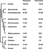
Fig. 1. The phylogenetic distribution of the major tick paralysis-inducing genera. Indicated are the number of species that are implicated in causing paralysis or toxicoses and the total number of species within the genus. The phylogenetic tree was compiled from data of cited sources (Klompen et al. 2000; Murrel, Campbell & Barker, 2001) and data on toxins from Gothe (1999).
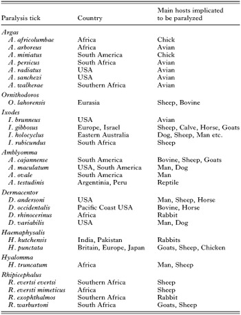
Table 1. Ticks that have been implicated in paralysis. Data were compiled from Gothe (1999). Definite confirmation of paralysis includes paralysis induced under experimental conditions, where numerous reports from various sources implicated the specific tick species or where the host recovered from paralysis symptoms after identification and removal of the tick
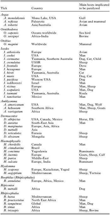
Table 2. Ticks that have been implicated in paralysis by case studies but for which no conclusive experimental data exist. Data were compiled from Gothe (1999)
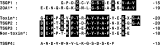
Fig. 2. N-terminal sequences obtained for the sand tampan toxins (TSGP2 and TSGP4) and a non-toxic homologue (TSGP3). The sequence for the non-toxic TSGP1 is also indicated, which shows sequence similarity to a highly abundant protein from O. moubata (20A1). The Toxin* and non-toxin* shown derive from sequences previously obtained (Neitz, 1976). Data were taken from Mans et al. (2001).
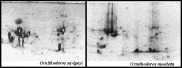
Fig. 3. Proteomes obtained for salivary gland extracts from O. savignyi and O. moubata. In each case, the salivary gland extracts are those from 10 ticks. Indicated are TSGP1-4 from O. savignyi. At least three highly abundant proteins in the SGE from O. moubata have similar molecular masses and iso-electric points as TSGP1-3.
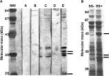
Fig. 4. Novel proteins associated with sweating sickness positive strains of H. truncatum. A. Western blot analysis of salivary gland extracts (SGE) obtained from positive and negative strains of H. truncatum. Lane A is naïve serum on SS+ SGE. Lane B is naïve serum on SS- SGE. Lane C is SS+ anti-sera on SS- SGE. Lane D is SS- anti-sera on SS- SGE. Lane E is SS+ anti-sera on SS+ SGE. B. SDS–PAGE analysis of SS- and SS+ SGE. Indicated are unique protein bands observed in the SS+ SGE.
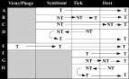
Fig. 5. Possible sources of tick toxins. A. Toxins (T) might derive directly from a pathogen or symbiotic organism living within the tick. B. A non-toxic (NT) component from a symbiotic organism is transformed by the tick to a toxic form. C. A non-toxic component from a symbiotic organism is transformed by the host to a toxic form. D. A non-toxic component from a tick is transformed by a symbiotic organism to a toxic form. E. A toxin could derive from a bacteriophage or virus that infects a tick symbiont or pathogen. F. A toxin might derive directly from the tick. G. The tick secretes a non-toxic component which is transformed within the host, to a toxin. H. A non-toxic component from the host is ingested by the tick and transformed to a toxic form before resecretion into the host.
 You have
Access
- 82
- Cited by