Case report
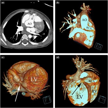
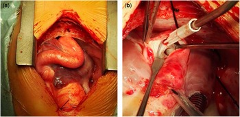
The patient was a 1-year-old Chinese boy, weighing 9 kg. He had a history of tachycardia for ~1 month. A physical examination revealed a diastolic murmur at intercostal space 3–4 along the left border of the sternum. An echocardiogram demonstrated an enlarged left ventricle (47 mm), with an inner orifice diameter of 15 mm at the right coronary artery and 2 mm at the left coronary artery. The right coronary artery fistula was found to drain into the left ventricle, and the diameter of the fistula orifice was 7.5 mm. Cardiac function was significantly compromised, and the ejection fraction (EF%) was 53%. A Multi-detector Computed Tomographic (MDCT) showed an enlarged right coronary sinus (Fig 1a and b) without obvious regurgitation and a tortuously enlarged right coronary artery with a fistula to the left ventricle (Fig 1c and d). A coronary artery fistula repair was performed. At surgery, the massive right coronary artery was observed to be tortuous (Fig 2a). The trunk of the right coronary artery was mobilised and tightened. After a longitudinal incision along the right coronary artery trunk, the fistula orifice was visible (Fig 2b) and closed using a running suture with 5/0 prolene. The patient was discharged in stable condition.
CT showing an enlarged right coronary sinus ( a ) and a giant beginning of the right coronary artery ( b ); volume-rendered images showing the fistula located at the bottom of the left ventricle ( c ) with the left ventricle enlarged ( d ). AO = aorta; LV = left ventricle; RCA = right coronary artery; RV = right ventricle.

Intraoperative appearance of the enlarged right coronary artery after a median sternotomy incision ( a ); the draining orifice of the coronary artery fistula into the left ventricle after a right coronary artery incision ( b ).

Concurrence of a coronary artery fistula and coronary artery ectasia is extremely rare.Reference Funabashi, Asano and Komuro 1 Most coronary artery fistulas are asymptomatic. Symptomatic coronary artery fistulas are rare in infants, and CT is useful for the diagnosis. We speculate that early operative intervention may avoid further development of coronary artery defects.
Acknowledgements
The authors acknowledge Dr Jijia Liu and Wangping Chen for their participation during surgical repair as assistants, and Dr Shuwen Yuan for providing these CT images.
Financial Support
This research received no specific grant from any funding agency or from commercial or not-for-profit organisations.
Conflicts of Interest
None.
Ethical Standards
The study has been approved by the ethics committee of Second Xiangya Hospital, Central South University. Written consent for publication has been obtained from the patient’s parents.