Hypoplastic left heart syndrome consists of several structural abnormalities in the left side of the heart. It is characterised mainly by hypoplasia or absence of the left ventricle and some degree of mitral (atresia or stenosis) or aortic valve underdevelopment.Reference Noonan and Nadas1 The hypoplastic left heart syndrome incidence has been estimated at 1 in 4000 to 6000 live births and its mortality rate for non-surgically treated individuals exceeds 90% over the first year.Reference Reamon-Buettner and Borlak2–5
Among the main factors in heart defects, those associated with mutations in genes responsible for regulating heart tissue development are considered the most important ones. Somatic mutations in one of these genes are associated with structural heart defects since even very low levels of these morphogenic regulators may result in reduced cardiomyocyte replication, ventricular hypoplasia, and endocardial abnormalities.Reference Reamon-Buettner and Borlak2–Reference Visoná, Benati and Monti4
Hypoplastic left heart syndrome may have a hereditary genetic cause,Reference Loffredo, Chokkalingam and Sill6, Reference Wessels, Berger and Frohn-Mulder7 possibly related to the connexin gene GJA1.Reference Chein8 Dasgupta et alReference Dasgupta, Martinez, Zuppan, Shah, Bailey and Fletcher9 found connexin-43 gene mutations in 8 of 14 children with hypoplastic left heart syndrome and in 1 child with a defective atrioventricular canal, but not in 46 healthy individuals and 6 children with other diagnoses requiring heart transplantation. It is important to highlight that six children with hypoplastic left heart syndrome did not show connexin-43 gene mutations, a fact consistent with the polygenic hypoplastic left heart syndrome nature.
Connexin-43 gene mutations affect phosphorylation sites in a similar way in two apparently unrelated diseases: hypoplastic left heart syndrome and visceroatrial heterotaxy syndrome. It is difficult to determine the exact onset of both. Hypoplastic left heart syndrome seems to be related to restricted blood flow at the end of embryogenesis, while visceroatrial heterotaxy syndrome probably starts earlier.Reference Chein8 Nevertheless, there are reports of two children with family histories of polysplenia presenting either hypoplastic left heart syndrome or visceroatrial heterotaxy syndrome.Reference Rose, Izukawa and Moës10 A newer study found a 10% incidence rate of left ventricular and mitral valve hypoplasia in patients with visceroatrial heterotaxy syndrome and polysplenia, as well as a 28% rate of aortic stenosis, conditions also seen in children with hypoplastic left heart syndrome.Reference Van Praagh, Geva and Friedberg11
Connexins may somehow play a role in HLHS development as such proteins belong to a family of phosphoproteins that act in gap junctions. This type of junctions consists of homologous protein groups that make up communicating canals, which in its turn apparently allow the passage of ions and low molecular weight molecules. Gap junctions thus coordinate intercellular communication permitting the flow of molecules lesser than 1000 daltons (e.g. ions, simple sugars, amino acids).5, Reference Beyer, Paul and Goodenough12
Connexins behave differently at each life period: embryonic development, neonatal period, and adulthood. Considering a normal heart, gap junction density increases during intrauterine life and connexin-43 expression level is higher in the neonatal period. In adulthood, heart maturation leads to a better metabolic and electrical coupling between cells and consequently to a fewer number of connexins into the membrane, as these are more concentrated in intercalated discs – sites more subject to stress.Reference Van Kempen, Vermeulen, Moorman, Gros, Paul and Lamers13
Hypoplastic left heart syndrome has been described since the 1950s by researchers who focus on heart defects. Although it is the most frequent and best-known defect affecting left heart outflow, its ultrastructural and molecular features are still under investigation. Moreover, only a few studies have investigated the relationship between connexin-43 and this syndrome, especially with human tissues because it is very difficult to obtain them.Reference Dasgupta, Martinez, Zuppan, Shah, Bailey and Fletcher9, Reference Lev14–Reference Sun, Wang, Wang, Zhang and Pu16
Given the scarcity of studies addressing the relationship between hypoplastic left heart syndrome and connexin-43, especially those with human tissues, this study aims to analyse the expression of connexin-43 in the cardiac muscle of children diagnosed with hypoplastic left heart syndrome by Western blot method and confocal laser scanning microscopy.
Methods
Sample and groups
Tissue samples were taken during corrective surgery to treat heart defects. A small quantity was removed from the right ventricle and processed for confocal scanning laser microscopy and Western blot technique analyses.
The control group (n = 8) consisted of children diagnosed with any type of heart defect that required surgery but with no relation to hypoplastic left heart syndrome, connexin-43, or its gene. The hypoplastic left heart syndrome group (n = 9) consisted of children presenting this disease singly, without any other associated congenital disease or syndrome.
Immunofluorescence confocal scanning laser microscopy
Five samples from each group were prepared to investigate the spatial distribution of the connexin-43 by confocal scanning laser microscopy. They were fixed with 2% paraformaldehyde in phosphate-buffered saline for 4 hours and embedded in Tissue-Tek. Twenty micrometer-thick slices were cut on a Leica CM1850 cryostat and transferred to silanized slides. These were then rinsed with phosphate-buffered saline, incubated in phosphate-buffered saline + 1% bovine serum albumin + 0.005% saponin for 30 minutes, and incubated with connexin-43 antibodies from Cell Signaling Technology in phosphate-buffered saline + saponin + bovine serum albumin for 2 hours. Following, they were incubated in anti-rabbit IgG conjugated to fluorescein isothiocyanate, diluted 1:500 in phosphate-buffered saline + saponin + 1% bovine serum albumin, for roughly 40 minutes. After incubation in the secondary antibody and rinsing, the samples were incubated in 4,6-diamidino-2-phenylindole, diluted 1:3000 in phosphate-buffered saline with 0.01% saponin, for 15 minutes. Finally, the samples were rinsed five times with phosphate-buffered saline and Milli-Q water and then coverslip-sealed with Fluoromont-G mounting medium.
The slices were blindly analysed by two observers using a BIO-RAD MRC-1024 confocal microscope.
Western blot
Four samples from control group and five from hypoplastic left heart syndrome group were analysed. The heart tissues of hypoplastic left heart syndrome and control samples were homogenized in lysis buffer containing protease inhibitors (phenylmethane sulfonyl fluoride or 0.1 mM Pefabloc, 1 µg/ml aprotinin, and 1 µg/ml leupeptin) according to the manufacturer’s instructions and an aliquot was taken for protein quantification. Samples containing 50 µg of protein were separated by SDS-polyacrylamide gel electrophoresis (10%) and transferred to a nitrocellulose membrane.
The membranes were incubated for 120 minutes in TSB-T (150 mM NaCl, 20 mM Tris, and 1% Tween 20, pH 7.4) containing 5% non-fat-dried milk to inhibit non-specific binding sites. The blots were then washed with TSB-T and probed with antibodies against anti-connexin-43 and anti-β-actin for overnight at 4 °C. The membranes were washed with TBS-T and incubated with peroxidase-conjugated monoclonal anti-rabbit IgG for anti-connexin-43 and peroxidase-conjugated monoclonal anti-mouse IgG for anti-β-actin for 120 minutes at room temperature.
The immunocomplexed peroxidase-labelled antibodies were visualized with an enhanced chemiluminescence kit according to the manufacturer’s instructions and were exposed to photographic film Kodak Dektol developer of the Department of Biochemistry of Paulista School of Medicine. The band densities were determined by densitometry analysis using the ImageJ software. The density values of the bands were normalized to the total β-actin present in each lane and were expressed as a percentage of a control sample with an intermediate value.
Statistical analysis and ethical issues
Data normality was verified by the Shapiro–Wilk test. The Mann–Whitney test was then applied to detect differences between the groups, with a p-value < 0.05 representing statistical significance.
This study was approved by the Ethics Committee of Federal University of São Paulo Research (protocol 0225/08).
Results
Microscopy analysis
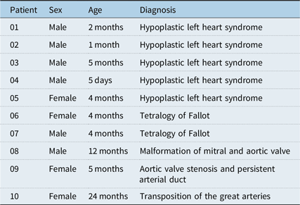
Patients’ characteristics from both groups are summarised in Table 1.
Patients’ characteristics: microscopy analysis

HLHS group: patients 1–5; control group: patients 6–10
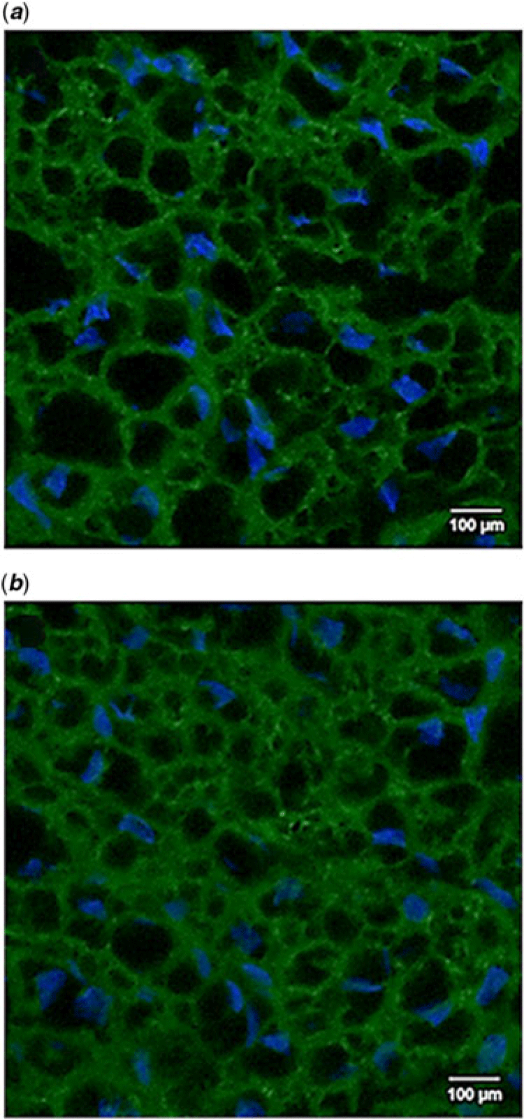
It was noticed no qualitative differences between the groups. All patients seem to present the same connexin-43 positioning and location pattern. Cells had one or two blue-stained nuclei surrounded by connexin-43 from the cell membrane, it clearly indicating the intercellular boundaries. They also had green-stained dots in the cytoplasm, especially near the nucleus. Connexin-43 expression in some control and hypoplastic left heart syndrome samples seems to be diffusely higher in certain regions. Figure 1 shows an image from each group to illustrate the above-mentioned information.
Confocal scanning laser microscopy photomicrographs of heart tissue samples from patients of HLHS (a) and control (b) group. Note the blue-stained cell nuclei surrounded by green-stained connexin-43 molecules located in the plasma membrane of these cells.

Western blot analysis
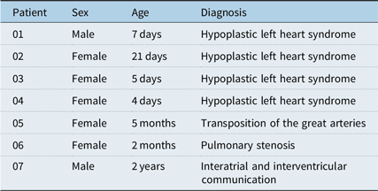
Patients’ characteristics from both groups are summarised in Table 2.
Patients’ characteristics: Western blot analysis

HLHS group: patients 1–4; control group: patients 5–7
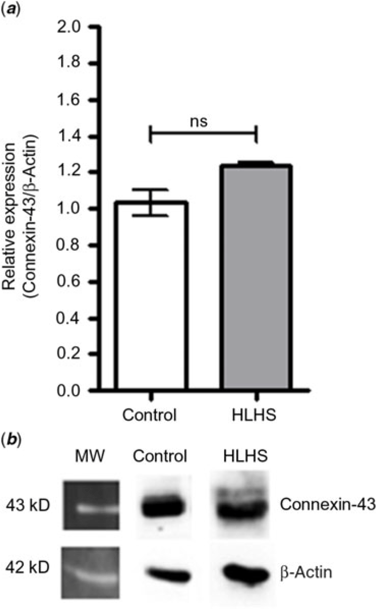
The mean connexin-43 expression from hypoplastic left heart syndrome and control groups was 1.239 (±0.2738) and 1.031 (±0.1270), respectively. There was no statistically significant difference between them (p = 0.0571) (Fig. 2).
Connexin-43 protein expression. (a) Graphic representation of the quantification of the bands. (b) Connexin-43 (43 kDa) and control β-actin protein expression (42 kDa) detected by Western blot analysis. MW = molecular weight; ns = no significance. (Mann–Whitney test p = 0.0571).

Discussion
Connexin-43 was detected in both groups by confocal scanning laser microscopy and Western blot analysis. It was expected, as the abundance of connexin-43 in heart tissue has been well described by many authors.Reference Weidmann17–Reference Strachan and Read19 In addition, no statistically significant difference was noted between both groups, probably because hypoplastic left heart syndrome has a multifactorial etiology. On the other hand, other heart diseases (e.g. aortic stenosis, hypertension, and even ischemia) may alter connexin-43 amount.Reference Goldfine, Walcott, Brink, Magid and Borer20A sample studied in the present study was from a patient with tetralogy of Fallot and, according to Kolcz et al,Reference Kolcz, Drukala, Bzowska, Rajwa, Korohoda and Malec21 newborns and infants with this disease may have a fewer connexin-43 amount. Thus, we believe this fact could be considered a study bias.
Confocal scanning laser microscopy showed that connexin-43 is located peripherally in the cell, that is, at the membrane. Connexin-43 in both study groups had this same spatial distribution, information confirmed by Sun et al,Reference Sun, Wang, Wang, Zhang and Pu16 who demonstrated its usual position between cells by staining the ventricular cardiomyocytes of neonatal rats with fluorescent antibodies. These authors confirmed their results submitting the samples to Western blot analysis, which was not possible in the present study. Moreover, unlike the above-mentioned authors, we did not use confocal microscopy to quantify protein expression, further limiting the comparison of both studies.
Goldfine et alReference Goldfine, Walcott, Brink, Magid and Borer20 studied connexin-43 in rabbits with dilated cardiomyopathy secondary to aortic insufficiency. Although aortic insufficiency was induced by surgery, the connexin-43 expression increased, a fact that did not occur in their control group and in our study. Even with not enough evidence, it is generally thought that protein content at the beginning of aortic insufficiency may be lower than the normal, but it is compensated later for by connexin-43 plasticity when the ventricle is enlarged. Western blot analysis showed that the amount of connexin-43 did not increase in any group, even in right ventricular hypertrophy, a common condition found mainly in hypoplastic left heart syndrome patients. In addition to aortic insufficiency, the same authors reported that connexin-43 expression also increases in animals with renovascular hypertension, aortic stenosis, and ischemic heart disease, that is, the amount of this protein increases in models with chronic heart volume overload. This situation was expected for the study sample, as hypoplastic left heart syndrome patients experience right ventricular volume overload during the fetal phase.
The heart tissue sections from both groups submitted to confocal microscopy suggest the connexin-43 presence on the entire surface of cardiomyocytes, in agreement with Kolcz et alReference Kolcz, Drukala, Bzowska, Rajwa, Korohoda and Malec21 and Peters et alReference Peters, Severs, Rothery, Lincoln, Yacoub and Green22 who suggested that human neonatal myocardium shows a random and irregular connexin-43 distribution on the entire surface of ventricular myocytes. During heart maturation, connexin-43 migrates to form the pattern seen in adults, in which they are highly organised in clusters within intercalated discs.Reference Severs, Coppen, Dupont, Yeh, Ko and Matsushita18, Reference Kolcz, Drukala, Bzowska, Rajwa, Korohoda and Malec21
Peters et alReference Peters, Severs, Rothery, Lincoln, Yacoub and Green22 stated that connexin-43 distribution may change until age 6 and demonstrated that almost all the connexin present in normal heart tissue of younger children is located in the intercalated discs, a pattern compatible with the normal distribution in adults. It suggests that the morphologic maturation of right ventricular tissue may occur earlier than it was believed.
The age-related changes in the connexin-43 distribution pattern are related to ventricular functionality and maturation.Reference Kolcz, Drukala, Bzowska, Rajwa, Korohoda and Malec21, Reference Sanchez-Quintana, Garcia-Martinez, Climent and Hurle23 Organisation of gap junctions in newborns is ideal for myocardial growth and adaptive remodelling. Hence, knowing the exact moment of connexin-43 redistribution may suggest the best time to repair surgically some defects, such as tetralogy of Fallot. Eliminating the right ventricular pressure load during childhood may prevent myocardial hypertrophy and fibrosis and improve the mechanical and electrical performance of the heart as well.Reference Sanchez-Quintana, Garcia-Martinez, Climent and Hurle23 Surgical repair in early life may influence myocardial maturation before the remodelling ability of the heart decreases with age.Reference Deanfield, McKenna and Hallidie-Smith24, Reference Pozzi, Trivedi, Kitchiner and Arnold25
In mature heart tissue, electrical impulses propagate faster and hardly passes transversally from one cell to another, mainly because of less amount of side-to-side gap-junction connections. The irregularly distributed junctions of immature myocardia result in slower impulse propagation.Reference Delmar, Michaels, Johnson and Jalife26, Reference Uzzaman, Honjo and Takagishi27
Conclusion
Within the limits of this study, it is suggested that cardiomyocytes of hypoplastic left heart syndrome children are similar in connexin-43 location, distribution, and structural and conformational patterns to those of children with heart defects not related to this protein and its genes.
Acknowledgements
None.
Financial Support
This research received no specific grant from any funding agency, commercial or not-for-profit sectors.
Conflicts of Interest
None.