Introduction
The molecular control of both totipotency and pluripotency is under intense investigation (Cockburn & Rossant Reference Cockburn and Rossant2010; Moura, Reference Moura2012; Yeo & Ng, Reference Yeo and Ng2013; Dang-Nguyen & Torres-Padilla, Reference Dang-Nguyen and Torres-Padilla2015; Rossant, Reference Rossant2016; Wu et al., Reference Wu, Yamauchi and Izpisua Belmonte2016; Silva et al., Reference Wu, Yamauchi and Izpisua Belmonte2017). These cellular states are controlled at different molecular levels (e.g. epigenetic, transcriptional, and post-transcriptional levels), but transcriptional control plays a pivotal role as most characterized genes so far are pluripotency-associated transcription factors (PATF) (Moura, Reference Moura2012; Rossant, Reference Rossant2016; Wu et al., Reference Wu, Yamauchi and Izpisua Belmonte2016). Several of such PATFs were identified as required for the maintenance of inner cell mass viability and pluripotency of embryonic stem (ES) cells (Nichols et al., Reference Nichols, Zevnik, Anastassiadis, Niwa, Klewe-Nebenius, Chambers, Schöler and Smith1998; Yeo & Ng, Reference Yeo and Ng2013; Goissis & Cibelli, Reference Goissis and Cibelli2014; Silva et al., Reference Wu, Yamauchi and Izpisua Belmonte2017).
Despite intense investigation of PATFs in humans and mice (Dang-Nguyen & Torres-Padilla Reference Dang-Nguyen and Torres-Padilla2015), limited information is available about these genes in other species (Berg et al., Reference Berg, Smith, Pearton, Wells, Broadhurst, Donnison and Pfeffer2011; Goissis & Cibelli, Reference Goissis and Cibelli2014; Takahashi et al., Reference Takahashi, Sakurai, Emura, Hashizume and Sawai2015; Moura et al., Reference Moura, Silva, Cantanhêde, Silva, Ferreira-Silva, Silva, Ramos-Deus, Pandolfi, Kido, Benko-Iseppon and Oliveira2018). Some reports have determined the temporal and cell-type specific expression of PATFs in goats, cattle, and buffaloes (He et al., Reference He, Pant, Schiffmacher, Bischoff, Melican, Gavin and Keefer2006; Sanna et al., Reference Sanna, Sanna, Mara, Pilichi, Mastinu, Chessa, Pani and Dattena2010; Rodríguez-Álvarez et al., Reference Rodríguez-Álvarez, Cox, Tovar, Einspanier and Castro2010; Anand et al., Reference Anand, Kumar, Singh, Shah, Chauhan, Manik, Singla and Palta2011; Moura et al., Reference Moura, Silva, Cantanhêde, Silva, Ferreira-Silva, Silva, Ramos-Deus, Pandolfi, Kido, Benko-Iseppon and Oliveira2018). However, few reports have investigated the expression of PATFs in sheep oocytes and embryos, while other reports have only detected PATFs at the blastocyst stage (Sanna et al., Reference Sanna, Sanna, Mara, Pilichi, Mastinu, Chessa, Pani and Dattena2010; Kim et al., Reference Kim, Sakurai, Bai, Bai, Sato, Nagaoka, Chang, Godkin, Min and Imakawa2013; Wang et al., Reference Wang, Lin and Yu2013). Moreover, even less information is available on PATF function and regulation in ruminant species (Berg et al., Reference Berg, Smith, Pearton, Wells, Broadhurst, Donnison and Pfeffer2011; Takahashi et al., Reference Takahashi, Sakurai, Emura, Hashizume and Sawai2015).
Therefore, this work aimed to determine the transcript availability of eight PATFs, namely CMYC, GLIS1, KLF5, NR0B1 (DAX1), OCT4 (POU5F1), RONIN (THAP11), ZFP281 (ZNF281), and ZFX during sheep and cattle preimplantation development.
Materials and methods
Ethical approval
All procedures were approved by the Ethics committee at the Universidade Federal Rural de Pernambuco (UFRPE), under the licence number 035/2016 (Process 23082.002437/2016).
Cattle and sheep embryo in vitro production
Cattle and sheep ovaries were collected at local slaughterhouses (Pernambuco, Brazil) and transported to the laboratory in 0.9% NaCl solution containing 1 μl ml–1 100× penicillin and streptomycin solution (Invitrogen) at 35°C, within 3 h after slaughter. Cumulus–oocyte complexes (COC) were aspirated from cattle and sheep ovaries using 10 ml syringes into a HEPES-buffered medium, consisting of TCM-199 with Hank’s salts supplemented with 2 mM l-glutamine, 10% fetal bovine serum (FBS), 25 IU ml–1 heparin, and 1 μg ml–1 gentamicin sulfate. Sheep COCs were retrieved from 2–6 mm follicles, while cattle COCs were obtained from 2–8 mm follicles, respectively. All cattle and sheep COCs that displayed three or more cumulus cell layers and homogeneous cytoplasm were destined for in vitro maturation (IVM), as described by Moura et al. (Reference Moura, De Sousa, Leme and Rumpf2008, Reference Moura, Silva, Cantanhêde, Silva, Ferreira-Silva, Silva, Ramos-Deus, Pandolfi, Kido, Benko-Iseppon and Oliveira2018).
The IVM medium for both cattle and sheep COCs was formulated with TCM-199 with Earle’s salts supplemented with 50 μg ml–1 sodium pyruvate, 2.6 mg ml–1 sodium bicarbonate, 5.0 μg ml–1 FSH, 5.0 μg ml–1 LH, 2 mM l-glutamine, 10% FBS, and 0.5 μg ml–1 gentamicin sulfate. Groups of 15–30 COCs were placed in 150 μl droplets of IVM medium covered with mineral oil and incubated under 5% CO2, with saturated humidity, at 38.5°C for 24 h.
The parthenogenetic activation (PA) of sheep eggs was carried out as previously described by Moura et al. (Reference Moura, De Sousa, Leme and Rumpf2008) and Bebbere et al. (Reference Bebbere, Bogliolo, Ariu, Fois, Leoni, Succu, Berlinguer and Ledda2010). Briefly, sheep COCs were denuded by initial treatment with 0.2% hyaluronidase and followed by gentle pipetting for 5 min. Furthermore, denuded sheep eggs were washed in embryo culture medium, composed of SOFaa supplemented with 5% FBS (SOFaaFBS). The PA was carried out with 5 μM ionomycin for 5 min, washed in SOFaaFBS and cultivated in 2 mM 6-DMAP for 3 h (Bebbere et al., Reference Bebbere, Bogliolo, Ariu, Fois, Leoni, Succu, Berlinguer and Ledda2010). Finally, activated eggs were washed extensively in SOFaaFBS before embryo culture (Moura et al., Reference Moura, De Sousa, Leme and Rumpf2008).
Cattle COCs were partially denuded after IVM by gentle pipetting before in vitro fertilization (IVF). Groups of 15–25 cattle COCs were initially washed in FERT–TALP medium containing 10 μg ml–1 heparin sulphate (IVF medium (IVFM)). Finally, COCs were placed in 100 μl IVFM droplets and covered with mineral oil. The IVF was carried out with frozen–thawed semen from bulls of proven fertility. A thawed 0.25 ml semen straw had its content deposited in a conical microtube containing 0.5 ml Percoll gradient (45%/90%) and centrifuged at 700 g for 7 min. The semen pellet was resuspended in IVFM and centrifuged at 700 g for 7 min. The supernatant was removed, and sperm concentration was determined for IVF. The IVF was performed with a final sperm concentration of 1.0 × 10⁶ sperm cells ml–1. Oocytes and sperm were co-cultured during IVF under 5% CO2, with saturated humidity, at 38.5°C for 18–20 h.
Groups of 15–20 cattle or sheep presumptive zygotes were washed once and placed into 150 μl SOFaaFBS droplets, on a cumulus cell monolayer, and covered with mineral oil. Cattle or sheep embryos were incubated in 5% CO2, under saturated humidity, at 38.5°C, for 7 days. Embryonic development was scored on day 3 (D3) for cleavage rates, on day 5 (D5) for morula development and day 7 (D7) for blastocyst development.
Total RNA extraction and cDNA synthesis
Pools were formed by 50–100 metaphase II eggs, 10–20 cleavage-stage embryos, 10–15 morulae or 10–15 blastocysts. Eggs or embryos were washed extensively in phosphate-buffered saline (PBS) without calcium and magnesium, snap frozen in liquid nitrogen (−196°C), and stored at −80°C. The total RNA extraction was performed using an RNeasy Micro Plus kit (Qiagen), according to manufacturer instructions. Total RNA was immediately used for cDNA synthesis.
The reverse transcription (RT) reaction was carried out using a QuantiTect® RT Kit (Qiagen). Samples were initially treated with the genomic DNA elimination reaction, containing 2 μl reaction buffer, 12 μl total RNA and at a final volume of 14 μl at 42°C for 2 min. The cDNA synthesis reaction was composed of 14 μl genomic DNA elimination reaction, 4 μl 5× quantiscript RT buffer, 1 μl RT primer mix, and 1 μl quantiscript RT in a final volume of 20 μl at 42°C for 30 min. The RT enzyme was inactivated by incubation at 95°C for 3 min. The cDNA was stored at −20°C.
Primer design
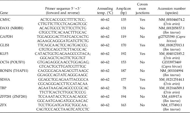
Primers were designed using Primer-BLAST (Ye et al., Reference Ye, Coulouris, Zaretskaya, Cutcutache, Rozen and Madden2012), and further selected by Primer 3 software (Rozen & Skaletsky, Reference Rozen and Skaletsky2000). Primers were expected to amplify 70–200 base pair amplicons, 40–60% GC content, 19–23 bases in size while avoiding primer–dimer formation, and preferably covering at least one exon junction (Table 1).
Primers for detection of pluripotency-associated transcription factors in sheep (Ovis aries) and cattle (Bos taurus) eggs and preimplantation embryos by reverse transcriptase quantitative PCR (RT-qPCR). Annealing and extension temperatures were 60ºC for sheep and 62ºC for cattle, respectively

Reverse transcriptase quantitative polymerase chain reaction (RT-qPCR)
The RT-qPCR was carried out in a LineGene 9660, model FQD-96 A (Bioer®), using the SYBR Green detection system. The RT-qPCR reactions were composed by 1 µl cDNA, 5 µl Go Taq qPCR Master Mix 2× (Promega®), 3.4 µl ultrapure H2O, and 0.6 µl primers (2.5 µM) in a final volume of 10 µl. Both oocyte and embryo cDNA samples were used as concentrated samples.
The RT-qPCR reactions were carried out with the following conditions: initial denaturation at 95°C for 2 min, followed by 40 cycles of 95°C for 15 s and 60–62°C for 30 s. Both annealing and extension at 60°C were used for reactions using sheep samples, while annealing and extension at 62°C were used for cattle samples. Melting curves were analyzed at 65–95°C for 20 min after 40 cycles. Transcript availability of each PATF was assessed by the quantification cycle (Cq) at the exponential phase of the RT-qPCR reaction.
Bioinformatics
Seed sequences from human (Homo sapiens) were used to search for candidate sequences in sheep (Ovis aries) and cattle (Bos taurus) for CMYC, GLIS1, KLF5, NR0B1 (DAX1), OCT4 (POU5F1), RONIN (THAP11), ZFP281 (ZNF281) and ZFX genes on UniProt (http://www.uniprot.org/). A tBLASTn was carried out using known mRNA seed sequences to search for additional PATFs candidates, and alignments with a cut-off e-value of <e–10 were considered hits (https://www.ncbi.nlm.nih.gov). Furthermore, protein translation was performed using ORF-Finder, to find the open reading frame of each candidate mRNA sequence (Rombel et al., Reference Rombel, Sykes, Rayner and Johnston2002). Protein molecular weight and isoelectric point predictions were performed using JVirGel 2.0 (Hiller et al., Reference Hiller, Grote, Maneck, Münch and Jahn2006). Protein subcellular location was performed using Cell-PLoc 2.0 (Chou & Shen, Reference Chou and Shen2008). The phenetic analysis was carried out using MEGA7 software using the neighbour-joining method (Kumar et al., Reference Kumar, Stecher and Tamura2016). All in silico analysis were carried out in February 2017.
Results
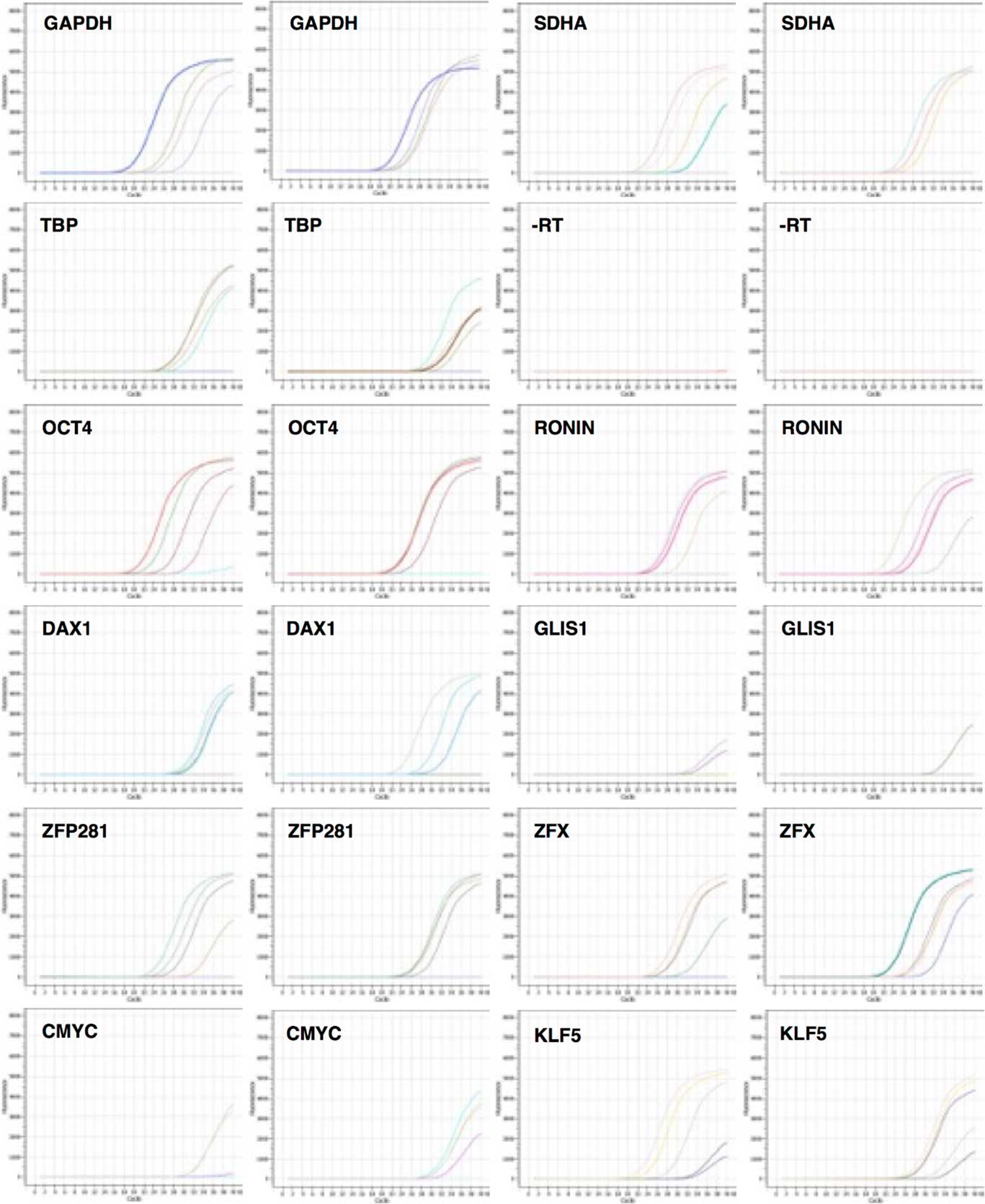
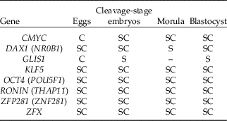
Cattle embryos were produced by IVF, while sheep embryos were produced by PA (Table S1). Housekeeping genes GAPDH, SDHA, and TBP were detected in all egg and embryo samples (Fig. 1). Gene transcripts of all candidate PATFs were found in sheep and cattle during preimplantation development (Fig. 1), with melting curves compatible with single amplicons (Fig. S1). Among all PATFs detected in both species, there were three distinct temporal expression patterns (Table 2). Firstly, five PATFs (KLF5, OCT4, RONIN, ZFP281, and ZFX) were detected in eggs and all embryonic developmental stages of both species (Table S2).
Amplification curves from reverse transcriptase quantitative PCR (RT-qPCR) of eight pluripotency-associated transcription factors in sheep (Ovis aries) and cattle (Bos taurus) eggs and during preimplantation development.

Summary of temporal expression of pluripotency-associated transcription factors in sheep (S) and cattle (C) eggs and preimplantation embryos as detected by reverse transcriptase quantitative PCR (RT-qPCR)

Secondly, the gene product of CMYC was detected in cattle eggs and embryos (Table 2). In sheep, CMYC was not detected in eggs but was indeed found in embryos at all developmental stages. Transcripts of NR0B1 were detected in all sheep samples but was not in cattle morula-stage embryos (Table 2). CMYC showed similar transcript availability throughout the preimplantation development of both species (Table S2).
Thirdly, GLIS1 showed a distinct temporal transcript availability than other PATFs (Table 2). This gene product was only detected in cattle eggs while, in sheep, it was detected in cleavage-stage embryos and blastocysts (Table S2).
In silico analysis of PATFs at the protein level displayed a mean of 3.37 sequences for each PATF in sheep (Table S3), mostly predicted sequences from the genome (not experimentally characterized). The retrieval in cattle led to one sequence for each PATF, except CMYC, with two different isoforms (Table S3). The CMYC, OCT4 and RONIN proteins were very similar between sheep and cattle, if not identical based on its size, isometric point and molecular weight (Table S3).
The other five PATFs showed such similarities among the cattle sequence and one sheep isoform, while other sheep isoforms displayed noticeable variation on all protein parameters (Table S3). Curiously, sheep had 13 different ZFX sequences, although protein sequence alignment suggested that only two different proteins are found (data not shown). A phenetic analysis of PATFs sequences showed that CMYC and ZFX are more closely conserved between sheep and cattle (Fig. S2). For the remaining PATFs, small ruminants clustered more closely, while cattle clustered with buffaloes (Fig. S2).
Discussion
The temporal expression of eight PATFs was determined in sheep and cattle during preimplantation development. Cattle embryos were produced by IVF and sheep embryos were produced by PA. The method used for in vitro embryo production affects PATF relative expression but does not influence temporal gene expression (Bebbere et al., Reference Bebbere, Bogliolo, Ariu, Fois, Leoni, Succu, Berlinguer and Ledda2010). OCT4 expression was lower in parthenogenetic blastocysts in comparison with IVF counterparts, although it was similar for cleavage and morula stages (Bebbere et al., Reference Bebbere, Bogliolo, Ariu, Fois, Leoni, Succu, Berlinguer and Ledda2010). Moreover, NANOG expression was similar within IVF and PA embryos at both morula and blastocyst stages (Bebbere et al., Reference Bebbere, Bogliolo, Ariu, Fois, Leoni, Succu, Berlinguer and Ledda2010). Therefore, it may be ruled out that the method of embryo production influenced the temporal transcript availability of PATFs in this study.
RONIN is a PATF detected throughout the preimplantation development of both species. This gene product was initially characterized as an essential factor for embryogenesis and mouse ES viability (Dejosez et al., Reference Dejosez, Krumenacker, Zitur, Passeri, Chu, Songyang, Thomson and Zwaka2008). Moreover, combined ectopic expression of RONIN, CMYC, and KLF4 allowed reprogramming of amniotic cells to pluripotency in mice and humans (Nagata et al., Reference Nagata, Toyoda, Yamaguchi, Hirano, Makino, Nishino, Miyagawa, Okita, Kiyokawa, Nakagawa, Yamanaka, Akutsu, Umezawa and Tada2009). At the genomic level, RONIN was identified as an inducer of protein synthesis and energy metabolism in mouse ES cells (Dejosez et al., Reference Dejosez, Levine, Frampton, Whyte, Stratton, Barton, Gunaratne, Young and Zwaka2010). In retinal cells, RONIN increases the expression of mitochondrial genes (Poché et al., Reference Poché, Zhang, Rueda, Tong, Mcelwee, Wong, Hsu, Dejosez, Burns, Fox, Martin, Zwaka and Dickinson2016).
This work is the first to describe the expression of ZFP281 in mammalian preimplantation embryos. The available information on ZFP281 was described in ES cells (Wang et al., Reference Wang, The, Chan, Chu, Rossbach, Kunarso, Allapitchay, Wong and Stanton2008; Fidalgo et al., Reference Fidalgo, Shekar, Ang, Fujiwara, Orkin and Wang2011). This gene was initially identified as a PATF due to its differential expression within ES cells and somatic cells (Wang et al., Reference Wang, Rao, Chu, Shen, Levasseur, Theunissen and Orkin2006, Reference Wang, The, Chan, Chu, Rossbach, Kunarso, Allapitchay, Wong and Stanton2008), and it forms protein complexes with NANOG, OCT4, and SOX2 (Wang et al., Reference Wang, Rao, Chu, Shen, Levasseur, Theunissen and Orkin2006, Reference Wang, The, Chan, Chu, Rossbach, Kunarso, Allapitchay, Wong and Stanton2008). Moreover, ZFP281 controls several downstream genes with these PATFs and also directly fine-tuning NANOG expression (Wang et al., Reference Wang, The, Chan, Chu, Rossbach, Kunarso, Allapitchay, Wong and Stanton2008).
GLIS1 displayed species-specific temporal expression in sheep and cattle preimplantation development. GLIS1 is one of the less investigated PATFs and was identified in a screen for genes involved in cellular reprogramming (Maekawa et al., Reference Maekawa, Yamaguchi, Nakamura, Shibukawa, Kodanaka, Ichisaka, Kawamura, Mochizuki, Goshima and Yamanaka2011). Its expression in mice is high in oocytes and zygotes, but low in ES cells, fibroblasts, and other somatic cell-types (Maekawa & Yamanaka Reference Maekawa and Yamanaka2011). This gene is expressed in cattle oocytes and embryos until the four-cell stage (Takahashi et al., Reference Takahashi, Sakurai, Emura, Hashizume and Sawai2015). GLIS1 knockdown caused embryonic arrest in cattle cleavage-stage embryos, possibly due to incomplete embryonic genome activation (Takahashi et al., Reference Takahashi, Sakurai, Emura, Hashizume and Sawai2015).
ZFX is expressed ubiquitously throughout preimplantation development in sheep and cattle. Previous studies have shown that ZFX expression during preimplantation development is an example of X-linked genes (Bernardi et al., Reference Bernardi, Cotinot, Payen and Delouis1996; Peippo et al., Reference Peippo, Farazmand, Kurkilahti, Markkula, Basrur and King2002). More recently, ZFX was demonstrated to be required for ES pluripotency in both mice and humans (Galan-Caridad et al., Reference Galan-Caridad, Harel, Arenzana, Hou, Doestsch, Mirny and Reizis2007; Harel et al., Reference Harel, Tu, Weisberg, Esquilin, Chambers, Liu, Carson, Studer, Reizis and Tomishima2012), although ZFX knockout embryos are capable of development up to the blastocyst stage and complete implantation (Luoh et al., Reference Luoh, Bain, Polakiewicz, Goodheart, Gardner, Jaenisch and Page1997; Galan-Caridad et al., Reference Galan-Caridad, Harel, Arenzana, Hou, Doestsch, Mirny and Reizis2007).
NR0B1 (DAX1) was expressed throughout embryonic development in both species, except cattle morula-stage embryos. NR0B1 has been detected in mouse oocytes and preimplantation embryos (Clipsham et al., Reference Clipsham, Niakan and McCabe2004; Stickels et al., Reference Stickels, Clark, Heider, Mattiske, Renfree and Pask2015), but it is not required for blastocyst formation (Yu et al., Reference Yu, Ito, Saunders, Camper and Jameson1998). NR0B1 contributes to the process of cellular reprogramming (Zhang et al., Reference Zhang, Liu, Ruan, Wang, Zhao, Wan, Liu, Zheng, Peng, Wu, He, Hu and Jian2014; Lujan et al., Reference Lujan, Zunder, Ng, Goronzy, Nolan and Wernig2015), although its requirement for ES pluripotency remains controversial (Khalfallah et al., Reference Khalfallah, Rouleau, Barbry, Bardoni and Lalli2009; Zhang et al., Reference Zhang, Liu, Ruan, Wang, Zhao, Wan, Liu, Zheng, Peng, Wu, He, Hu and Jian2014).
KLF5 is a PATF detected for the first time in eggs and embryos of both sheep and cattle. This gene is ubiquitously expressed during mouse preimplantation development and is required for pluripotency (Ema et al., Reference Ema, Mori, Niwa, Hasegawa, Yamanaka, Hitoshi, Mimura, Kawabe, Hosoya, Morita, Shimosato, Uchida, Suzuki, Yanagisawa, Sogawa, Rossant, Yamamoto, Takahashi and Fujii-Kuriyama2008; Parisi et al., Reference Parisi, Cozzuto, Traction, Passaro, Ciriello, Aloia, Antonini, De Simone, Pastore and Russo2010). The role of KLF5 in pluripotent cells is through direct regulation of other PATFs and silencing of developmental regulators that trigger cellular differentiation (Parisi et al., Reference Parisi, Cozzuto, Traction, Passaro, Ciriello, Aloia, Antonini, De Simone, Pastore and Russo2010).
The expression of CMYC differed in eggs as it was detected only in cattle, as previously described by Miles et al. (Reference Miles, McDaneld, Wiedmann, Cushman, Echternkamp, Vallet and Smith2012). Although CMYC was found throughout the preimplantation development of both species, another report did not detect CMYC at the morula-stage in cattle, but only in blastocysts (Madeja et al., Reference Madeja, Sosnowski, Hryniewicz, Warzych, Pawlak, Rozwadowska, Plusa and Lechniak2013).
In silico analysis at the protein level gave some insights into PATFs conservation within cattle and sheep. The strategy of function prediction by homology (Lee et al., Reference Lee, Redfern and Orengo2007), showed high homology at the protein level for all PATFs, suggesting conserved function within ruminants. Curiously, sheep showed multiple isoforms for each PATFs, with significant protein variation. The fact that some sheep PATFs isoforms show structural differences suggests that this species may hold particularities on the PATF-mediated control of pluripotency. Experimental validation of PATFs sequence and function may further exclude redundant sequences in sheep and may identify paralogous sequences with novel properties. The phenetic analysis also suggests that PATFs may have conserved roles within these species.
New computational tools have been described to evaluate pluripotent cells (Bian & Cahan Reference Bian and Cahan2016; Kurum et al., Reference Kurum, Benayoun, Malhotra, George and Ucar2016). The combined application of these approaches to experimental data on PATFs may accelerate our ability to dissect totipotency and pluripotency control during ruminant preimplantation development.
In conclusion, temporal expression of PATFs differs between sheep and cattle, suggesting possible species-specific regulation during preimplantation development.
Acknowledgements
This work was supported by the FACEPE funding (Grant number APQ-1101-5.05-15).
Conflict of interest
The authors declare no conflict of interest.
Author contributions
P.G.C. Silva, M.T. Moura, R.L.O Silva, A.M Benko-Iseppon and M.A.L. Oliveira conceived the study. P.G.C. Silva, M.T. Moura, R.L.O. Silva, P.S. Nascimento, J.B. Silva, J.C. Ferreira-Silva, L.F. Cantanhêde and M.S. Chaves performed the study. P.G.C. Silva, M.T. Moura, R.L.O Silva, A.M Benko-Iseppon and M.A.L. Oliveira performed analysis and interpretation of the data. P.G.C. Silva, M.T. Moura, R.L.O. Silva, A.M. Benko-Iseppon and M.A.L. Oliveira wrote the manuscript with suggestions from all authors.
Supporting Information
Additional Supporting Information is available online at the publisher’s website.
Figure S1. Melting values from reverse transcriptase quantitative PCR (RT-qPCR) for eight pluripotency-associated transcription factors in sheep (Ovis aries) and cattle (Bos taurus) eggs and preimplantation embryos.
Figure S2. Phenetic analysis of eight pluripotency-associated transcription factors in 10 selected species. Sheep (Ovis aries) and cattle (Bos taurus) sequences are outlined.
Table S1. Cattle (Bos taurus) and sheep (Ovis aries) in vitro embryo production efficiency
Table S2. Parameters from reverse transcriptase quantitative PCR (RT-qPCR) reactions for eight pluripotency-associated transcription factors in sheep (Ovis aries) and cattle (Bos taurus) eggs and preimplantation embryos
Table S3. Protein size, isometric point, molecular weight, and subcellular localization for eight pluripotency-associated transcription factors in sheep (Ovis aries) and cattle (Bos taurus)
Supplementary materials
To view supplementary material for this article, please visit https://doi.org/10.1017/S0967199418000175