Issues of nomenclature
The word “heterotaxy” is derived from the greek words “heteros” and “taxis”, meaning no more than other than normal arrangement. The heterotaxy syndromes are characterized by the presence of abnormal symmetry of the organs and the veins, as well as discordance between the arrangement of the various organs, and between the various segments of the heart. This results in a high incidence of cardiac anomalies of extreme heterogeneity, with symmetrical lungs and disorganized abdominal organs. Characteristically, there are splenic abnormalities, such as absence of the spleen, so-called asplenia, or multiple spleens, known also as polysplenia. In a minority of patients, nonetheless, the spleen can be a solitary structure.
The body normally has paired and unpaired structures. The unpaired structures are usually lateralized, while the paired structures are usually bilaterally symmetrical and mirror imaged, a fine example being the hands. These paired structures are the essence of bodily isomerism.1 In patients with visceral heterotaxy, the lungs usually exhibit isomerism, so that there are either morphologically right lungs bilaterally, or morphologically left lungs on both sides. For example, in right isomerism the main bronchuses are bilaterally short and eparterial, whereas in left isomerism they are long and hypoarterial. With respect to the heart, the most constant cardiac feature that determines sidedness is the anatomy of the pectinate muscles of the atrial appendages.2
Although everyone agrees on the use of the word heterotaxy, there is ongoing controversy as to the issue of cardiac isomerism. Although we recognize that we are unable to settle this controversy in this review, understanding both sides of the issue is important, since they are widely reflected in the literature. Those that support the concept of cardiac isomerism divide the patients with heterotaxy into two groups, namely those with isomerism of the right or left atrial appendages.2, 3 Those that oppose the idea of cardiac isomerism divide the patients into two general groups based on the state of the spleen, and recognize the syndromes of asplenia or polysplenia.4 In general, right isomerism and asplenia, as well as left isomerism and polysplenia, have been used as synonyms. Although this would be true in most cases, Uemura and associates5 have demonstrated that among 82 autopsy cases with isomerism of the right atrial appendages in which the splenic state was known, the spleen was absent in only 65, with 14 having a solitary spleen, and three multiple spleens. Likewise, among 41 autopsied cases known to have isomerism of the left atrial appendages with known splenic state, only 36 had multiple spleens, with three having a solitary spleen, and the spleen being absent in two.
Thus, although these general groupings are useful in helping us to anticipate what associated lesions to expect, and to search for, the precise cardiac anomalies in each specific patient need to be diagnosed using the various diagnostic modalities. From the point of view of the clinician diagnosing the living patient, the state of the spleen can be currently be ascertained with greater ease than the extent of the pectinate muscles within the atrial chambers.
Cardiac anomalies in heterotaxy
Although the cardiac malformations show considerable variability in the setting of heterotaxy, they tend to occur in two major patterns, those associated with absence of the spleen, and those coexisting in patients with multiple spleens. The patterns summarized below are based on a study of 109 pathologic cases from the Cardiac Registry at the Boston Children's Hospital.6
Asplenia syndrome
According to the venoarterial connections, the atrial arrangement was deemed to be usual in half the cases, mirror imaged in three-tenths, but ambiguous in the remainder. The heart itself was left sided in two-thirds. Bilateral superior caval veins were found in 71% of cases, and the coronary sinus was absent in 95% of cases.
Although interruption of the inferior caval vein was not seen, a separate and contralateral hepatic vein is present 28% of the time. Totally, anomalous pulmonary venous connection to a systemic vein is present in 64% of cases. A common atrioventricular junction guarded by a common valve is seen in 69% of cases, with two separate atrioventricualr orifices in 21%, respectively. In terms of ventriculo-arterial connections, 82% have double outlet right ventricle, while the connections are discordant in 9%. Only 45% of cases have two well-developed ventricles, with 42% having either left ventricular hypoplasia or an absent left ventricle, and 13% having right ventricular hypoplasia or an absent right ventricle. The ventricular topology is right handed in 62%, and left handed in the remainder. Obstruction to the pulmonary outflow tract is present in 96% of cases, whereas subaortic obstruction is extremely rare.
Polysplenia syndrome
According to the venoarterial connections, the atrial arrangement was thought to be usual in 78%, and mirror imaged in the remainder, with no ambiguous cases. The heart was left sided in two-thirds of cases. Interruption of the inferior caval vein is present in 80% of cases, and 50% have bilateral superior caval veins. The pulmonary venous connections were normal in 61%, and to the right-sided atrium with abnormal attachment of the interatrial septum in 22%. A common atrioventricular junction guarded by a common valve was found in 33% of cases, and by separate right and left valves in 32%. There was double outlet right ventricle in 37%, but discordant ventriculo-arterial connections in only 2%. Two well-developed ventricles were seen in 63% of cases, left ventricular hypoplasia in 24%, and right ventricular hypoplasia in 11% of cases. Obstruction to the subpulmonary outflow tract was present in 43%, and subaortic obstruction in 22%.
Similar patterns were observed in another large series of 183 patients with visceral heterotaxy reported by Uemura and associates,5 but the latter workers, of course, divided their cohort into groups with right or left isomerism.
Conduction system
The anatomy of the conduction system in the heterotaxy syndromes is variable, and is dependent on whether there is right or left isomerism, two atrioventricular valves or a common atrioventricular junction, and on the presence of right hand or left hand, ventricular topology. Knowledge of the location of the conduction system, particularly of the sinus node is important in order to avoid damage during the cavopulmonary anastomosis or the Fontan operation. In a detailed study on the conduction system in relation to cardiac morphology in heterotaxy, by Smith and associates,7 valuable conclusions were made for the surgeon. The arrangements of the sinus nodes is exclusively dependent on the type of isomerism. In right isomerism, there are bilateral sinus nodes, while in left isomerism the sinus nodes are hypoplastic or absent. The nodes in right isomerism are located in the venoatrial grooves bilaterally, even in the presence of only one superior caval vein. The surgeon should avoid these areas during the performance of the cavopulmonary anastomoses or during the Fontan operation. In left isomerism, things are less clear, as the sinus nodes, when present, are closer to the atrioventricular junction. The surgeon is well advised to respect the venoatrial junctions and the atrioventricular vestibules.
In left isomerism, there is usually a common atrioventricular junction and the ventricular loop determines the atrioventricular conduction axis. With right hand topology, there is typically a solitary atrioventricular node located posteroinferiorly at the meeting point of the ventricular septum and the atrioventricular junction. With left hand topology, in contrast, there are usually two atrioventricular nodes, irrespective of the type of isomerism. Although the atrioventricular conduction can be damaged during biventricular repair, this is much less likely with during cavopulmonary anastomosis or the Fontan operation, especially if the procedures are extracardiac.
Surgical considerations
The success of surgery for patients with the heterotaxy syndromes depends on a complete and precise understanding of the cardiac anatomy and physiology in each patient. This includes an accurate knowledge of the systemic and pulmonary venous drainage; the size, anatomy and function of the atrioventricular valves; the ventricular morphology and size; the types of atrioventricular connections; the size, anatomy and function of the arterial valves; and the anatomy and disposition of the specialized conducting tissues. Furthermore, precise knowledge of the systemic and pulmonary circulations is also essential.
The surgical options will depend on the precise constellation of anatomic cardiac malformations, including anomalies of systemic and pulmonary venous connections. Biventricular repair is extremely unlikely in patients with asplenia or right isomerism, due to the much higher occurrence of functionally univentricular physiology and complex intracardiac lesions, such as common atrioventricular junction with double outlet right ventricle or discordant ventriculo-arterial connections.5, 6, 8 Pacifico and associates9 have reported biventricular repair for these lesions involving a modified Senning or Mustard operations. Recently, Tchervenkov and associates10 reported a technique of translocation of the ventricular septal defect to achieve anatomical biventricular repair for these lesions when the aorta completely arises from the right ventricle and the ventricular septal defect has no subaortic extension. In patients with polysplenia syndrome, or left isomerism, although biventricular repair is more likely, functionally univentricular palliation still predominates.11–13 One and a half ventricle repair, with a concomitant cavopulmonary shunt, has been reported for some patients with right ventricular hypoplasia.14
Due to the frequent occurrence of multiple cardiac anomalies, and the presence of significant ventricular hypoplasia, the majority of patients will undergo functionally univentricular palliation, and ultimately be managed by creation of the Fontan circulation. The presence of a common atrioventricular junction, with the potential for regurgitation across the common valve with a negative impact on ventricular function, implies that the type and timing of surgical palliation in early life is extremely important. Furthermore, an accurate and complete understanding of the systemic and pulmonary venous anatomy is an absolute prerequisite for the successful creation of the Fontan circulation. Indeed, in the presence of significant ventricular dysfunction, or unrepairable regurgitation across a common atrioventricular valve, transplantation may be the only surgical option.
Functionally univentricular palliation
For the patient with a functionally univentricular heart, there are numerous hurdles before the systemic and pulmonary circulations can successfully be separated. Most patients will undergo three surgical stages. The importance of initial palliation cannot be overstated, since it will have a great impact in allowing the patient to become an ideal candidate for the eventual creation of the Fontan circulation. Furthermore, the high incidence of totally anomalous pulmonary venous connection in patients with asplenia or right isomerism makes initial palliation in these patients even more challenging.
Initial palliation in early life
The goal of initial surgical palliation is to achieve the highest possible survival in an optimal state for the eventual creation of the Fontan circulation. Achieving this goal requires the establishment of adequate but not excessive flow of blood to the lungs, an unobstructed systemic circulation, correction and prevention of pulmonary venous obstruction, prevention of pulmonary arterial distortions or stenoses, prevention of pulmonary vascular obstructive disease, preservation of normal systolic and diastolic ventricular function, and preservation of atrioventricular and arterial valvar function. These goals are not easy to achieve. Furthermore, the decision as to the type and timing of initial palliation is also not always easy. This is dependent on the extent of flow of blood to the lungs on initial evaluation, be it decreased and inadequate, excessive, or adequate but not excessive, the latter representing the balanced situation. In the presence of inadequate flow the patient is severely cyanotic, and requires creation of a systemic-to-pulmonary arterial shunt. Recently, one of us (C.I.T.) has successfully used an “hourglass” transannular autologous pericardial patch, instead of a shunt, to regulate the flow of blood to the lungs, and protect the distal pulmonary arterial bed. In the presence of excessive flow, the patient will present with mild if any cyanosis, and congestive heart failure. Banding of the pulmonary trunk will be needed in the absence of subaortic stenosis, or a Damus–Kaye–Stansel anastomosis and a creation of a systemic-to-pulmonary arterial shunt in the presence of subaortic stenosis. The presence of an obstructed aortic arch will require a Norwood type procedure. In the uncommon situation of balanced flow to the lungs, nothing needs to be done initially. This is usually the situation in the presence of moderate, but not severe, obstruction to the pulmonary outflow tract.
In the absence of totally anomalous pulmonary venous connection, patients with asplenia or right isomerism may require creation of a systemic-to-pulmonary arterial shunt due to the high incidence of obstruction within the pulmonary outflow tract. In the patients with multiple spleens or left isomerism, the majority entering the functionally univentricular route will also require initial palliation.11 Subaortic obstruction, extremely rare in those with asplenia or right isomerism, occurs in about one-fifth or more of those with polysplenia or left isomerism. These patients may be best treated with the Norwood operation.15
Pulmonary venous obstructive abnormalities
Pulmonary venous obstructive abnormalities are most commonly seen in the form of totally anomalous pulmonary venous connection, and rarely due to divided atrium, or failure of incorporation of the pulmonary veins into the atrial chambers. A majority of patients with asplenia or right isomerism have totally anomalous pulmonary venous connection. This places those in need of initial palliation during the newborn period at very high risk. The mortality of repair of totally anomalous pulmonary venous connection with functionally univentricular palliation has been reported to be between 33% and 95%.8, 16–18
Bidirectional cavopulmonary anastomosis
In the last 15 years, an intermediate stage has become the norm between initial palliation and creation of the Fontan circulation. This consists of directing the systemic venous return from the superior caval vein(s) into the pulmonary circulation, achieved with the bidirectional cavopulmonary anastomosis, or Glenn shunt, or the hemi-Fontan operation. Patients with heterotaxy surviving neonatal palliation, or presenting in late infancy, are candidates for the superior cavopulmonary anastomosis. Since preservation of optimal atrioventricular valvar and ventricular function are important to ensure that the patient remains a good candidate for creation of the Fontan circulation, an early cavopulmonary anastomosis should be considered in order to reduce the volume load on the functionally single ventricle, particularly in the presence of a common atrioventricular junction. Concomitant repair of the common atrioventricular valve, should it be moderately or severely regurgitant, may be necessary at the time of the cavopulmonary anastomosis. Bilateral bidirectional cavopulmonary anastomoses are usually necessary in the majority of the patients, due to the high incidence of bilateral superior caval veins. While some series of bidirectional cavopulmonary anastomosis have found heterotaxy to be a significant risk factor for mortality,19 others have not found this to be the case.20
In some ways, the second procedure is the “clean-up” stage, which offers the chance to correct associated abnormalities prior to creation of the Fontan circulation. At this stage, the surgeon should repair any stenoses in the pulmonary arteries, take care of residual or recurrent pulmonary venous obstruction, and deal with subaortic stenosis either directly or using the Damus–Kaye–Stansel anastomosis. Recurrent or residual obstruction in the aortic arch will also need attention, as will repair of the atrioventricular valve in the presence of moderate or severe regurgitation.
Kawashima operation
Many patients with heterotaxy, almost exclusively those with left isomerism, will have interruption of the inferior caval vein, with azygos continuation, usually into the contralateral superior caval vein, which is typically left sided. Although such interruption has not been seen in patients with right isomerism or asplenia, it is present in up to four-fifths of those with left isomerism or polysplenia.5, 6 This results in a special physiological situation, since the superior caval vein receiving the inferior caval venous return through an azygos connection carries a much higher volume of blood than is usual. The performance of the cavopulmonary anastomosis in these patients would result in a much greater proportion of the systemic venous return being diverted into the pulmonary circulation, excluding only the hepatic venous return. This was reported by Kawashima and associates,21 who described their procedure as a total cavopulmonary shunt. Many now refer to this procedure as the Kawashima operation. The superior caval vein receiving the azygos or hemi-azygos continuation of the interrupted inferior caval vein is anastomosed to the pulmonary artery as a bidirectional cavopulmonary anastomosis. Since most patients have bilateral superior caval veins, a concomitant contralateral Glenn shunt is also performed. Although most of the systemic venous return is directed to the pulmonary arteries, the hepatic drainage continues to drain into the atrium. A frequent late complication of this procedure has been the eventual development of pulmonary arteriovenous fistulas, and progressive arterial desaturation. Regression of these has been reported following reoperation to divert the hepatic venous return to the pulmonary circulation.22, 23 Uemura and associates24 have reported the use of an intra-atrial Goretex tube to divert the hepatic venous drainage to the pulmonary arteries.
The optimal timing of the Kawashima operation, and the subsequent completion of the Fontan circulation, remains to be determined. Due to the greater amount of systemic return that is diverted into the pulmonary circulation, the optimal time might be expected to be later than for a Glenn procedure, more around 1 year of age. It is also questionable as to whether the Kawashima operation provides definitive palliation. The development of pulmonary arteriovenous malformations, as well as the development of collateral channels between the higher pressure caval venous circulation and the lower pressure hepatic veins, will often result in progressive cyanosis. This will mandate completion of the Fontan circulation in order to direct the hepatic venous return to the pulmonary circulation, usually accomplished with an intra-atrial tube graft.
Fontan operation
The Fontan operation in patients with heterotaxy presents several technical challenges due to abnormalities in cardiac position and abnormal hepatic and pulmonary venous drainage. Due to this, it is difficult to generalize as to the optimal type of Fontan connection. Although the debate continues between the supporters of the lateral tunnel Fontan and the extracardiac Fontan, in some situations either one of these techniques is suboptimal. For example, it may not be possible to create a baffle within the atriums without causing pulmonary venous obstruction using the lateral tunnel technique, due to an abnormal position of some of the pulmonary veins or because of the location of the atrial anastomosis created during repair of totally anomalous pulmonary venous connection in early life. Likewise, the existence of separate inferior caval veins and hepatic venous return in some patients may preclude the ability to perform an extracardiac Fontan operation. In these situations, the complete intra-atrial conduit, or the combination of intra-atrial and extra-atrial conduits, may be indicated. Although the routine use of a fenestration is controversial, most will agree that it is indicated in patients with heterotaxy undergoing the Fontan operation.
Traditionally, patients with heterotaxy have been considered to be at high risk for the Fontan operation. This is well illustrated in a retrospective review of Fontan operations from the Mayo clinic.25 In their experience prior to 1987, among 500 patients undergoing the Fontan operation, only one-tenth had heterotaxy. This proportion had almost doubled in the 339 patients undergoing the Fontan operation since 1987. Also, the earlier period was associated with mortality of 43% for those with heterotaxy, whereas since 1987 the mortality was only 14.5%. The improvement was partially due to the use of the intra-atrial tube to prevent pulmonary venous obstruction. The presence of atrioventricular valvar regurgitation, and the need for valvar repair or replacement, was associated with a higher mortality.
Likewise, in a series of 500 consecutive patients from Boston Children's Hospital,26 heterotaxy was a risk factor for early failure. The use of a complete intra-atrial tube between the inferior caval vein and the cavopulmonary anastomosis to avoid pulmonary venous obstruction, and the addition of an intra-atrial fenestration, has resulted in decreased mortality.27, 28 In the most recent reports of the Fontan operation for heterotaxy, a further improvement in outcome was noted.29, 30
The improved outcome following the Fontan operation for heterotaxy is probably multifactial. It can be attributed to the introduction of an intermediate stage, such as the bidirectional Glenn and Kawashima procedures; the correction of concomitant disease such as pulmonary venous anomalies, atrioventricular valvar regurgitation and pulmonary arterial stenoses and distortions, the individualized Fontan technique with the use of the intraatrial tube graft when appropriate; and the use of Fontan fenestration. Despite the significantly improved outcome after the Fontan operation for heterotaxy, nonetheless, there remain several serious long-term complications.31 These include sinus nodal dysfunction, pulmonary venous obstruction, atrioventricular valvar regurgitation, and recurrent or persistent cyanosis due to intrahepatic shunting and the development of pulmonary arteriovenous malformations.
Overall outcome
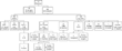
The significant challenges in the surgical treatment of patients with visceral heterotaxy are evident by the poor long term survival reported by the Hospital for Sick Children in Toronto (Fig. 1).8, 11 This series offers a glimpse into the natural history of patients with asplenia, with only 1 of 22 patients surviving without surgical treatment. The high surgical mortality is partially explained by the high incidence of pulmonary venous obstruction and major atrioventricular valvar abnormalities. Biventricular repair was performed in only 4 of 69 patients (5.8%) undergoing surgery. Only 13 patients in this series underwent the Fontan operation, with four deaths (30.7%). The overall pathways for treatment and outcomes for patients with right isomerism are illustrated in Figure 2.

Figure 1. Kaplan–Meier survival curve for 91 patients with right isomerism. Dashed lines represent 95% confidence limits. Reproduced with permission from: Hashmi A, Abu-Sulaiman R, McCrindle BW, Smallhorn JF, Williams WG, Freedom RM. Management and outcomes of right atrial isomerism: a 26-year experience. J Am Coll Cardiol 1998; 31: 1120–1126.

Figure 2. Flow chart showing surgical interventions and outcome in 91 patients with right isomerism. CPS: cavopulmonary shunt; Bivent: biventricular; CVS: cardiovascular surgery; PAB: pulmonary artery band; PVR: pulmonary vein repair; S-P: systemic-to-pulmonary artery; transplant: heart transplantation. Reproduced with permission from: Hashmi A, Abu-Sulaiman R, McCrindle BW, Smallhorn JF, Williams WG, Freedom RM. Management and outcomes of right atrial isomerism: a 26-year experience. J Am Coll Cardiol 1998; 31: 1120–1126.
Although long-term survival was better for those with left isomerism in the same institution, the mortality was still significant.11 The pathways for treatment and outcomes are illustrated in Figure 3. The long-term survival following biventricular repair at 1, 5, 10 and 15 years was 80%, 71%, 66% and 63%, respectively; whereas for functionally univentricular palliation for the same intervals, it was 73%, 61%, 53% and 48%, respectively (Fig. 4).

Figure 3. Flow chart of surgical interventions and outcome in 163 patients with left isomerism, comprising *nine patients with biventricular atrioventricular connections, 23 with functionally univentricular hearts, and 22 with virtually normal hearts; **four patients had Fontan-type surgery without prior interventions. Fontan-type includes bidirectional cavopulmonary shunt, right-atrium-to-pulmonary-artery anastomosis and hepatic-vein-to-pulmonary-artery rerouting. Reproduced with permission from: Gilljam T, McCrindle BW, Smallhorn JF, Williams WG, Freedom RM. Outcomes of left atrial isomerism over a 28-year period at a single institution. J Am Coll Cardiol 2000; 36: 908–916.

Figure 4. Kaplan–Meier survival curve for 163 patients with left isomerism, of whom 22 patients had an otherwise structurally normal heart, 71 had a heart suitable for biventricular repair, and 70 were suitable for functionally univentricular palliation. Survivors are denoted by dots. Vertical bars represent 95% confidence limits. Reproduced with permission from: Gilljam T, McCrindle BW, Smallhorn JF, Williams WG, Freedom RM. Outcomes of left atrial isomerism over a 28-year period at a single institution. J Am Coll Cardiol 2000; 36: 908–916.
Transplantation
Transplantation of the heart, or the heart and lungs, is offered when there are no other surgical options. In patients with visceral heterotaxy and a functionally univentricular heart, this could be any time along the treatment track before the Fontan operation, or for patients with a failing Fontan circulation. Cardiac transplantation will be indicated in the presence of poor ventricular function, and/or unrepairable regurgitation across the atrioventricular junctions, in the absence of pulmonary vascular obstructive disease. The presence of systemic venous anomalies, such as bilateral superior caval veins without a bridging vein, particularly in the presence of interruption of the inferior caval vein and azygos continuation, or separate return into the atriums of the inferior caval vein and the hepatic veins, will present important but not insurmountable technical challenges. Transplantation of the heart and lungs will be the only option in the presence of pulmonary vascular obstructive disease, or the presence of pulmonary venous obstruction, most likely after repair of totally anomalous pulmonary venous connection, precluding the Fontan operation as well as cardiac transplantation.
There are few published reports on transplantation in heterotaxy. Larsen and associates32 reported on experience with 29 patients with heterotaxy considered at high risk for surgical palliation or correction. Of these patients, 20 had undergone previous surgery. There were no perioperative deaths, and the survival of the graft at 1, 5 and 10 years was 86%, 68% and 50%, respectively.
Conclusions
Cardiac surgery for patients with visceral continues to present unique challenges, and requires precise understanding of the extremely heterogeneous variations in cardiac anomalies. Although biventricular repair is possible even in complex forms of biventricular atrioventricular connections, most patients will undergo functionally univentricular palliation culminating with creation of the Fontan circulation. Furthermore, although the results of functionally univentricular palliation are constantly improving, the presence of totally anomalous pulmonary venous connection and atrioventricular valvar regurgitation continue to be associated with higher mortality. Overall, patients with asplenia or right isomerism are much less amenable to biventricular repair, and have a poorer overall survival. Improved understanding of the heterotaxy syndromes and of the various surgical options, combined with detailed knowledge of the individual cardiac anatomy and physiology, will result in timely and appropriate surgical treatment. We sincerely hope that this review will, in small way, contribute to this goal.