Introduction
Pseudo-aneurysms of the petrous internal carotid artery (ICA) secondary to skull base osteomyelitis are uncommon. They do, however, present risks in an already ill patient of significant morbidity and mortality due to emboli from thrombus formation within the sac, or rupture resulting in a potentially catastrophic haemorrhage.
We present two cases of petrous ICA pseudo-aneurysms due to skull base osteomyelitis, and discuss their clinical presentation and management. In both patients, the pattern of cranial nerve involvement was not the one we would expect from skull base osteomyelitis. We think this finding could be an alarm sign for clinicians to look for other complications in these patients, such as ICA aneurysm.
Case reports
Case one
A 75-year-old diabetic male presented, having undergone several previous mastoidectomies performed in other centres for chronic otitis media with cholesteatoma. He underwent a further revision left-sided modified radical mastoidectomy in February 2017, and was found to have extensive recurrent cholesteatoma again. During this procedure, he underwent obliteration of the mastoid cavity, including closure of a post-aural mastoid cutaneous fistula. Two months following this procedure, he developed severe left-sided otalgia with bloody otorrhoea. In addition, he developed dysphonia, and miosis and ptosis of the left eye.
Microscopy demonstrated the presence of a large aural polyp within the mastoid cavity. The histology was consistent with a benign inflammatory lesion. Direct flexible laryngoscopy demonstrated left vocal fold palsy in the paramedian position. His facial nerve function was graded as House–Brackmann I.
A high-resolution computed tomography (CT) scan of the temporal bones demonstrated features consistent with his prior surgical interventions and bony erosion involving the jugular foramen, and soft tissue change consistent with skull base osteomyelitis. No organism was cultured.
The patient was commenced on a six-week course of intravenous piperacillin with tazobactam, and given topical treatment of ciprofloxacin and dexamethasone eardrops. Microdebridement was performed frequently.
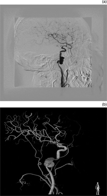
An intracranial magnetic resonance imaging (MRI) scan revealed a large pseudo-aneurysm of the left internal carotid artery (ICA) involving its entry into the skull base. A CT angiogram confirmed this finding (Figure 1).
(a & b) Digital subtraction angiographic images following left carotid injection, showing narrowing of the left distal cervical internal carotid artery and two focal pseudo-aneurysmal out-pouchings at the junction of the cervical–petrous carotid artery.

The patient underwent temporary balloon occlusion of the left ICA, which demonstrated good collateral circulation. The patient underwent endovascular coil occlusion of the left ICA the following day. The procedure was well tolerated, with no clinical evidence of new neurological deficits.
Case two
A 60-year-old diabetic male with a 6-month history of a chronically discharging right ear that was recalcitrant to topical treatment presented with progressive dysphagia and dysphonia. Microscopic examination revealed a central tympanic membrane perforation, with granulations involving the posterior rim of the perforation and extending into the mesotympanum. Multiple cranial nerve palsies were clinically evident, involving the right glossopharyngeal, vagal and hypoglossal nerves. His facial nerve function was graded as House–Brackmann I.
An MRI scan of the brain with gadolinium demonstrated inflammatory soft tissue enhancement of the right lateral skull base encasing the petrous portion of the right internal carotid artery (ICA). Pseudomonas aeruginosa was isolated on culture.
The patient was commenced on a six-week course of intravenous Tazocin®. Ciprodex® was applied topically and microdebridement was conducted frequently.
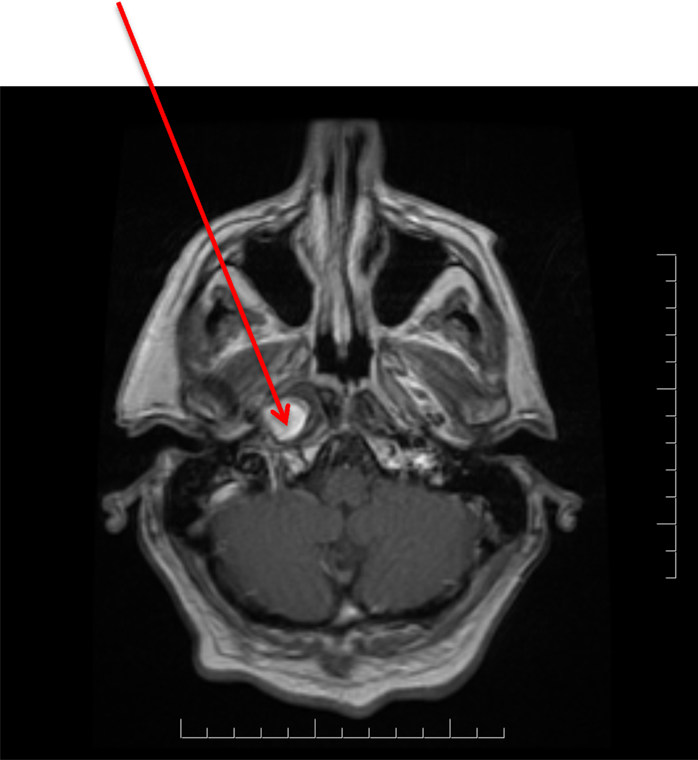
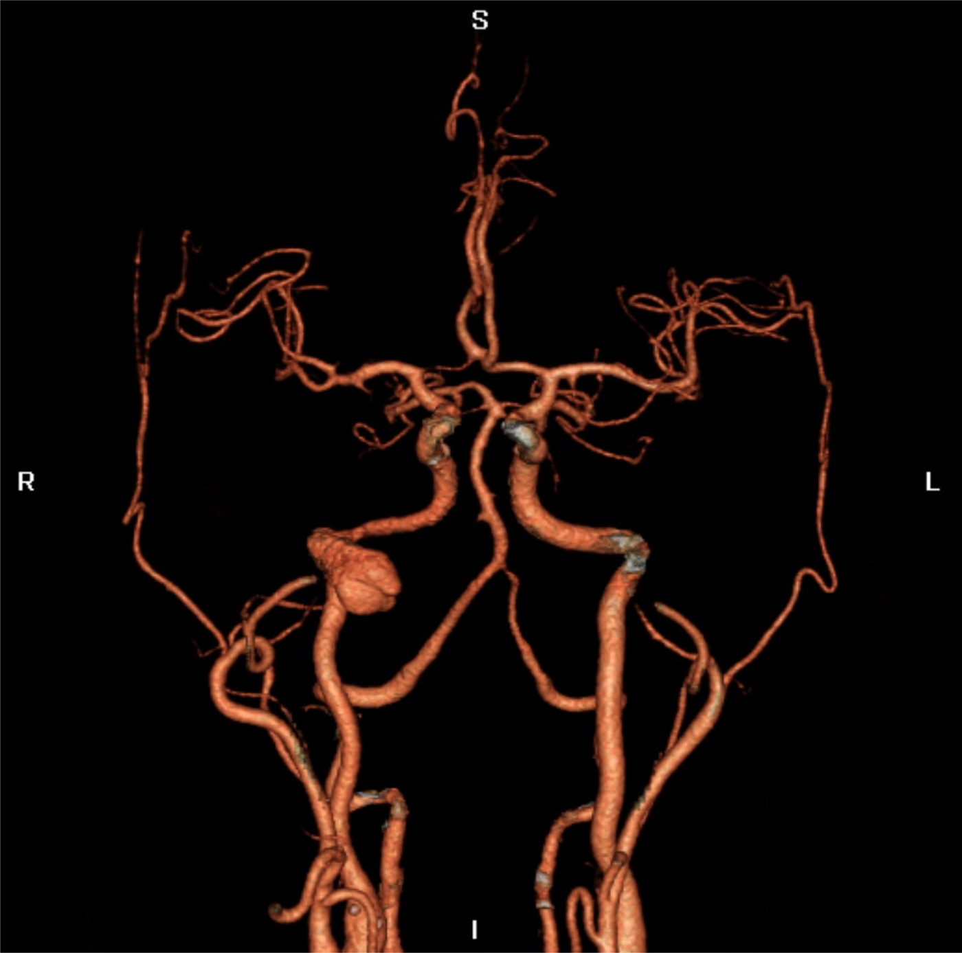
The patient's follow-up MRI scan, carried out six-months later, demonstrated marked improvement in prior soft tissue inflammation, but now revealed the presence of a pseudo-aneurysm involving the right ICA within its horizontal petrous portion (Figure 2). This finding was confirmed with CT angiography (Figure 3).
Axial, T1-weighted magnetic resonance imaging with gadolinium scan, demonstrating a pseudo-aneurysm (arrow) involving the distal right cervical internal carotid artery near the junction with the petrous segment.

Computed tomography angiogram, demonstrating a right junctional cervical–petrous internal carotid artery pseudo-aneurysm. S = superior; R = right; L = left; I = inferior

The patient was immediately recommenced on a further two-month treatment course of oral ciprofloxacin. The pseudo-aneurysm was coiled by the interventional neuroradiology division, after it was demonstrated that there was adequate collateral circulation following a balloon test occlusion.
Discussion
Skull base osteomyelitis usually occurs in immunocompromised patients, most commonly in the setting of diabetes. P aeruginosa is the causative organism in the majority of cases. Cranial nerve involvement occurs from progression of the osteomyelitis along the skull base. The facial nerve (VIIth cranial nerve) seems the most commonly involved cranial nerve, in up to 60 per cent of cases, primarily because of its proximity to the osteomyelitis within the external auditory canal.Reference Mani, Sudhoff, Rajagopal, Moffat and Axon1, Reference Chandler2 Medial progression of osteomyelitis to the jugular foramen and hypoglossal canal can result in palsies of the glossopharyngeal nerve, vagus nerve, spinal accessory nerves and hypoglossal nerve.Reference Rowlands, Lekakis and Hinton3 In one study involving 103 patients with skull base osteomyelitis, 60 patients (58 per cent) developed cranial nerve palsies; 58 of these 60 patients (97 per cent) developed facial nerve palsy, while 27 (45 per cent) had multiple cranial nerve palsies (typically involving the IX–XIIth cranial nerves).Reference Corey, Levandowski and Panwalker4 In that series, the facial nerve palsy preceded other cranial nerve palsies in 55 cases (92 per cent).
• Petrous internal carotid artery aneurysm is a rare complication of skull base osteomyelitis
• Facial nerve is the most commonly involved cranial nerve, preceding other cranial nerve palsies in skull base osteomyelitis
• Lower cranial nerve involvement, with minimal or no facial nerve involvement, should raise suspicion of pathologies other than skull base osteomyelitis, such as aneurysm or jugular thrombosis
• One should be mindful of this potentially fatal complication in patients with lower cranial nerve palsies, especially with intracranial thromboembolic events or Horner's syndrome
Pseudo-aneurysms of the intra-petrous portion of the internal carotid artery (ICA) are rare. They arise as a result of weakening of the adventitia associated with surrounding inflammation. While many petrous ICA aneurysms are asymptomatic, they can produce a wide range of clinical signs and symptoms. Various combinations of single or multiple cranial nerve deficits have been reported.Reference Tanaka, Patel, Shrier and Coniglio5 Compression or stretching of the sympathetic caroticotympanic plexus from aneurysms in this location can result in Horner's syndrome,Reference Mangat, Nayak and Chandna6 as identified in case one. Ruptures of the petrous ICA aneurysm may present with massive haemorrhage from the ear, epistaxis via the Eustachian tube and neurological deficit associated with stroke, provided the haemorrhage does not prove lethal in the first instance.Reference Costantino, Russell, Reisch, Breit and Hart7
Both our cases presented with lower cranial nerve palsy with minimal facial nerve involvement. This pattern of cranial involvement is unusual for skull base osteomyelitis, in which facial nerve involvement usually precedes other cranial nerve involvement.
Considering their fatal complications, one should be cognisant of carotid artery aneurysms, particularly when a recalcitrant case of malignant external otitis presents with medial progression of skull base osteomyelitis at the level of the jugular foramen and/or hypoglossal canal, especially when it occurs in association with Horner's syndrome and minimal facial nerve involvement. These findings should alert clinicians to search for other causes of cranial nerve involvement, including ICA pseudo-aneurysm or jugular vein thrombosis,Reference Oliveira, Trigo, Castanho, Marques and Almeida8 and not simply attribute them to skull base osteomyelitis. In suspected cases, vascular imaging of the skull base in the form of a CT or magnetic resonance angiogram should be arranged.
The management of petrous aneurysms is challenging. Currently, treatment options for petrous ICA aneurysm include conservative management with serial imaging or surgery. Open surgical intervention is a well-described treatment option, but a similar result can be achieved with a less invasive endovascular occlusion.Reference Bien, Cress, Nguyen, Westagate and Nanda9 More recently, endovascular techniques have become more appealing. These include endovascular ICA balloon occlusion or endovascular coil placement, with sacrifice of the vessel following balloon occlusion testing to ensure there is sufficient contralateral blood supply.Reference Bien, Cress, Nguyen, Westagate and Nanda9, Reference McGrail, Heros, Debrum and Beyerl10
Conclusion
Petrous internal carotid artery aneurysms are a rare complication of skull base osteomyelitis. One should be mindful of this potentially fatal complication in patients with lower cranial nerve palsies, with or without facial nerve involvement, especially in the presence of intracranial thromboembolic events or Horner's syndrome.
Competing interests
None declared