

Introduction
‘Oboyerite’ was one of four new tellurium oxysalt minerals described from the Grand Central mine, Tombstone, Cochise County, Arizona, USA by Williams (Reference Williams1979), the others being ‘girdite’, fairbankite and winstanleyite. Only winstanleyite has a known crystal structure (Bindi and Cipriani, Reference Bindi and Cipriani2003), while Kampf et al. (Reference Kampf, Mills and Rumsey2017) recently discredited ‘girdite’ as a mixture. Kampf et al. (Reference Kampf, Mills and Rumsey2017) showed that the powder X-ray diffraction (PXRD) data for ‘girdite’ reported by Williams were mostly consistent with ottoite (Pb2TeO5; Kampf et al., Reference Kampf, Housley, Mills, Marty and Thorne2010) and that “wet chemical analyses were conducted on an impure mixture of phases, which appears to mainly correspond to the same material he [Williams] described as the new mineral oboyerite”. At the time of discrediting ‘girdite’, ‘oboyerite’ was noted to also “appear to be a mixture” (Kampf et al., Reference Kampf, Mills and Rumsey2017). ‘Oboyerite’ was described by Williams (Reference Williams1979) as tiny milk-white spherules of fibrous material perched on opal, closely associated with jarosite and fairbankite. ‘Oboyerite’ was noted to closely resemble ‘girdite’. Williams gave the ideal formula of oboyerite as H6Pb6(Te4+O3)3(Te6+O6)2·2H2O based upon wet chemical analyses and water determination using the Penfield method. He also provided powder data, but was unable to determine a unit cell, although he conjectured that ‘oboyerite’ was triclinic based upon a polarised-light optical study. A year later, Roberts (Reference Roberts1980) indexed the ‘oboyerite’ powder pattern using a triclinic unit cell with parameters a = 12.249(8), b = 15.113(6), c = 6.868(3) Å, α = 116.45(4), β = 98.58(4) and γ = 85.82(4)°. Optical determinations in S−Se melts yielded the indices of refraction α = 2.24 and γ = 2.26. The specific gravity measured by the Thoulet method was 6.4(6) g/cm3. No crystal structure has been reported for ‘oboyerite’, and no synthetic Pb–Te–O compound has a similar formula. One of the minerals shown here to be a component of ‘oboyerite’, plumbotellurite, was first reported from the Zhana–Tyube Au deposit, Akmola Region, Kazakhstan (Spiridonov and Tananaeva, Reference Spiridonov and Tananaeva1982), as orthorhombic Pb2+Te4+O3. Back (Reference Back1990) determined that plumbotellurite is not orthorhombic, but is identical to the monoclinic form of Pb2+Te4+O3, α-Pb2+Te4+O3; however, the reporting of plumbotellurite as orthorhombic has remained in some crystallographic databases. Weil et al. (Reference Weil, Shirkhanlou, Füglein and Libowitzky2018) noted that although three synthetic polymorphs of Pb2+Te4+O3 have been described, none of these matched the two minerals with the formula Pb2+Te4+O3, plumbotellurite and fairbankite. The discreditation of ‘oboyerite’ provides an ideal opportunity to clarify the identity of plumbotellurite. Together these results allow for a better understanding of the Pb–Te–O system, potentially paving the way for future thermodynamic study of lead–tellurium oxysalts and their formation environments (Grundler et al., Reference Grundler, Brugger, Etschmann, Helm, Liu, Spry, Tian, Testemale and Pring2013; Brugger et al., Reference Brugger, Liu, Etschmann, Mei, Sherman and Testemale2016).
Oboyerite specimens analysed
No formal type specimens of ‘oboyerite’ were designated in the published paper (Williams, Reference Williams1979), but the submission for the new mineral to the Commission on New Minerals and Mineral Names indicated that the designated type specimens were to be deposited in the Natural History Museum, London (NHM) and the Geological Museum of the University of Arizona, USA. Online, a number of different institutions claim type material, with the Smithsonian Institution the sole institution listed on www.mindat.org, while the IMA Commission on Museums Catalogue of Type Mineral Specimens claims that types were to be preserved at the Smithsonian Institution, the NHM in London and the Muséum National d'Histoire Naturelle in Paris. These specimens can all be traced directly to the author, Sid Williams, but based on the official new-mineral submission, which ultimately has precedence, only those at the NHM in London and the University of Arizona should be considered type specimens.
The original published paper indicates that there were two ‘fist-sized’ specimens that contained ‘oboyerite’, but significantly, only one was used to determine the characteristics and therefore, to the best of our knowledge, this specimen was the original holotype sample. It is also significant that this specimen was the only one found containing fairbankite and, therefore, is the original holotype for both ‘oboyerite’ and fairbankite.
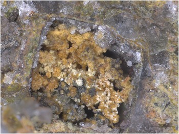
Other studies (e.g. Kampf et al., Reference Kampf, Mills, Rumsey, Dini, Birch, Spratt, Pluth, Steele, Jenkins and Pinch2012) have shown that Sid Williams was not meticulous when it came to the designation of type material. The NHM, London type specimen (BM 1980,540) has with it a label in the handwriting of Williams indicating that, as we should expect, it is a type for both fairbankite and ‘oboyerite’, however the dimensions are 30 mm × 20 mm × 10 mm: therefore it can only be a part of the original fist-sized holotype. PXRD analysis of the sample when it was registered at the NHM, London in 1979 failed to identify ‘oboyerite’ and optical study during this work also failed to identify any material that resembled the description given in the original paper. Material most similar to the visual description was extremely small cream-coloured wispy aggregates associated with jarosite and chlorargyrite rather than opal (Fig. 1). Being unsuitable for further PXRD or single-crystal XRD, a fragment was mounted into a probe block and analysed using a Cameca SX100 microprobe (Core Research Laboratories, NHM, London). This material was determined to be an Fe-rich winstanleyite, the associated mineralogy of which fits with that in the original winstanleyite description, but is not mentioned as a mineral present on the fist-sized ‘oboyerite’ type sample, which we consider a consequence of the very small size of these aggregates.
A cavity formed from a completely replaced galena crystal on the BM 1980,540 specimen. The cream coloured wispy material most resembling ‘oboyerite’ is in fact Fe-rich winstanleyite. Associated yellowish minerals are chloragyrite and jarosite, while opal lines the cavity. Scale bar = 500 μm.

In a final attempt to locate ‘oboyerite’, the entire sample was studied in a JEOL JT1500 variable-pressure scanning electron microscope (SEM) in low vacuum mode. No crystals were found that resembled the physical form, the associated mineralogy or the chemical determination of ‘oboyerite’.
On the basis of these analyses and the presence of a substance with a PXRD pattern matching fairbankite on the specimen, it was concluded that, although once a part of the fist-sized holotype, after splitting, no ‘oboyerite’ was retained on this specific piece. From henceforth, it should be recorded that the NHM, London specimen is a ‘dead type’ for the species ‘oboyerite’.
As no type ‘oboyerite’ material could be obtained from the type NHM, London sample, some of the other samples that could be traced directly to Williams were studied. The cotype specimen at the Geological Museum of the University of Arizona was assessed, but although minute white balls of material were visible on the specimen and attempts were made to remove them for analysis, none were suitable for an accurate PXRD determination (University of Arizona, personal communication).
Consequently, the major specimen used for analysis in this study is housed in the collection of the Natural History Museum of Los Angeles County (NHMLAC, catalogue number 67241) and was donated for study by Robert A. Jenkins. It is a 40 mm × 25 mm × 15 mm fragment from a specimen that was acquired by Mr. Jenkins from Williams and that is accompanied by a label from Williams. It is believed that this specimen was studied by Williams during his descriptive work on ‘oboyerite’, even though it was not specifically designated as a type (R. A. Jenkins, personal communication). This is consistent with Williams’ treatment of other worked samples (e.g. Kampf et al., Reference Kampf, Mills, Rumsey, Dini, Birch, Spratt, Pluth, Steele, Jenkins and Pinch2012; Reference Kampf, Mills and Rumsey2017). The matrix is massive quartz with minor feldspar and the secondary phases, including ‘oboyerite’, occur on fracture surfaces and in small vugs.
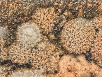
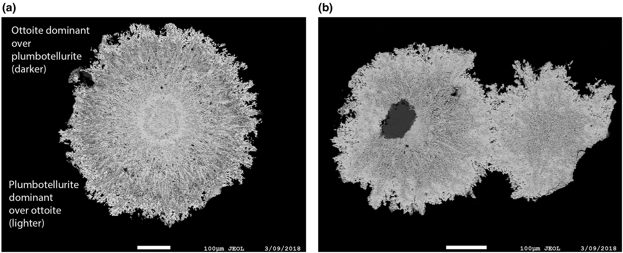
The ‘oboyerite’ occurs on the specimen as cream-coloured spherules with exterior surfaces covered by spiky crystal terminations and interiors composed of radiating white needles (Fig. 2). The outer portions of the interiors are transparent with resinous lustre, while the cores are opaque and dull to silky white. Other secondary phases identified on the specimen are burckhardtite (dull white balls and tan lacy growths), cerussite (colourless prisms), emmonsite (olive rounded crystals) and schieffelinite (white pearly plates). Two SEM images of spherule cross-sections are shown in Fig. 3.
‘Oboyerite’ spherules on matrix from NHMLAC specimen 67241, field of view 1.7 mm. Note that the spherules are composed of many radiating whitish and yellowish crystals, rather than being uniformly smooth.

Back-scatter SEM images showing a cross-section of ‘oboyerite’ grains from NHMLAC 67241: (a) cross-section of a single spherule; (b) cross-section of two fused spherules, one of which is nucleated around a grain of emmonsite.

Another specimen from Williams housed at the National Museum of Natural History, Washington, DC (specimen number 162210e) was also analysed by PXRD, providing a pattern very similar to those provided by the NHMLAC specimen 67241 (see next section), but was not used for further analysis. Several other specimens, notionally containing ‘oboyerite’ and traceable to Williams, also came to our attention during the discreditation process. It seems likely that Williams considered these to have a ‘type status’ and distributed them as such, leading to the greater than expected number of institutions and some private individuals that have specimens attributed as ‘type oboyerite’.
Plumbotellurite sample
The sample of plumbotellurite (α-Pb2+Te4+O3) used in this study was a synthetic sample stored in the collections of the Royal Ontario Museum, specimen number M47098. The synthetic plumbotellurite was synthesised by and identified as identical to natural plumbotellurite using XRD methods (Back, Reference Back1990).
Powder XRD
Four portions of ‘oboyerite’ spherules from the NHMLAC specimen 67241 were used for powder XRD analysis, each of which gave an identical pattern to that reported by Williams (Reference Williams1979). Williams (Reference Williams1979) also examined these spherules in thin-section to check their phase purity, and determined that ‘oboyerite’ was triclinic by using optical methods to complement the powder pattern, which did not index using the Ito method. Roberts (Reference Roberts1980) indexed this cell as a = 12.249(8), b = 15.113(6), c = 6.868(3) Å, α = 116.45(4), β = 98.58(4) and γ = 85.82(4)°.
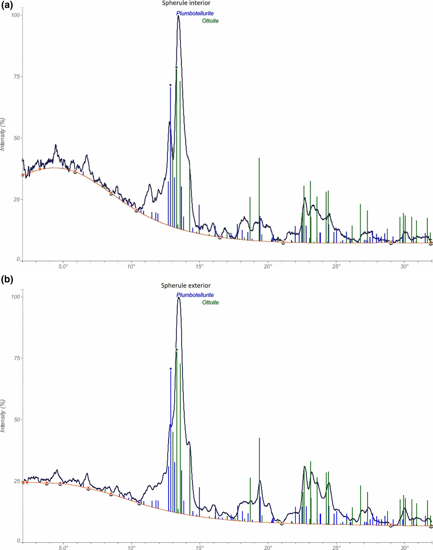
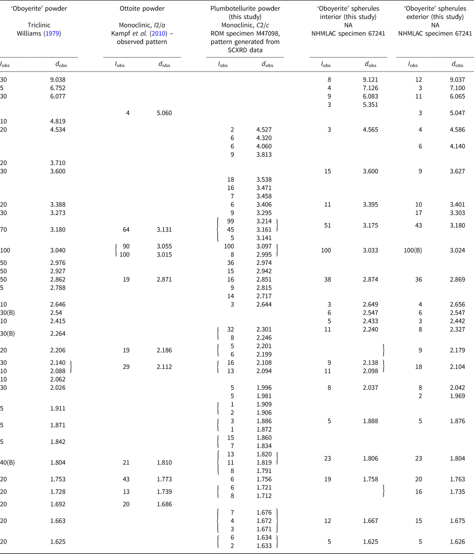
Detailed analysis of the ‘oboyerite’ powder pattern shows that ottoite (Pb2+2Te6+O5) and plumbotellurite (Pb2+Te4+O3) are the major constituents of ‘oboyerite’ spherules (see Table 1). As part of our investigation, powder X-ray patterns of the exterior and interior of ‘oboyerite’ spherules were collected. The exterior and interior patterns are broadly similar, both corresponding to a mixture of ottoite [monoclinic, I2/a, a = 7.5353(6), b = 5.7142(5), c = 10.8981(12) Å and β = 91.330(6)°; Kampf et al., Reference Kampf, Housley, Mills, Marty and Thorne2010] and plumbotellurite, but in different ratios (see Fig. 4). Other component minerals are also present in more minor amounts, including unidentified phases which generated the three highest d values. The interior powder pattern appears to correspond better with plumbotellurite and the exterior with ottoite (see Table 1).
Powder patterns of (a) interior and (b) exterior ‘oboyerite’ spherules (NHMLAC catalogue number 67241), with patterns for ottoite and plumbotellurite provided for reference. Patterns collected using MoKα radiation. The patterns of interior and exterior are broadly similar, but plumbotellurite is slightly more dominant in the interior of the spherule.

Powder X-ray diffraction data (d, Å) for ‘oboyerite’ (original pattern), with matched ottoite and plumbotellurite peaks, along with patterns recorded from the interior and exterior of the ‘oboyerite’ spherules in this study.

Notes: Williams’ equipment did not allow for determination of powder peaks with d values below 1.60, thus any peaks in the four comparison patterns which have d < 1.60 Å are not shown. All unmatched peaks with I < 5% for ottoite and plumbotellurite are not included in the table. SCXRD – single crystal X-ray diffraction. B = broad.
This result suggests that ‘oboyerite’ is formed from an initial crystallisation of plumbotellurite, but after initial radial plumbotellurite crystal growth occurs, the conditions became more oxidising and a greater proportion of the Te4+ that was being deposited with Pb as plumbotellurite was oxidised to Te6+, leading to ottoite precipitation instead. The nucleus of the ‘oboyerite’ grains may not necessarily be a Pb–Te–O mineral – in one cross-section emmonsite was found at the centre of the spherule (see Fig. 3b).
Most tellingly, the broad, highest intensity peak in ‘oboyerite’ (d value reported by Williams = 3.040 Å) is nearly identical to the highest intensity peaks for ottoite (observed d values of 3.055 and 3.015 Å) and plumbotellurite (calculated d value of 3.097 Å). The same near-match with ottoite was seen in the strongest intensity line in the powder pattern of ‘girdite’ (d value = 3.054 Å). Thus, it seems likely that the highest intensity peak in ‘oboyerite’ is a combination peak incorporating intensity from both ottoite and plumbotellurite. It is thus very likely that the material used for the powder pattern determination of ‘oboyerite’ by Williams (Reference Williams1979) was an exterior portion with plumbotellurite greater than ottoite.
Single-crystal XRD
Oboyerite
A single-crystal study of ‘oboyerite’ was undertaken on the MX2 beamline at the Australian Synchrotron, part of ANSTO. A small spherule of ‘oboyerite’ from the NHMLAC specimen 67241 was crushed and a transparent fragment of material (~10 μm × 10 μm × 10 μm) from near the edge of the spherule was placed on a resin mount. Data were collected at 100 K by a Dectris EigerX 16M detector and monochromatic radiation with a wavelength of 0.71073 Å.
After processing the data using XDS (Kabsch, Reference Kabsch2010), XPREP (Bruker, 2001) and SADABS (Bruker, 2001), 1096 reflections were found with an R int of 0.0657. The monoclinic unit cell a = 10.86, b = 5.67, c = 7.52 Å and β = 91.4° was found to fit the data, which is almost identical to that of ottoite (with a and c swapped). A small contraction in axis lengths was observed due to the low temperature (100 K) used for the crystal structure analysis.
Plumbotellurite
A 20 μm × 20 μm × 50 μm crystal of synthetic plumbotellurite was also analysed on the MX2 beamline at the Australian Synchrotron, part of ANSTO, using the same initial analysis procedure described above. 18,440 reflections were found with an R int of 0.0292. The structure solution was carried out by direct methods using SHELXT (Sheldrick, Reference Sheldrick2015a). Structure refinement by full-matrix least-squares was implemented by SHELXL (Sheldrick, Reference Sheldrick2015b), converging to R 1(all) = 0.0205. The unit-cell parameters determined were a = 26.442(5), b = 4.6010(9), c = 17.890(4) Å, β = 107.01(3)° and V = 2081.3(8) Å3, in agreement with those reported previously for synthetic α-Pb2+Te4+O3 (Zavodnik et al., Reference Zavodnik, Ivanov and Stash2008). The orthorhombic parameters reported for plumbotellurite, a = 8.41, b = 13.72 and c = 9.18 Å have V = 1059.2 Å3 (Spiridonov and Tananaeva, Reference Spiridonov and Tananaeva1982), which is approximately half of the true unit-cell volume of plumbotellurite, V = 2081.3(8) Å3. Additionally, the powder pattern reported for plumbotellurite closely matches that generated from the crystal structure, indicating that α-Pb2+Te4+O3 is identical to plumbotellurite.
The crystallographic information file for plumbotellurite has been deposited with the Principal Editor of Mineralogical Magazine and is available as Supplementary material (see below).
Chemical analyses
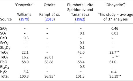
The use of bulk chemical methods is fraught with difficulty when analysing crystals whose sizes do not exceed 1 mm, because mixtures of different minerals are likely to be analysed by accident. The chemical analyses for ‘oboyerite’ reported by Williams (Reference Williams1979) and those for ottoite by Kampf et al. (Reference Kampf, Housley, Mills, Marty and Thorne2010) and plumbotellurite by Spiridonov and Tananaeva (Reference Spiridonov and Tananaeva1982) are shown in Table 2, along with the average compositional data determined in this study.
Chemical analytical data (in wt.%) for ‘oboyerite’, ottoite and plumbotellurite.

# ‘Oboyerite’ concentrations are from the near-perfect spherule (NHMLAC 67241) shown in Fig. 3a.
* The low total for ottoite has been discussed previously in Kampf et al. (Reference Kampf, Housley, Mills, Marty and Thorne2010)
** Reported here as entirely TeO2 given that there is a variable ratio of ottoite/plumbotellurite in the analyses, resulting in a lower total than the true value.
n.a. = not analysed; – = not detected.
Three fragments of ‘oboyerite’ from the NHMLAC specimen 67241 were embedded in resin and then polished to reveal cross sectional surfaces. One of these was close to a perfect spherule, the second a fusion of two spherules, and the third an irregularly shaped fragment of two half-spherules. Quantitative chemical analyses of a crystal cluster from the specimen were performed on a Cameca SX100 microprobe (Core Research Laboratories, NHM, London), in wavelength-dispersive spectroscopy mode, operated at 12 kV with 10 nA probe current, 5 μm beam diameter and PAP matrix correction. The standards used were: synthetic fayalite (Si), baryte (S), Cu metal (Cu), sphalerite (Zn), Ag metal (Ag), tellurium dioxide (Te) and vanadinite (Pb). Results (reported in Table 2) are recorded to only one decimal place for Te and Pb, as the low values of voltage and current used to reduce potential dehydration during analysis input less energy into the sample and, consequently, give lower counting statistics (and errors slightly greater than ± 0.1 wt.%) for these elements. Pb and Te are the only major elements, with Si the most prevalent trace (though this may be due to matrix effects, or minute amounts of associated opal). Traces of S, Cu, Zn and Ag were occasionally detected, while Ca, As, Se and Sb were sought, but not detected. Despite the differences in the powder patterns on the interior and exterior of the ‘oboyerite’ grains and the presence of a rim of ottoite (brighter contrast in back-scatter mode) forming the exterior of the spherules (see Fig. 3), Pb:Te ratios of the fibrous material did not vary significantly for interior and exterior analyses. Most of the rather fibrous material radiating out from the centre of the spherules thus shows two intergrown phases. The fibrous nature of the material accounts for some of the relatively low totals – totals below 90 wt.% (as calculated with TeO2) were discarded. To confirm that the low totals were due to the nature of the sample rather than a controllable error, the emmonsite grain in the centre of one ‘oboyerite’ fused spherule was analysed. After correcting for H2O content, the average total was 101.08 wt.%. The Pb:Te ratios are rather variable, with extreme values near 1:1 (minimum 1.06) corresponding to near-pure plumbotellurite, and values near 2:1 (maximum 1.88) to near-pure ottoite. Analysis totals for near-pure ottoite tended to be higher than those for near-pure plumbotellurite, indicating that the ottoite either forms more uniform grains, or perhaps simply polishes better than plumbotellurite. Intermediate values (average value 1.31) result when two intergrown phases were analysed simultaneously – as is likely to have occurred on a bulk scale in the initial description of ‘oboyerite’.
The ideal ratio of Pb:Te in ‘oboyerite’ is reported to be 1.2:1, while in ottoite it is 2:1 and in plumbotellurite 1:1. As shown by PXRD, both of these minerals are likely to have been accidentally analysed in the description of ‘oboyerite’, with a predominance of plumbotellurite. These two lead–tellurium oxysalts have differing Te valences, which explains why ‘oboyerite’ was described as a mixed-valence mineral. A mixture of three parts plumbotellurite to two parts ottoite gives a hypothetical composition of 3Pb2+Te4+O3·2Pb2+2Te6+O5 = Pb2+7(Te4+O3)3(Te6+O5)2, a formula differing from ‘oboyerite’ (H6Pb2+6(Te4+O3)3(Te6+O6)2·2H2O) in terms of major elements by only one Pb atom. 0.3 wt.% of Ca was also identified in ‘oboyerite’ (Williams, Reference Williams1979), possibly from Ca substitution for Pb, but considering that our analyses contain no Ca, we suspect it is from the inclusion of a small amount of calcite in the bulk analyses. The H2O analysed by Williams may have come from the minor emmonsite noted in the cores above.
Conclusions
Oboyerite
Williams (Reference Williams1979) based his description of ‘oboyerite’ on data obtained from at least two and probably more different phases, including ottoite and plumbotellurite, which dominate the mixture. The mixed-valence formula of ‘oboyerite’ appears likely to have been developed from the simultaneous analysis of a lead tellurite (plumbotellurite) and a lead tellurate (ottoite) with possibly minor calcite and emmonsite. As the criteria which serve to make a mineral unique (Dunn, Reference Dunn1990) were not met by ‘oboyerite’, ‘oboyerite’ has been discredited (IMA-CNMNC Proposal 19-D; Miyawaki et al., Reference Miyawaki, Hatert, Pasero and Mills2019).
The examples of the discredited minerals ‘girdite’ and ‘oboyerite’, both reported as mixed-valence Pb–Te oxysalts, show that bulk methods are completely unsuitable for the analysis of microscale mineral intergrowths. These sort of characterisations become even more problematic when more than one specimen is used to characterise the same mineral. Following the discreditations of ‘girdite’ and ‘oboyerite’, the number of remaining valid mixed-valence Te oxysalt minerals is just four. Carlfriesite and tlapallite have known crystal structures (Missen et al., Reference Missen, Kampf, Mills, Housley, Spratt, Welch, Coolbaugh, Marty, Chorazewicz and Ferraris2019), but tlalocite (Williams, Reference Williams1975) and yecoraite (Williams and Cesbron, Reference Williams and Cesbron1985) do not. The latter two minerals will be the subject of future study to determine whether they are truly mixed-valence Te minerals and not mixtures.
Plumbotellurite
When first described, plumbotellurite was reported as orthorhombic; however, Back (Reference Back1990) showed that plumbotellurite is actually monoclinic, C2/c, the α-Pb2+Te4+O3 of Mariolacos (Reference Mariolacos1969) and Zavodnik et al. (Reference Zavodnik, Ivanov and Stash2008). The current study reiterates the conclusion presented by Back (Reference Back1990). It is also worth noting that of the three synthetic forms of Pb2+Te4+O3 (tetragonal, monoclinic and triclinic), only the monoclinic form is known as a mineral (Christy et al., Reference Christy, Mills and Kampf2016). The triclinic synthetic form of Pb2+Te4+O3 (γ-Pb2+Te4+O3) has different unit-cell parameters to those reported for fairbankite (Williams, Reference Williams1979). Meanwhile, an ongoing study on fairbankite based on the NHM, London type (as described above) by some of the authors has indicated that fairbankite is indeed triclinic (a = 7.0205(3), b = 10.6828(6), c = 14.4916(8) Å, α = 75.161(5), β = 81.571(4), γ = 83.744(4)° and V = 1036.35(9) Å3), but with unique unit-cell parameters. More surprisingly, the early results indicate that the formula for fairbankite is also incorrect and it is not in the Pb2+Te4+O3 family of polymorphs.
Acknowledgements
Equipment used in this study at Museums Victoria has been partly funded by The Ian Potter Foundation grant ‘tracking tellurium’ to SJM. Equipment at the Natural History Museum of Los Angeles County was funded by the John Jago Trelawney Endowment. Support funding has been provided to OPM by an Australian Government Research Training Program (RTP) Scholarship, a Monash Graduate Excellence Scholarship (MGES) and a Monash–Museums Victoria Scholarship (Robert Blackwood). We thank Eric Fritz and Hexiong Yang (University of Arizona) for their assistance in examining their ‘oboyerite’ specimen. We thank Joël Brugger (Monash University) for his insightful comments. This research was undertaken in part using the MX2 beamline at the Australian Synchrotron, part of ANSTO, and made use of the Australian Cancer Research Foundation (ACRF) detector. The authors acknowledge use of facilities within the Monash Centre for Electron Microscopy.
Supplementary material
To view supplementary material for this article, please visit https://doi.org/10.1180/mgm.2019.63