Introduction
The volcanogenic exhalation sulfate mineralization from fumaroles of the Tolbachik volcano is renowned for its rich mineral diversity (Vergasova and Filatov, Reference Vergasova and Filatov2016; Siidra et al., Reference Siidra, Nazarchuk, Zaitsev, Lukina, Avdontseva, Vergasova, Vlasenko, Filatov, Turner and Karpov2017a,Reference Siidra, Lukina, Nazarchuk, Depmeier, Bubnova, Agakhanov, Avdontseva, Filatov and Kovruginb). Many fumarolic sulfate minerals contain alkali elements (K and/or Na) and chloride ions. Exhalative mineral assemblages from fumaroles of the First and Second scoria cones, Great Fissure Tolbachik eruption 1975–76 are well-known for their exceptional diversity of Cu-containing minerals. In addition to Cu, condensates of the 1975–76 eruption are also highly enriched in Zn (Menyailov et al., Reference Menyailov and Nikitina1980a,Reference Menyailov, Nikitina and Shaparb). Zinc impurities are very common in the copper fumarolic minerals. A variety of zinc minerals sensu stricto (i.e. minerals with independent crystallographic positions of Zn) are known from fumaroles of the First and Second scoria cones: sofiite, Zn2(SeO3)Cl2 (Semenova et al., Reference Semenova, Rozhdestvenskaya, Filatov and Vergasova1992), prewittite, KPb1.5ZnCu6(SeO3)2O2Cl10 (Shuvalov et al., Reference Shuvalov, Vergasova, Semenova, Filatov, Krivovichev, Siidra and Rudashevsky2013), chubarovite, KZn2(BO3)Cl2 (Pekov et al., Reference Pekov, Zubkova, Pautov, Yapaskurt, Chukanov, Lykova, Britvin, Sidorov and Pushcharovsky2015a), mellizinkalite, K3Zn2Cl7 (Pekov et al., Reference Pekov, Zubkova, Yapaskurt, Lykova, Belakovskiy, Vigasina, Sidorov, Britvin and Pushscharovsky2015b), flinteite, K2ZnCl4 (Pekov et al., Reference Pekov, Zubkova, Yapaskurt, Britvin, Vigasina, Sidorov and Pushcharovsky2015c), cryobostryxite, KZnCl3·2H2O (Pekov et al., Reference Pekov, Zubkova, Britvin, Yapaskurt, Chukanov, Lykova, Sidorov and Pushcharovsky2015d), zincobradaczekite, NaZn2Cu2(AsO4)3 (Pekov et al., Reference Pekov, Lykova, Koshlyakova, Belakovskiy, Vigasina, Turchkova, Britvin, Sidorov and Scheidl2016a), zincomenite, ZnSeO3 (Pekov et al., Reference Pekov, Zubkova, Yapaskurt, Britvin, Chukanov, Lykova, Sidorov and Pushcharovsky2016b) and pharmazincite, KZnAsO4 (Pekov et al., Reference Pekov, Yapaskurt, Belakovskiy, Vigasina, Zubkova and Sidorov2017). The first sulfate Zn mineral hermannjahnite CuZn(SO4)2 was described only recently from the new fumaroles of the Fissure Tolbachik Eruption 2012–13 and fumaroles of the Great Fissure Tolbachik Eruption 1975–76 (Siidra et al., Reference Siidra, Nazarchuk, Agakhanov, Lukina, Zaitsev, Turner, Filatov, Pekov, Karpov and Yapaskurt2017c).
Herein we report on the chemical composition, structure and properties of belousovite, KZn(SO4)Cl. The mineral is named in honour of Dr Alexander Borisovich Belousov (Александр Борисович Белоусов) (b. 1962), Institute of Volcanology, Russian Academy of Sciences, Petropavlovsk-Kamchatskiy, Kamchatka, Russia, in recognition of his contributions to volcanology (Belousov et al., Reference Belousov, Belousova and Voight1999, Reference Belousov, Belousova and Nechayev2013, Reference Belousov, Belousova, Edwards, Volynets and Melnikov2015). Both the mineral and the mineral name were approved by the International Mineralogical Association Commission on New Minerals, Nomenclature and Classification (IMA2016-047, Siidra et al., Reference Siidra, Nazarchuk, Zaitsev, Lukina, Kayukov, Vergasova, Filatov, Karpov and Shilovskikh2016). Type material is deposited at the Mineralogical Museum, St. Petersburg State University, St. Petersburg, Russia (catalogue no. 1/19665).
Occurrence and association
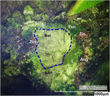
Belousovite occurs as a product of fumarolic activity. It was found in July, 2015 in the Yadovitaya fumarole, Second scoria cone, Northern Breakthrough (North Breach), Great Fissure eruption, Tolbachik volcano, Kamchatka, Russia. The Second scoria cone is located ~18 km SSW of the active shield volcano Ploskiy Tolbachik (Fedotov and Markhinin, Reference Fedotov and Markhinin1983). Associated minerals are kamchatkite, langbeinite, euchlorine, anglesite and zincite (Fig. 1). Langbeinite forms intimate intergrowths with belousovite. Belousovite is a fumarolic mineral that is deposited directly from volcanic gas emissions as a sublimate. The temperature of gases at the sampling location was ~250°C. Belousovite is soluble in H2O at room temperature and transforms slowly into hydrate in humid air. For this reason all the samples recovered were packed hermetically immediately after collecting and isolated to avoid any contact with the atmosphere.
Belousovite (Bel, centre) in association with kamchatkite (Kam), langbeinite (Lan), euchlorine (Euc), anglesite (Ang) and Zincite (Zin).

Physical properties
Belousovite occurs as irregularly-shaped grains and in the form of microcrystalline masses up to 0.1 cm wide (Fig. 1). Belousovite is colourless; the streak is white; and the lustre is vitreous. The mineral is transparent in individual grains and translucent in aggregates. Belousovite is brittle with perfect cleavage on (100). The fracture is uneven. Hardness and density could not be measured due to the very small size of individual grains and the porosity of the aggregates. The calculated density based on the empirical formula of the holotype is 2.89 g cm–3. Belousovite is optically biaxial (+), α = 1.532(2), β = 1.544(2) and γ = 1.570(2) (589 nm) with 2V (calc.) = 69.4°. Belousovite appears colourless and non-pleochroic under the microscope. The Gladstone-Dale compatibility index, 1 – (Kp/Kc) = 0.020, is excellent (Mandarino, Reference Mandarino2007).
Chemical composition
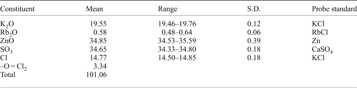
Four crystals (65 × 30, 30 × 10, 15 × 10 and 10 × 10 µm) of belousovite checked previously by single-crystal X-ray diffraction for quality were mounted in epoxy resin and polished without water and oil using a BUELHER CarbiMet P1000, MicroCut P2500 and ChemoMet I. The mineral was analysed using a Hitachi S-3400N scanning electron microscope equipped with an Oxford Instruments X-Max 20 Energy Dispersive Spectrometer (EDS) and INCA Wave 500 wavelength dispersive spectrometer (WDS). The conditions for EDS were: accelerating voltage of 20 kV, electron beam current of 1.0 nA and defocused beam (up to 15 µm). The current was measured using a Faraday cup. X-ray acquisition time was 30 s and X-ray processing time ‘5’ for analysis. System calibration was performed on CoKα-Kβ. X-ray matrix correction was carried-out automatically by the Oxford Instruments AZtec Tru-Q including an exponential (XPP, Pouchou and Pichoir, Reference Pouchou, Pichoir, Heinrich and Newbury1991) and pile-up correction routine. The results of six spot analyses are reported in Table 1. The empirical formula based on O + Cl = 5 atoms per formula units is K0.97Rb0.01Zn1.00S1.01O4.03Cl0.97. The ideal formula is KZn(SO4)Cl.
Chemical composition (wt.%) of belousovite.

S.D. – standard deviation.
The energy-dispersive spectrum shows that K, Zn, S, O and Cl are the major elements in the mineral, together with small amounts of Rb (Fig. S1, see supplementary material section below). We assumed that the mineral with potassium as a major element and minor rubidium may also contain sodium as a minor component, however, there is significant overlapping of ZnLα (12.254 Å, 1.0120 keV) and Lβ (11.983 Å, 1.0350 keV) over NaKα (11.910 Å, 1.0410 keV) and Kβ (11.617 Å, 1.0670 keV) so it is not feasible to measure low quantities of Na in the presence of Zn with the ED spectrometer. Instead we performed WD scanning in the range 11.500–12.500 Å and found only two lines belonging to Zn (TAP crystal, 10 nA beam current, scan speed = 0.25 Å per minute) (Fig. S2). Therefore, we are confident that the mineral does not contain Na. An attempt to use wavelength-dispersive analysis lead to strong damage of the mineral surface (Figs S3 and S4) so the mineral was only analysed by energy-dispersive analysis.
X-ray crystallography
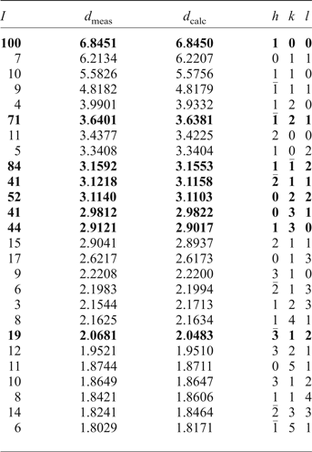
Powder X-ray diffraction data were collected using a Rigaku R-AXIS Rapid II single-crystal diffractometer equipped with cylindrical image plate detector using Debye-Scherrer geometry (CoKα radiation, d = 127.4 mm). Data (in Å) are given in Table 2. Unit-cell parameters refined from the powder data (Britvin et al. Reference Britvin, Dolivo-Dobrovolsky and Krzhizhanovskaya2017) are as follows: monoclinic, space group P21/c, a = 6.90(1) Å, b = 9.65(2) Å, c = 8.18(2) Å, β = 96.5(2)°, V = 541(1) Å3 and Z = 4.
Powder X-ray diffraction data for belousovite.

The eight strongest lines are shown in bold.
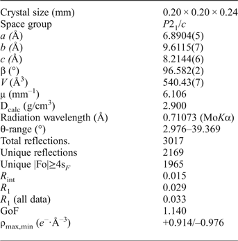
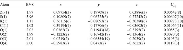
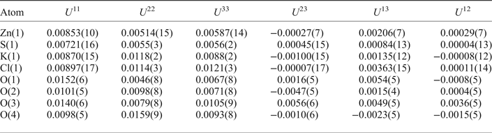
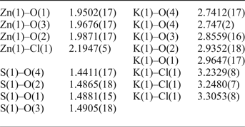
A transparent isometric crystal fragment of belousovite was mounted on a thin glass fibre for X-ray diffraction analysis using a Bruker APEX II DUO X-ray diffractometer with a micro-focus X-ray tube operated with MoKα radiation at 50 kV and 40 mA. The data were integrated and corrected for absorption using a multiscan type model implemented in the Bruker programs APEX and SADABS (Bruker-AXS, 2014). More than a hemisphere of X-ray diffraction data was collected. The refinement in the P21/c space group converged to R 1 = 0.029 (Table 3). The final model included anisotropic displacement parameters for all atoms. The final atomic coordinates and anisotropic displacement parameters are given in Tables 4, 5 and selected interatomic distances in Table 6. Lists of observed and calculated structure factors have been deposited with the Principal Editor of Mineralogical Magazine and are available as Supplementary material (see below).
Crystallographic data and refinement parameters for belousovite.

Coordinates and isotropic displacement parameters (Å2) of atoms and bond-valence sums* (BVS) in belousovite.

* Expressed in valence units (vu) and calculated using the parameters from Brese and O'Keeffe (Reference Brese and O'Keeffe1991).
Anisotropic displacement parameters (Å2) of atoms in belousovite.

Selected interatomic distances (in Å) in belousovite.

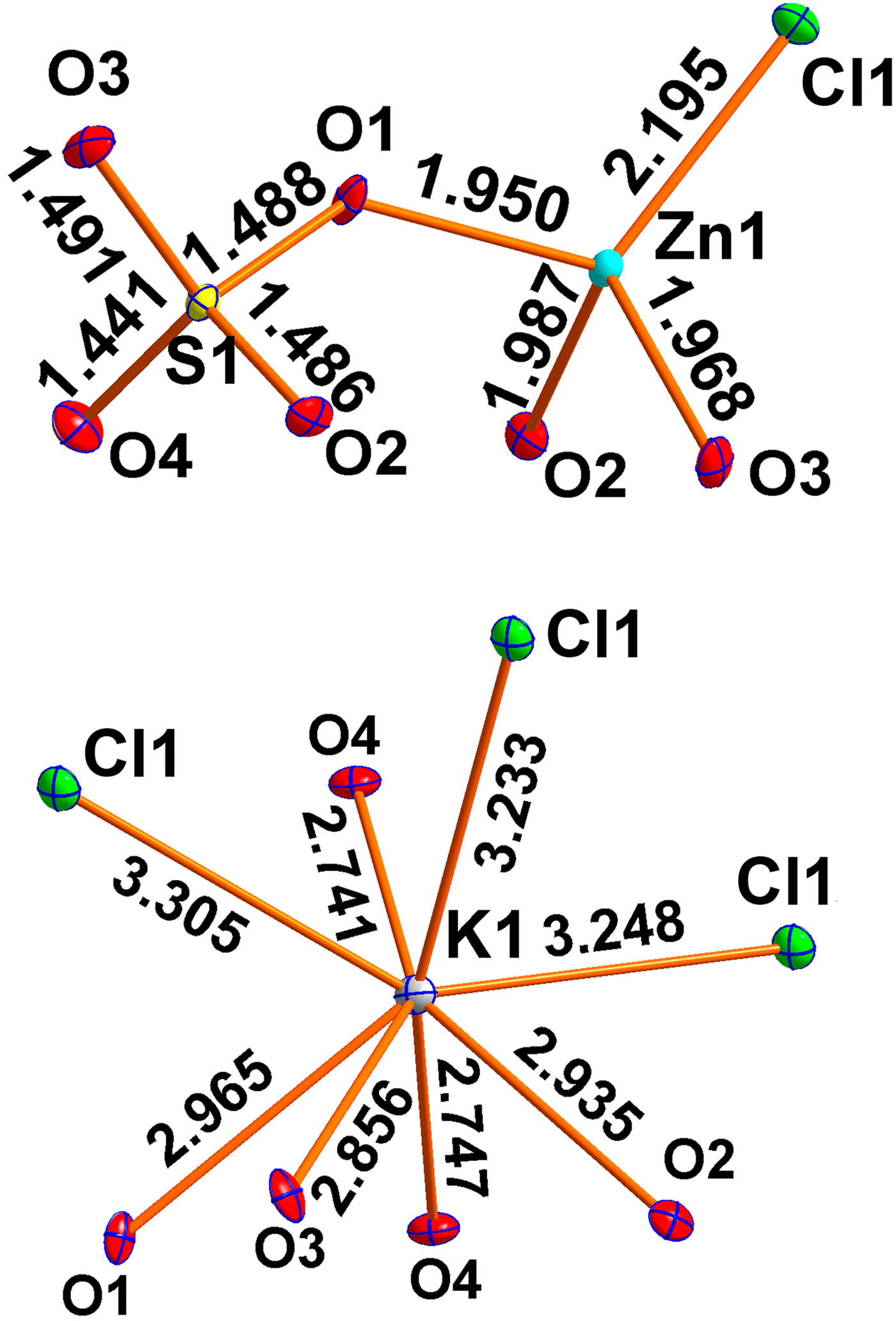
The structure of belousovite contains one K+, one Zn2+ and one S6+ site (Fig. 2, Table 4).
Coordination of atoms in belousovite.

Each of the K+ cations forms four K–O bonds with the K–O bond lengths in the range of 2.747(2)–2.9647(17) Å and three K–Cl bonds with the K–Cl bond lengths in the range of 3.2329(8)–3.3053(8) Å. The Zn2+ cation is in a tetrahedral environment. It is coordinated by three oxygens and a chloride anion. The Zn–Cl distance of 2.1947(5) Å is significantly longer than those of the Zn–O bonds (1.9502(17)–1.9871(17) Å). One symmetrically independent S6+ cation forms rather symmetrical SO4 tetrahedra. The individual S–O distances are in the range of 1.4411(17)–1.4905(18) Å, which is in good agreement for well-refined sulfate structures (1.394–1.578 Å; Hawthorne et al., Reference Hawthorne, Krivovichev, Burns, Alpers, Jambor and Nordstrom2000).
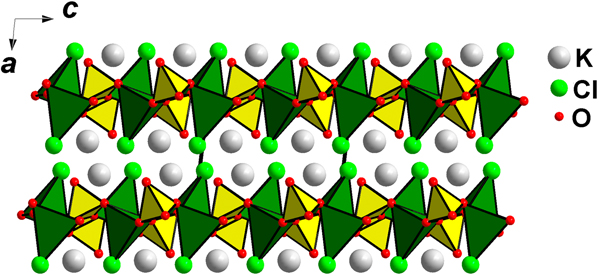
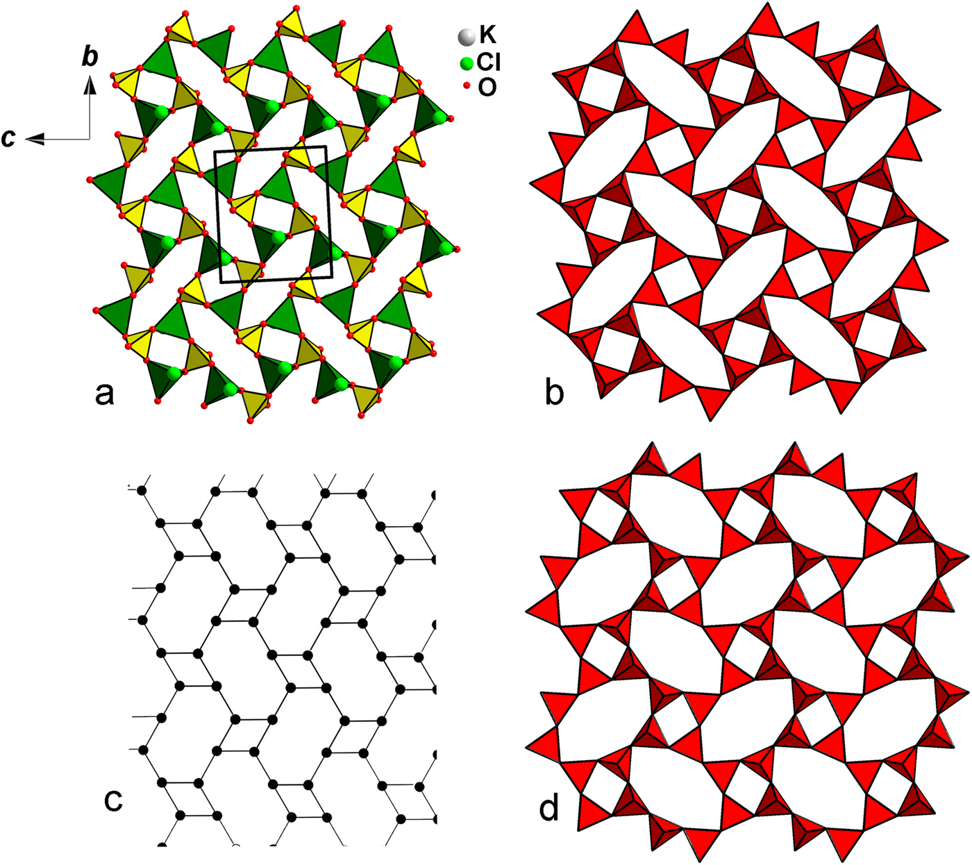
The structure of belousovite consists of infinite [ZnSO4Cl]– layers and K+ ions (Fig. 3). [ZnSO4Cl]– layers (Fig. 4a) are parallel to the bc plane and are interconnected via K+ cations.
General projection of the crystal structure of belousovite along the b axis (ZnO3Cl = green, SO4 = yellow).

(a) General projection of the [Zn(SO4)Cl]– layer formed by SO4 (yellow) and ZnO3Cl (green) tetrahedra in belousovite. (b) [Si4O10]4– layer formed by the linkage of 4-membered rings resulting in 8-membered rings in the crystal structure of apophyllite. (c) Nodal representation, identical for [Zn(SO4)Cl]– layers in belousovite and [Si4O10]4– layers in apophyllite. (d) The [Si4O10]4– layer in cavansite has the ‘…up–down…’ orientations of the Si–Ot bonds in 4-membered rings similar to Zn–Cl and S–Ot bonds orientations in belousovite. See the text for details.

Discussion
The crystal structure of belousovite is isotypic with previously reported synthetic RbZnSO4Cl and TlZnSO4Cl (Bosson, Reference Bosson1976). In order to investigate whether the topological structure of the layers in belousovite has any similarities with other minerals and synthetic compounds one has to examine their nodal representations (Fig. 4c), where each black node represents tetrahedron (SO4 or ZnO3Cl). Two nodes are connected by an edge if the corresponding polyhedra share one common anion. This type of topology (linkage of 4-membered rings creating 8-membered rings) is known in silicate crystal chemistry as the topology of the [Si4O10]4– layers (Fig. 4b) in the minerals of the apophyllite group (Dunn et al. Reference Dunn, Rouse, Norberg and Peacor1978; Dunn and Wilson, Reference Dunn and Wilson1978; Liebau, Reference Liebau1985). The ‘…up–down…’ orientations of the Si–Ot (Ot = terminal oxygen atom in a silicate group) bonds relative to the plane of the layer in apophyllite are different from Zn–Cl and S–Ot bond orientations in belousovite. All silicate tetrahedra in each 4-membered ring are oriented in the same direction in apophyllite whereas in belousovite each 4-membered ring consists of two pairs of tetrahedra oriented in opposite directions. Observed ‘…up–down…’ orientations of tetrahedral groups in 4-membered rings in belousovite are similar to those in the [Si4O10]4– layer in cavansite (Fig. 4d) (Zubkova et al. Reference Zubkova, Pekov, Pushcharovsky and Chukanov2009).
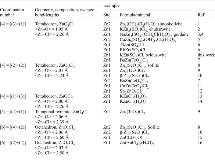
One of the interesting structural features of belousovite is the presence of mixed-ligand ZnO3Cl coordination polyhedra. Table 7 gives a review of observed Zn–O–Cl stereochemistries in minerals and synthetic materials. There are in total six stereochemically different mixed-ligand coordinations. The ZnO3Cl tetrahedra, as in belousovite, are the most common, whereas ZnO4Cl tetragonal pyramids are very rare. Tetrahedra of ZnO3Cl were reported recently in the structure of another exhalative mineral, chubarovite, KZn2(BO3)Cl2 (Pekov et al., Reference Pekov, Zubkova, Pautov, Yapaskurt, Chukanov, Lykova, Britvin, Sidorov and Pushcharovsky2015a), also found in the sublimates of active fumaroles at the Second and First scoria cones of the Northern Breakthrough of the Great Fissure Tolbachik eruption. The topology of layers in chubarovite is completely different from that of belousovite. The crystal structure of the fumarolic selenite–chloride sofiite, Zn2(SeO3)Cl2 (Semenova et al., Reference Semenova, Rozhdestvenskaya, Filatov and Vergasova1992) contains two different mixed-ligand coordinations of Zn: octahedron ZnO4Cl2 and tetrahedron ZnO2Cl2. Protonated Zn(OH)3Cl tetrahedra were described previously in the structures of gordaite NaZn4(SO4)(OH)6Cl(H2O)6 (Adiwidjaja et al., Reference Adiwidjaja, Friese, Klaska and Schlueter1997) and simonkolleite Zn5(OH)8Cl2(H2O) (Hawthorne and Sokolova, Reference Hawthorne and Sokolova2002).
Mixed-ligand ZnOmCln coordination polyhedra in minerals and inorganic compounds.

References: (1) Hawthorne and Sokolova (Reference Hawthorne and Sokolova2002); (2) Pekov et al. (Reference Pekov, Zubkova, Pautov, Yapaskurt, Chukanov, Lykova, Britvin, Sidorov and Pushcharovsky2015a); (3) Adiwidjaja et al. (Reference Adiwidjaja, Friese, Klaska and Schlueter1997); (4) Zhu et al. (Reference Zhu, Seff, Witzke and Nasdala1997); (5) Burns et al. (Reference Burns, Roberts and Nikischer1998); (6) Bosson (Reference Bosson1976); (7) Jiang et al. (Reference Jiang, Feng and Mao2006); (8) Semenova et al. (Reference Semenova, Rozhdestvenskaya, Filatov and Vergasova1992); (9) Johnsson and Törnroos (Reference Johnsson and Törnroos2003a); (10) Johnsson and Törnroos (Reference Johnsson and Törnroos2007); (11) Johnsson and Törnroos (Reference Johnsson and Törnroos2003b); (12) Jo et al. (Reference Jo, Kim, Lee, Shim and Ok2010); (13) Suesse and Brehler (Reference Suesse and Brehler1964); (14) Brehler and König (Reference Brehler and König1969); (15) Follner and Brehler (Reference Follner and Brehler1970); (16) Jones et al. (Reference Jones, Schelbach, Schwarzmann and Thoene1988).
Prewittite, KPb1.5Cu6Zn(SeO3)2O2Cl10 (Shuvalov et al., Reference Shuvalov, Vergasova, Semenova, Filatov, Krivovichev, Siidra and Rudashevsky2013), has a chemical composition somewhat similar to belousovite. But Zn2+ cations form pure chloride ZnCl4 tetrahedra in the structure of prewittite.
Belousovite, KZn(SO4)Cl, reported herein is a new and unique mineral species, and the first sulfate compound and mineral containing K, Zn and Cl in the one mineral. The discovery of belousovite expands our knowledge on mineralogy and geochemistry of zinc mixed-ligand species in volcanic fumarolic environments.
Acknowledgements
We are grateful to Ian Graham, Igor Pekov, Peter Leverett and Daniel Atencio for valuable comments. This work was financially supported by the Russian Science Foundation through the grant 16-17-10085. Technical support by the SPbSU X-ray Diffraction and Geomodel Resource Centres is gratefully acknowledged.
Supplementary material
To view supplementary material for this article, please visit https://doi.org/10.1180/minmag.2017.081.084