Introduction
Carotid blowout is the most feared sequalae of major head and neck surgery. Despite modern endovascular rescue techniques, only just over a third of these patients (36.6 per cent) will survive one year, with 60 per cent of patients suffering significant morbidity, including a 10.8 per cent risk of peri-operative stroke.Reference Pathak, Viallet and Nason1,Reference Liang, Guedes, Duvvuri, Singh, Chaer and Makaroun2 The risk of carotid blowout is increasingly recognised as the proportion of surgical procedures performed in a salvage setting following prior (chemo)radiotherapy expands, and with free flap reconstructive techniques enabling more aggressive ablative resections. A pharyngocutaneous fistula is of particular concern, as an artery exposed to the tryptic enzymatic effects of continuous saliva places the vessel wall at significant risk of desiccation and breakdown.
We present a simple method of protecting the great vessels at the initial surgery, utilising the adjacent sternal heads of the sternocleidomastoid muscle.
Technical note
This surgical technique can be utilised in any primary or salvage laryngectomy procedure, with or without pharyngeal resection, as long as the sternocleidomastoid muscle has been preserved, if oncologically feasible.
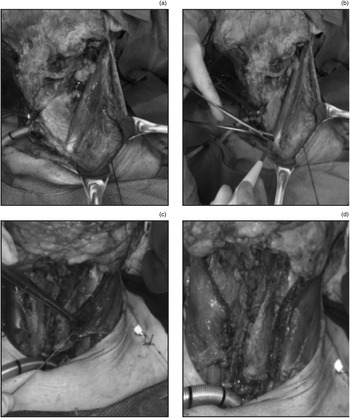
A laryngectomy is carried out in the standard manner. Following meticulous primary closure or free flap inset, myotomy and release of the sternal heads of the sternocleidomastoid is performed (Figure 1a, b). This release is already commonly conducted to prevent a deep-lying stoma and to aid functional speech rehabilitation. The superiorly based muscular flaps are then rotated medially and approximated to the prevertebral fascia with size 2-0 Vicryl® (polyglactin 910) sutures to cover the lower third of the vascular axis (Figure 1c, d), ensuring there is sufficient laxity in the muscular bridge not to impede flow in the great vessels. Care must be taken to prevent compression of the pedicle in cases requiring free flap reconstruction of the pharynx.
Sternal head sternocleidomastoid flap. (a) Left sternal head of sternocleidomastoid identified and (b) divided utilising monopolar cautery. (c) Following pharyngeal closure, the sternal head is rotated medially as a flap over the great vessels and (d) secured to the prevertebral fascia.

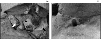
In the index case presented, a 63-year-old male with a heavy smoking and alcohol history underwent a total laryngectomy for a recurrent tumour–node stage T4N0 transglottic squamous cell carcinoma, having completed primary radiotherapy in 2018. Following salvage laryngectomy with bilateral neck dissection, the sternocleidomastoid flap procedure was performed to cover the patient's left great vessels, but omitted on the right side because of concerns about the position and local pressure on the pedicle of the radial forearm free flap utilised for patch closure of the pharynx. Clinical evidence of a salivary leak on post-operative day 4 was noted following persistent, intractable vomiting, mandating a return to the operating theatre for exploration and washout. A pharyngeal dehiscence was noted, with a large volume salivary leak and exposed right carotid artery, requiring pedicled pectoralis muscle flap closure to protect the vascular axis. However, on the left side, the vessels remained protected by healthy tissue, adequately covered by the medialised sternocleidomastoid sternal head flap and negating the need for any additional tissue coverage (Figure 2a). Following discharge, the patient remains free of disease, with satisfactory healing of the pharyngocutaneous fistula at his most recent clinic review (Figure 2b).
Appearance of a sternocleidomastoid flap following salvage laryngectomy. A large volume post-operative salivary leak mandated an operative neck exploration, where a significant pharyngeal wound dehiscence was noted. (a) In the left side of the neck, the sternocleidomastoid sternal head flap remained in its rotated position, ensuring adequate coverage of the great vessels, which were protected from the salivary fistula (black arrow). A sternocleidomastoid flap procedure was not performed on the right side in light of the nearby free flap pedicle (arrowhead).The free-lying sternal head is visibly retracted (white arrow), with exposure of the carotid medially requiring subsequent pectoralis major flap coverage. (b) Neck appearance at the most recent out-patient review, demonstrating successful healing.

The senior author and team have been utilising this technical modification since 2019. Since then, the illustrated case was the only patient requiring a pectoralis major flap for vessel coverage in a non-protected exposed vessel. In all other fistula cases explored, the sternocleidomastoid flap was appropriately positioned to protect the major vessels, and no further pedicled flap or salivary diversion techniques other than drains were required. To date, there have been no episodes of actual or threatened carotid blowout in this patient cohort (n = 22).
Discussion
Despite its versatility, the sternocleidomastoid flap has failed to be incorporated into regular head and neck reconstructive practice. The limitations of its mobility and rotational reach, concerns about oncological safety in the setting of salvage neck dissection, and the perception of a variable and unreliable blood supply have resulted in the sternocleidomastoid muscle flap being relegated to secondary uses.
Comprehensive anatomical studies have delineated the sternocleidomastoid blood supply into three clear sections, with the superior third supplied by occipital artery branches, and the middle third from either the superior thyroid artery or the external carotid artery.Reference Leclère, Vacher and Benchaa3 The lower third does indeed have a more variable supply, although in almost 75 per cent of cases it arises from the suprascapular artery. Use of the distal portion is reported as the most susceptible to perfusion compromise, but typically in relation to external skin paddle insufficiency when utilised as a myocutaneous flap.Reference Wei, Liu, Zhao, Zhu, Li and Wang4
Conley and GullaneReference Conley and Gullane5 published an important review of potential applications of the sternocleidomastoid flap 30 years ago; these included lower buccal and mandible reconstruction when used as a soft tissue or composite flap, anterior floor of mouth repair, and even assisting lower facial reanimation. These authors also advocated use of a sternocleidomastoid muscle flap to protect the carotid and innominate artery following laryngotracheal mediastinal resection, recognising the importance of adequate healthy tissue coverage of the great vessels to prevent delayed major arterial haemorrhage.
This thought was echoed in a more recent technical suggestion by Pathak et al.,Reference Pathak, Viallet and Nason1 who recognised the need for great vessel coverage, as the use of organ-preservation modalities resulted in increasing rates of salvage surgery and a subsequently greater risk of wound breakdown and salivary fistulae. Their recommendation was to secure the whole medial border of the sternocleidomastoid down to buccopharyngeal and prevertebral fascia. However, we found this technique less useful because variable muscle fibrosis following radiotherapy limits the mobility of the sternocleidomastoid, and often prevents it from reaching medially, resulting in a high-tension closure. This technique also fails to address the sternal heads, potentially resulting in a deep-lying stoma and limiting rehabilitation potential.
A more complicated variation of the inferior sternocleidomastoid flap involves extending the flap to allow anteromedial rotation to cover the upper two-thirds of the vascular axis, with a corresponding platysmal muscle flap to cover the caudal section of the vessels.Reference González-García, Moreno-García, Moreno-Sánchez and Román-Romero6 We feel the inferior third is the most important area of the vessels to cover, as it is the most dependent part of the neck and has the most consistent and continual contact with any potential saliva leak. The inferior third therefore deserves the most robust regional muscular coverage with a sternocleidomastoid flap rather than a thin platysmal flap. Finally, thinning the overlying skin flaps by separating platysma in a post-chemoradiation neck increases the risk of subsequent devascularisation and skin necrosis.
We have demonstrated a marked reduction in the requirement for secondary vascularised tissue coverage of the great vessels to prevent catastrophic haemorrhage by utilising the sternocleidomastoid flap. A large review conducted at Emory University (in Atlanta, Georgia, USA) demonstrated that 17 pedicled pectoralis major flaps were required in 47 patients suffering a pharyngocutaneous fistula following total laryngectomy for wound breakdown.Reference Mclean, Nicholas, Duggal, Chen, Grist and Losken7 This was corroborated in a review by Dr Wax and colleagues, who noted that, over a 10-year study period, use of the pedicled pectoralis major flap was a secondary procedure in 38 per cent of cases after a free flap complication, with the procedure solely performed for great vessel exposure in 3 of 20 patients.Reference Schneider, Wu and Wax8 This common usage of the pedicled pectoralis major flap is advocated in other major centre reviews.Reference Liu, Gullane, Brown and Irish9 We believe the direct illustration of the benefits of the described simple technique modification prompt consideration in head and neck patients undergoing laryngectomy.
In conclusion, we describe a simple modification of a technique utilising the sternocleidomastoid sternal head(s), which are released as a routine step during laryngectomy. These sternal heads provide a local flap to robustly protect the inferior great neck vessels with healthy tissue. Their use reduces the probability of requiring further regional flap surgery in the event of a salivary fistula, and helps to prevent catastrophic haemorrhage.
Acknowledgements
The authors would like to thank the Royal Society of Medicine Ellison-Cliffe Travelling Fellowship, the Colledge Family Memorial Fellowship Fund and the HCA International Foundation Grant for their support of the International Fellowship at the University of Alabama at Birmingham during the study.
Competing interests
None declared