Introduction
Considerable controversy remains regarding the aetiology of the lateral cleft cyst, sinus or fistula since it was first described in 1785;Reference Golledge and Ellis1 however, it is widely believed to be branchiogenic in origin.Reference Glosser, Pires and Feinberg2 Some authors have rejected the branchiogenic theory in favour of the growing evidence for a cystic lymph node origin.Reference Golledge and Ellis1, Reference Bhaskar and Bernier3, Reference Chandler and Mitchell4 Lateral cervical cysts are reported to be aetiologically distinct from sinuses and fistulae, and many mechanisms are proposed to be involved in their pathogenesis.Reference Golledge and Ellis1, Reference Glosser, Pires and Feinberg2, Reference Shinkwin, Whitfield and Robson5 There is little detailed published information about the incidence or prevalence of branchial anomalies; however, it is an uncommon condition, which partly explains the ongoing controversy surrounding its aetiology.
Papillomaviridae, or the human papillomaviruses (HPVs), are now a well-recognised cause of oropharyngeal cancers.Reference D'Souza, Kreimer, Viscidi, Pawlita, Fakhry and Koch6 In a recent summary by the National Cancer Institute State of the Science, Adelstein et al. discussed the continuing evolution of our understanding of the significance of HPV infection in the setting of head and neck cancer.Reference Adelstein, Ridge, Gillison, Chaturvedi, D'Souza and Gravitt7 They went on to suggest that HPV may serve as a future therapeutic target.Reference Adelstein, Ridge, Gillison, Chaturvedi, D'Souza and Gravitt7 Human papillomavirus involvement in benign head and neck papillomas is also well known. Thus, it is reasonable to consider whether HPV infection may play a role in other head and neck diseases.
Case report
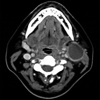
A 42-year-old woman presented with a 1-month history of an enlarging left lateral neck mass. She also suffered from recurrent bronchitis, gastric reflux, dyslipidaemia and type 2 diabetes mellitus, which was treated with oral hypoglycaemic drugs. Examination identified a palpable left lateral neck mass approximately 3 cm in diameter. A computed tomography scan with contrast showed a hypodense lesion of dimensions 20 mm × 24 mm × 30 mm at the division of the internal and external carotid arteries anteromedial to the left sternocleidomastoid muscle (Figures 1 and 2). Ultrasonography showed this to have an avascular cystic structure. Under ultrasound guidance, 10 ml of purulent fluid was aspirated. Cytological analysis revealed abundant neutrophil polymorphs, squamous cells, anucleate squames, histiocytes and proteinaceous material, consistent with an inflamed branchial cyst. No malignant cells were seen. An aspirate of the lesion was positive for p16INK4A but negative for HPV.
Coronal computed tomography scan showing a hypodense lesion in the left neck.

Axial computed tomography scan showing a hypodense lesion in the left neck.

The patient underwent panendoscopy, during which a papillomatous lesion was noted in the left hypopharynx. The pharyngeal lesion was biopsied, and then the cystic neck mass was removed via en bloc left level two neck dissection. No cutaneous tracts or deep extensions from the neck mass were noted during the operation. The biopsy specimen was sent for histopathological, p16INK4A and HPV analysis. The patient's recovery was uneventful.
The pharyngeal lesion was diagnosed as a squamous cell papilloma with typical histopathology. Histological analysis of neck mass sections showed a benign cyst lying either within or adjacent to a lymph node, predominantly lined by bland stratified squamous cell epithelium and containing keratin-like material and sparse inflammatory cells, consistent with a benign branchial cyst. The surrounding connective tissue contained nine lymph nodes, all of which showed benign reactive changes. Both the squamous cell papilloma and the branchial cyst were positive for p16INK4A expression and HPV infection. Immunohistochemical analysis showed occasional p16INK4A-positive cells within the squamous cell papilloma epithelium and the branchial cyst wall.
Ethical approval for publishing the case report was obtained from the Austin Health Office for Research on behalf of the Austin Health Human Research Ethics Committee.
Discussion
At least five theories exist regarding the aetiology of lateral cervical cysts, sinuses and fistulae: these are thought to derive from the remnants of embryological structures or the cystic transformation of lymph nodes. However, it is possible that multiple mechanisms may be responsible.Reference Golledge and Ellis1
The ‘Branchial Theory’, described by Ascherson in 1832, holds that branchial apparatus anomalies result from entrapped remnants of branchial cleft components which have failed to obliterate during normal development. He proposed the ‘Pre-cervical Sinus Theory’, which claimed that branchial apparatus anomalies arise from trapped cervical sinus remnants retained during embryogenesis. The ‘Vestigial Remnant Theory’, an expansion of the first two theories, proposes that any part of the branchial apparatus (such as the cleft or pouch) or the cervical sinus can give rise to a lateral cervical cyst, sinus or fistula if it does not obliterate. Alternative theories suggest that a persistent thymopharyngeal duct could lead to cyst formation.Reference Golledge and Ellis1
The ‘Inclusion Theory’ states that lateral cervical cysts, sinuses and fistulae are derived from the cystic transformation of lymph nodes induced by epithelium entrapped within them.Reference Golledge and Ellis1 Thus, branchial or parotid gland epithelium, which develops at the same time as the lymph nodes, is entrapped or translocated postnatally from the palatine tonsils.Reference Golledge and Ellis1, Reference Bhaskar and Bernier3 However, there are a number of problems with this theory: lymph nodes are recognised inclusions within the parotid tissue but few branchial cysts are located in this area; salivary gland inclusions are rare in the areas that branchial cysts are commonReference Little and Rickles9; and cystic transformation of lymph nodes is not recognised elsewhere in the body.Reference Maran and Buchanan10 Most of the support for the ‘Inclusion Theory’ comes from histopathological studies.Reference Golledge and Ellis1 In a review of branchial anomaly aetiology, Maran and Buchanan support the ‘Inclusion Theory’ because most branchial cysts, sinuses or fistulae contain lymphoid epithelium in their walls, they are found in structures not derived from the branchial apparatus, and embryological evidence is sparse for the branchial theory.Reference Maran and Buchanan10 Histopathological analysis in this case supports the ‘Inclusion Theory’ as the aetiology for branchial anomalies because it shows a cystic mass lined by stratified squamous epithelium containing lymphoid tissue.
In the current case, immunohistochemical staining showed p16INK4A expression in the papilloma epithelium and the wall of the cystic neck lesion. Similar p16INK4A staining patterns have been identified in branchial cleft cysts.Reference Pai, Erickson, Pourmand and Kong11 Epithelial p16INK4A expression can result from HPV infection, and is used as a surrogate marker of this infection. Human papillomavirus E7 gene products cause functional inactivation of the retinoblastoma protein, which is a negative regulator of p16INK4A, resulting in p16INK4A over-expression.Reference Adelstein, Ridge, Gillison, Chaturvedi, D'Souza and Gravitt7
The ‘Inclusion Theory’ holds the branchial cyst to be the cystic transformation of a lymph node. If this is correct, then it is plausible that oral mucosa infected with HPV could infect local lymphatic drainage structures. In the present case, the branchial cyst was found in the draining lymphatic basin of the papilloma. There seems to be a clear link between p16INK4A over-expression and HPV positivity in the papilloma and in the branchial cyst, which supports the ‘Inclusion Theory’.
If branchial cysts were truly derived from branchial apparatus, then to be infected with p16INK4A they would have to directly communicate with, or drain, HPV-infected epithelium. This case presents a branchial cyst with no apparent communication with a mucosal surface. Thus, this case does not support an embryological origin for the branchial cyst. It is worth noting that the most commonly infected site in the pharynx or oral cavity (as indicated by p16INK4A positivity) is the tonsils, and that type II branchial fistulae and sinuses are traditionally considered to communicate with the tonsil. Thus, communication between a subclinical sinus or fistula tract and HPV-infected epithelium cannot be ruled out. However, in this case, the papilloma was situated well posterior to the tonsil, in an area not traditionally considered to be of branchial apparatus origin.
Human papillomavirus and p16INK4A testing is complex and in Adelstein et al., Gravitt stresses that the test results are greatly influenced by virus type, sample quality and detection method.Reference Adelstein, Ridge, Gillison, Chaturvedi, D'Souza and Gravitt7 Pai et al. found 6 out of 21 branchial cleft specimens to be positive for p16INK4A expression; polymerase chain reaction identified HPV DNA in 7 out of 19 specimens. However, testing by in situ hybridisation showed that HPV was absent in all samples.Reference Adelstein, Ridge, Gillison, Chaturvedi, D'Souza and Gravitt7 In Adelstein et al., Westra points out that HPV positivity (as determined by the polymerase chain reaction) does not usually show whether there is a biologically meaningful HPV infection; instead, it detects DNA from numerous strains of HPV. There is a discrepancy of 25 per cent between p16INK4A analysis and in situ hybridisation (for HPV16) when large numbers of head and neck squamous cell carcinomas are tested: often, p16INK4A expression is positive and HPV in situ hybridisation results are negative owing to non-HPV 16 strains causing p16INK4A over-expression and to p16INK4A expression being an imperfect surrogate marker.Reference Adelstein, Ridge, Gillison, Chaturvedi, D'Souza and Gravitt7 Interestingly, p16INK4A can be detected in normal tissues, and its expression increases with age because of its role in cellular senescence.Reference Nielsen, Stemmer-Rachamimov, Shaw, Roy, Koh and Louis12 The discrepancies between p16INK4A and HPV testing of the FNA and tissue samples are most likely accounted for by sampling and testing factors. These data highlight some of the current limitations of p16INK4A and HPV testing, as well as the need to improve the accuracy of HPV testing and its application.
• Branchial apparatus anomalies may arise from epithelium-induced cystic lymph node transformation (‘Inclusion Theory’) or from branchial apparatus remnants
• Concurrent oropharyngeal squamous cell papilloma and branchial cleft cyst were positive for p16INK4A and Human papillomavirus (HPV) DNA
• This case supports the ‘Inclusion Theory’
• HPV infection may play a much larger role in head and neck disease than previously thought
• The standard of care for branchial cyst management should include panendoscopy
The oropharyngeal papilloma was noted by panendoscopy at the time of operation and not at pre-operative nasendoscopy. This raises the question of whether the standard of care for the management of branchial cysts should include panendoscopy. Panendoscopy might help further answer the question of aetiology; however, its clinical importance may be minimal because it is unlikely to alter the management strategy. The immunohistochemical data in the present case, indicating an oropharyngeal papilloma and branchial cyst, provide elegant but circumstantial support for the ‘Inclusion Theory’ of branchial anomaly aetiology. This case also raises the possibility that HPV infection may play a much larger role in head and neck disease than previously known; however, there are clearly limitations to HPV testing.