INTRODUCTION
Helminth parasites have been long-time human companions. The impact of cestode parasites on humans is considerable, with species such as Echinoccocus spp. being of particular concern in regions of South America, Asia and sub-Saharan Africa (Moro and Schantz, Reference Moro and Schantz2009). In addition, taeniasis (e.g. neurocysticercosis), diphyllobothriasis and other cestode infections can be endemic (and under-reported) in developing regions of the world (Cabello, Reference Cabello2007; Barton et al. Reference Barton-Behravesh, Mayberry, Bristol, Cardenas, Mena, Martíenez-Ocaña, Flisser and Snowden2008). Increases in immigration and vacations to tropical destinations suggest that health-care providers in westernised society may need to become more vigilant of cestode parasites. So while the trematode Schistosoma spp. and the nematodes (e.g. hookworms and filarial worms) have the limelight by virtue of their often obvious and immense impact on human health, infection with cestodes remains a real, and possibly re-emerging, health concern. Thus, research directed towards understanding cestode-host relationships is warranted for at least three reasons: (1) to identify candidate vaccine molecules and strategies to treat disease in humans and their domesticated animals (zoonotic transmission to humans being a concern); (2) to define the intricacies of the mammalian immune system in response to large, extra-cellular helminth parasites; and, (3) to identify cellular and molecular events that are evoked in response to infection with cestodes that could be mimicked to treat other diseases. Indeed, as documented below, analyses of helminth-rodent models has the potential to reveal immunological responses mobilised by the host to combat infection with helminth parasites that could be used as the basis for the development of novel therapies to treat inflammatory and autoimmune diseases.
HYMENOLEPIS DIMINUTA-INFECTED RODENTS AS A MODEL SYSTEM
The genus Hymenolepis includes ~400 species, but only three have been the focus of significant research attention: H. diminuta, H. nana (the ‘dwarf’ tapeworm is a common gut-lumen dwelling parasite in rodents) and H. microstoma (resides in the common bile duct and hepatic ducts of rodents). While there will be some commonality in the host responses to these helminths, the basis of the host-parasite relationship is specificity and so the following commentary focuses on H. diminuta only (Smyth reports that there are five strains of H. diminuta in use, which could result in strain-specific subtle differences between studies (Smyth, Reference Smyth1994)). Although H. diminuta would not be considered a common parasite of humans, infections, particularly in children, have been noted in developing nations such as Peru (Personal communication Dr. J. Innacone, University Ricardo Palma, Lima).
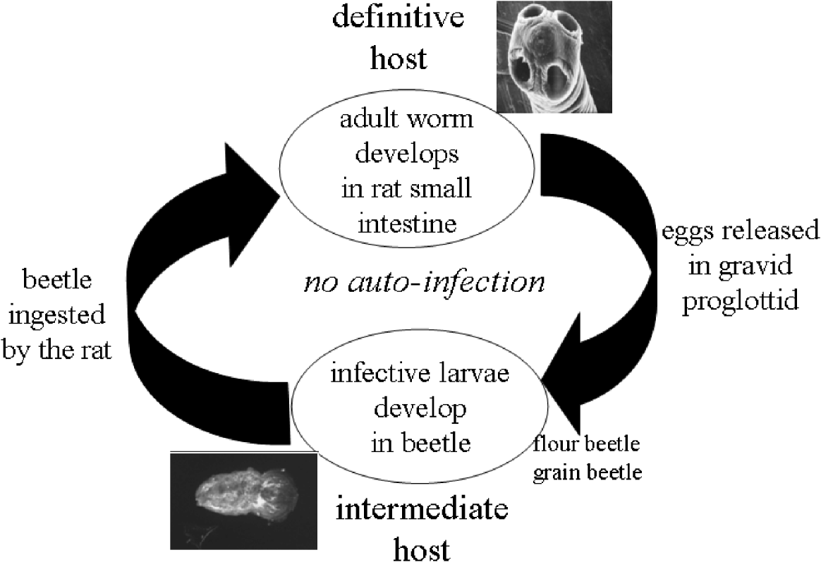
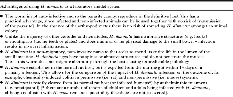
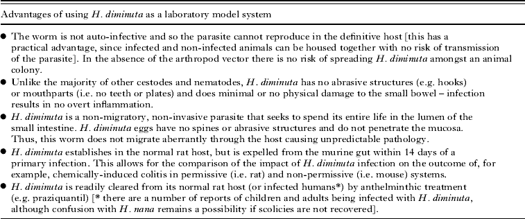
The lifecycle of H. diminuta is indirect, relatively simple and easily maintained in the laboratory (Fig. 1). Adult worms reside in the small intestine of the definitive host, typically rats, and release embryonated eggs in gravid proglottids into the environment in the hosts' faeces. When consumed by an insect intermediate host (typically grain (Tenebrio molitor) or flour beetles (Tribolium confusum) have been the preferred laboratory intermediary host species) the eggs hatch and the oncosphere penetrates the haemocoel where they develop into the infective larval cysticercoid stage; a process that takes approximately 10–14 days. When a rat ingests infected beetles, the cysticercoids pass through the stomach and exposure to trypsin and bile salts in the duodenum triggers their excystment. The worms attach to the small intestine and develop to sexual maturity within 2–3 weeks. H. diminuta is distinguished from related hymenolepids by the lack of a protruding rostellum and the absence of hooks or abrasive structures on the scolex. The advantages of using H. diminuta as a model tapeworm parasite in laboratory studies are presented in Table 1, and perhaps the most important of which is that the helminth establishes a patent infection in rats but is expelled from mice (see below).
Diagrammatic representation of the life-cycle of H. diminuta. The scanning electron micrograph shows the scolex of the adult worm (kindly provided by Dr. D. W. Halton, Emeritus Professor, The Queen's University of Belfast, N. Ireland) and the insert is a cysticercoid that has been retrieved from the haemocoel of T. confusum and immunostained with antibodies directed against serotonin.

Hymenolepis diminuta as a model parasite for the analysis of cestode-host interactions
THE IMMUNE RESPONSE TO INFECTION WITH H. DIMINUTA
Much of our knowledge relating to the immune response of the rat and mouse hosts to infection with H. diminuta comes from pioneering research in the 1970s and 1980s by Hopkins, Andreassen and colleagues (Hopkins et al. Reference Hopkins, Subramanian and Stallard1972a; Andreassen et al. Reference Andreassen, Bennet-Jenkins and Bryant1999).
Patent infections of H. diminuta establish in the rat host. It has long been known that the rat gut has a certain ‘carrying capacity’ for H. diminuta infections such that rats experimentally infected with 20–100 cysticercoids will, upon autopsy, 6–12 weeks later, typically harbour <10 worms. A prevailing view has been that this is due to inter-worm competition for space and nutrients. Indeed, when a range of infectivity is noted, total worm biomass tends to be very similar between the infections, i.e. when there are fewer worms present each individual is on average larger that those obtained from rats harbouring greater numbers of H. diminuta. However, there is also abundant evidence showing that rats are capable of mounting an immune response against H. diminuta, albeit of insignificant magnitude or character to accomplish complete expulsion of the worm burden.
The fact that appropriate immune effector systems are in place was aptly shown in rats infected with the nematode, Trichnella spiralis, in which H. diminuta undergo destrobilization (a survival trait) followed by expulsion (Silver et al. Reference Silver, Dick and Welch1980). If rats were given H. diminuta cysticercoids at the time of nematode expulsion, the tapeworm did not establish (Christie et al. Reference Christie, Wakelin and Wilson1979). Thus, as a bystander consequence of the immune and enteric response to infection with T. spiralis, H. diminuta was successfully expelled or failed to establish in the rat small intestine. Similarly, an immunological memory response to H. diminuta in rats has been shown: infection with 100 cysticercoids, followed by anthelminthic clearance, led to an enhanced expulsion response to secondary infection with 100 cysticercoids, although some worms did establish (Andreassen and Hopkins, Reference Andreassen and Hopkins1980). This protective response in the rat rapidly wanes following eradication of the primary infection.
Increases in molecules and cells that are typically associated with the spontaneous expulsion of gastrointestinal helminths such as IgE, IgG, mast cells and eosinophils (Andreassen et al. Reference Andreassen, Bennet-Jenkins and Bryant1999), and more recently T helper-2 (TH2) type cytokine mRNA (Webb et al. Reference Webb, Hoque and Dimas2007) have all been documented in H. diminuta-infected rats (rat strain-specific differences have also been noted). Despite this, H. diminuta establishes in the rat small intestine and one is left with the unsatisfactory and vague conclusion that as an essential element of the enigmatic host-parasite interaction, the rat fails to mobilize the appropriate orchestrated immune response involving all the components required to expel the worm (or that H. diminuta can specifically ablate or undermine the key effector mechanism(s) that would accomplish its eviction from the rat gut). In addition, once the worm establishes and grows to maturity it has the potential to suppress or re-direct the immune response of its rat host via the release of excretory or secretory (E/S) products (discussed below). How such E/S products would move from the gut lumen across the epithelial barrier layer (while retaining its immunosuppressive activity) and modulate the components of the mucosal or systemic immune system has not been determined. Furthermore, given the small biomass of the excysted worms it is unclear how much immunomodulator would be liberated and if this would make a substantial contribution to worm establishment. Thus, one can envisage how E/S products from the adult worm would help maintain an infection once established, but it is more challenging to gauge the contribution of E/S products to the establishment of infection.
Most strains of mice eradicate a primary infection of H. diminuta within 8–14 days and it is only in the severely immunocompromised mouse that H. diminuta can survive to maturity and produce gravid proglottids with functional eggs: mature worms can be obtained from mice treated with corticosteroids (McKay et al. Reference McKay, Halton, McCaigue, Johnston, Fairweather and Shaw1990b) or broad spectrum immunsuppressive drugs (Hopkins et al. Reference Hopkins, Subramanian and Stallard1972b). Mice generate a potent immunological memory against H. diminuta, such that secondary infection 28 days after a primary infection results in complete loss of the worms within two-four days (McKay et al. Reference McKay, Halton, McCaigue, Johnston, Fairweather and Shaw1990b). This memory is retained for at least eight months and clearly indicates a role for lymphocytes in the response against H. diminuta (Hopkins, Reference Hopkins1982). Athymic mice show delayed expulsion of H. diminuta, suggesting that T cells are dispensable for the spontaneous rejection of H. diminuta from the mouse gut (Andreassen et al. Reference Andreassen, Hindsbo and Vienberg1982) and that other compensating mechanisms can be called upon when T cells are absent. However, this does not undermine an active and important role for T cells when they are present (Isaak et al. Reference Isaak, Jacobson and Reed1975). In addition, T cell depletion is not 100% in these mice and so a small but important population of T cells may still have been available in the athymic mice to contribute to the expulsion of H. diminuta. More unequivocal data would be forthcoming with the use of the RAG knock-out mouse which is completely devoid of T and B lymphocytes.
There is a clear increase in TH2 type cytokines in mice infected with H. diminuta. This has best been shown by stimulation of spleen or mesenteric lymph node cells that show a time-dependent increase in, for example, IL-4, IL-5, IL-9 and IL-13 following infection (Isaak, Reference Isaak1983; Palmas et al. Reference Palmas, Bortoletti, Gabriele, Wakelin and Conchedda1997; Persaud et al. Reference Persaud, Wang, Reardon and McKay2007). Local gut levels also reveal a skewing towards expression of TH2 cytokines, but this has been assessed by RT-PCR for mRNA and not protein measurements (Persaud et al. Reference Persaud, Wang, Reardon and McKay2007) (the TH2 response in rats is dependent on the size of the infection and data on enteric levels of cytokine mRNA expression need to be complemented by the measurement of bioactive proteins (Webb et al. Reference Webb, Hoque and Dimas2007)). A dominant and strong TH2 response would be predicted to accompany worm expulsion since the cytokines produced will mobilise B cells and antibody production, eosinophils, mast cell and goblet cells, all of which would contribute to successful eradication of a lumen-dwelling gut parasite. Indeed, analysis of worms retrieved from C57/Bl6 mice 8–12 days post-infection (it can in fact be difficult to find any worms at this stage) shows that they are stunted, destrobilated, and have severe damage to the tegument which can be coated with immunoglobulin (Befus and Threadgold, Reference Befus and Threadgold1975; McCaigue and Halton, Reference McCaigue and Halton1987). Furthermore, we showed that mice genetically engineered to lack signal-transducer and activator of transcription (STAT)-6 do not expel five cysticercoid infections of H. diminuta, and that the worms produced gravid proglottids that when fed to beetles resulted in infective cysticercoids (McKay and Khan, Reference McKay and Khan2003). These STAT-6 KO mice did not show the typical goblet cell hyperplasia that accompanies expulsion of H. diminuta (see below). STAT-6 is a critical intracellular molecule that mediates, to a large extent, IL-4- and IL-13-driven transcriptional events, highlighting the need for at least one of these molecules if H. diminuta is to be expelled from the mouse gut.
In the light of these findings it is surprising that mice lacking CD4 (i.e. CD4+ T cells and hence TH2 cells), were found to expel H. diminuta with kinetics very similar to those of wild-type time-matched controls (Persaud et al. Reference Persaud, Wang, Reardon and McKay2007). There are other sources of TH2 cytokines, including natural killer cells and mast cells, and a novel IL-4, IL-5 and IL-13 producing non-T/non-B cell has been postulated following helminth infection (Fallon et al. Reference Fallon, Ballantyne, Mangan, Barlow, Dasvarma, Hewett, McIlgorm, Jolin and McKenzie2006) which could compensate for the lack of CD4+ T cells. However, the CD4 gene deletion can be leaky and so analysis of H. diminuta in major histocompatibility class (MHC) II mice that are also devoid of CD4+ T cells is warranted.
Humoral (i.e. elevations in IgE and IgG) and cell-mediated (i.e. mast cell hyperplasia and eosinophila) immune events have been demonstrated in H. diminuta infected mice and have been implicated in the successful eradication of the helminth (Andreassen et al. Reference Andreassen, Bennet-Jenkins and Bryant1999). However, all of these potential effector mechanisms have to one degree or another also been shown in rats infected with H. diminuta (noting strain specific findings (Andreassen et al. Reference Andreassen, Bennet-Jenkins and Bryant1999; Starke and Oaks, Reference Starke and Oaks2001)). So the value of any single effector cell or molecule needs to be assessed in the context of the overall host response to H. diminuta, both locally and systemically.
There does appear to be a very close relationship between intestinal goblet cell hyperplasia and the expulsion of H. diminuta from C57/Bl6 mice (McKay et al. Reference McKay, Halton, McCaigue, Johnston, Fairweather and Shaw1990b). The increase in goblet cell number and theca size matches exactly the peak time of expulsion of H. diminuta following primary and secondary infections and this goblet cell, and hence mucus, response was not observed in mice treated with corticosteroids or those lacking STAT-6 in which the worms can establish (McKay and Khan, Reference McKay and Khan2003). Moreover, while there was an increase in goblet cells in the ileum of rats 10 days after infection with H. diminuta this did not persist as the helminth established in the rat.
Finally, it is intuitive to accept that given the size of helminth parasites that an effective anti-worm response is mediated by the adaptive immune system, with a coordinated multi-cellular approach that likely targets destruction of the outer protective layers of the worm and their organs of attachment. Certainly helminths are too large for ingestion by phagocytes. However, the possibility that helminths may express pathogen-associated molecular patterns (hPAMP) that would allow for a rapid response and the attraction of pertinent effector cells is intriguing (Smith et al. Reference Smith, Mangan and Fallon2009). Currently there is no information in this area relating to H. diminuta.
OTHER ENTERIC CHANGES THAT MAY ALTER THE HOST-PARASITE RELATIONSHIP
It is tempting to focus solely on the immune response to H. diminuta, to identify factors that are critical to regulating (e.g. cytokines) or potentially mediating (e.g. eosinophils) expulsion from the gut. Even though the gut is the largest repository of immune cells in the human body it is also a complex, multi-cellular and dynamic environment in which the immune cells are juxtaposed to a variety of other cell types. Thus, one must raise the possibility that other enteric cells participate in the anti-H. diminuta effect in the mouse.
There is a voluminous literature illustrating the two-way communication between nerves and immune cells and the reciprocal cross-regulation of each system. Similarly, changes in the expression of neurotransmitters, neurotransmitter receptors and neuronal activity in the gut of parasitised rodents have been presented (McKay and Fairweather, Reference McKay and Fairweather1997). We documented increases in gut levels of neuropeptides that would be considered pro-inflammatory or immunostimulatory (i.e. substance P) and decreases in those that have been associated with suppression of immune activity (i.e. vasoactive intestinal peptide (VIP)) in the small intestine of mice infected with H. diminuta (McKay et al. Reference McKay, Halton, Shaw, Johnston, Fairweather and Buchanan1991). However, the functional consequences of these, and concomitant alterations in enteroglucagon, neurotensin, and peptide histidine isoleucine (PHI) were not defined. In addition, immunocytochemical detection of glial-derived neurotropic factor (GDNF) revealed increased numbers of GDNF+ cells in the small intestine of H. diminuta – infected rats; the functional significance of this is not known (Starke-Buzetti and Oaks, Reference Starke-Buzetti and Oaks2008). Alterations in enteric neuronal activity have been shown in tissue from parasitised rats, such as diminished neurotransmitter (acetylcholine (ACh) or noradrenaline) (Collins et al. Reference Collins, Blennerhassett, Blennerhassett and Vermillion1989) release and this could have implications for a ‘washer/sweeper’ (i.e. increased water movement into the gut and increased peristalsis) event that is often generated in response to enteric infection (Wood, Reference Wood1993).
Analysis of active jejunal epithelial ion transport revealed a two-fold increase in baseline short-circuit current (indicates net active electrogenic ion transport) 11 days post-infection with H. diminuta and a reduction in changes in active ion transport evoked electrical field stimulation that depolarizes nerves to release neurotransmitters (typically ACh and VIP) to evoke active chloride secretion at eight and 11 days post-infection (Persaud et al. Reference Persaud, Wang, Reardon and McKay2007). The former could reflect increased water movement into the gut and the latter suggests an enteric neural abnormality in the infected mice. Thus, discrete changes in the enteric nervous system can be found in H. diminuta-infected rats, and other changes correlated with the kinetics of worm rejection from mice. While it is tempting to link these events to modulation of the host immune response to infection, it must be acknowledged that this is supposition at this stage of our understanding of the H. diminuta-mouse relationship.
The enteric enteroendocrine cell occupies a strategic position with its apical surface in contact with the luminal contents so that it can sense this environment and transduce signals via the paracrine or haemocrine release of serotonin and other messenger molecules. For instance, serotonin (5-HT)-positive enteroendocrine cells have been implicated in enhancing the diarrhoea associated with Vibrio cholera infection (Lundgren, Reference Lundgren1998). Analysis revealed a positive correlation between an increase in number of 5-HT+ cells and the peak time of H. diminuta rejection from mice following primary and secondary infections (McKay et al. Reference McKay, Halton, Johnston, Fairweather and Shaw1990a). Activation of these cells and the release of their contents would be expected to make the gut more inhospitable for H. diminuta by increasing water efflux into the lumen and by activating serotoninergic nerves that could mediate a variety of changes in the gut. In support of this, it has recently been shown that Balb/c mice that generate a TH2 response and effectively clear the nematode, Trichuris muris, also have increases in enteric 5-HT+ enteroendocrine cells, whereas the AKR mouse that is susceptible to infection generates a TH1 response and no concomitant 5-HT+ cell hyperplasia (Motomura et al. Reference Motomura, Ghia, Wang, Akiho, El-Sharkawy, Collins, Wan, McLaughlin and Khan2008).
Finally, the intestine, and the colon in particular, is home to a complex ecosystem of bacteria. This commensal flora is an essential component of the normal gut and serves many beneficial roles including digestion of complex carbohydrates and the generation of vitamins and butyrate, the latter being an important energy source for the colonocyte. Little is known of how infection with H. diminuta might affect the normal gut flora or vice versa: only a single study is available that indicates a reduction in the numbers of aerobic bacteria in rats infected with H. diminuta (numbers of anaerobic bacteria were unaffected) (Dwinell et al. Reference Dwinell, Bass, Schaefer and Oaks1997).
SUPPRESSION OF HOST IMMUNE RESPONSES BY H. DIMINUTA
The ability to evade or suppress host immunity is a prerequisite for a successful parasite. Indeed, there are numerous examples of crude extracts or partially characterized fractions of helminth parasites modifying immune responses in their hosts' (Fallon and Alcami, Reference Fallon and Alcami2006; Harnett and Harnett, Reference Harnett and Harnett2008; Johnston et al. Reference Johnston, MacDonald and McKay2009a). Somatic extracts or E/S products from a plethora of helminth parasites when added to immune cells in vitro exert an immunosuppressive effect (e.g. inhibiting stimulated cytokine responses). When administered to mice, these helminth-derived fractions affect a variety of immune cell types and skew CD4+ T cell responses away from TH2 events (i.e. those that are anti-helminthic) and towards TH1 events. Furthermore, helminth-derived products have been used to reduce inflammation in murine models of disease (Harnett and Harnett, Reference Harnett and Harnett2006; Motomura et al. Reference Motomura, Wang, Deng, El-Sharkawy, Verdu and Khan2009). However, to date only a few immunoregulatory/immunosuppressive helminth-derived molecules have been purified and structurally characterized (van der Kleij et al. Reference van der Kleij, Latz, Brouwers, Kruize, Schmitz, Kurt-Jones, Espevik, De Jong, Kapsenberg, Golenbock, Tielens and Yazdanbakhsh2002; Goodridge et al. Reference Goodridge, Stepek, Harnett and Harnett2005). Thus, helminth parasites are an untapped source of immunoregulatory molecules that could serve as blueprints for novel drugs.
We have found that a PBS-soluble, high-molecular weight (>50 KDa, as defined by spin-column separation) fraction of adult H. diminuta or E/S products from the adult worm can suppress T cell and macrophage activation in vitro. Proliferation and IL-2 production by murine spleen cells and human peripheral blood mononuclear cells evoked by treatment with the mitogen, concanavalin A (conA), or the bacterial superantigen, Staphylococcus aureus enterotoxin B (SEB) were dose-dependently (1–1000 μg/mL) inhibited by the H. diminuta high molecular weight fraction (HdHMW) (Wang and McKay, Reference Wang and McKay2005). This inhibition of immune cell activity was not due to a cytotoxic effect of the HdHMW. In the same in vitro system, the HdHMW enhanced production of the immunoregulatory cytokine IL-10, while suppressing IL-4 synthesis; the latter would limit TH2 events. These effects of HdHMW were wholly or partially blocked by boiling, indicating the protein nature of the molecule(s) responsible for the immunosuppression. Moreover, the worm extract had an IL-12-like molecule as defined by its ability to bind in ELISA and detection on immunoblots, and if bioactive this molecule would promote TH1-type responses that would antagonize TH2-driven anti-helminth events. Recent analyses have revealed that the HdHMW significantly reduces lipopolysaccharide and poly I:C (toll-like receptor (TLR) 4 and 3 ligands, respectively) production of TNFα, IL-1β and IL-6 by murine or human macrophages, while leaving the ability of the cells to phagocytose bacteria intact. In this instance, boiling and digestion of the HdHMW with trypsin or proteinase K had a minimal impact on the suppression of macrophage activity and lead to the speculation that the bioactive molecule was a glycoprotein (M. J. G. Johnston, personal communication). In addition, H. diminuta can release cGMP and this could contribute to immunosuppression by the worm (Zimmerman et al. Reference Zimmerman, Brownfield, DeVente, Bass and Oaks2008).
Initial observations showed that splenocytes retrieved from mice treated with the HdHMW (1 mg ip.) were hypo-responsive to in vitro challenge with conA (Wang and McKay, Reference Wang and McKay2005). Subsequently it has been found that daily injections of this dose of the HdHMW, over a 3-day period, significantly reduced the severity of dinitrobenzene sulphonic acid (DNBS)-induced colitis in mice (M. J. G. Johnston, personal communication). These studies provide proof-of-principle data in favour of drug development from fractions of H. diminuta, and yet clearly much remains to be done to: (1) identify, purify and characterize the immunoregulatory molecules in the HdHMW, (2) identify the in vivo target cells, and (3) define the molecular mechanisms of action that underlie the immunosuppressive activity of HdHMW.
HYMENOLEPIS DIMINUTA AND THE MODULATION OF CONCOMITANT DISEASE
Juxtaposing the facts that infection with helminth parasites evokes immune responses in humans and mice that are orchestrated by TH2-type cytokines and that TH2 events suppress TH1-driven immunity, the hypothesis arises that infection with helminth parasites has the potential to protect an individual from diseases or immunopathology in which TH1 events predominate. Indeed, one can postulate that infection with parasitic helminths is a component of the hygiene hypothesis given the disparity between global regions in which helminth infections are endemic and the increasing incidence of autoimmune and inflammatory disorders in westernised societies (Bach, Reference Bach2002). During the last decade, we and others have employed murine models of inflammatory bowel disease (IBD), diabetes, asthma and multiple sclerosis, and found that infection with helminth parasites can reduce the severity of these conditions (Maizels, Reference Maizels2005; Elliott et al. Reference Elliott, Summers and Weinstock2007; McKay, Reference McKay2009).
Addition of dextran-sodium sulphate (DSS) to the drinking water of mice results in a predicable inflammation that is restricted to the colon (Reardon et al. Reference Reardon, Sanchez, Hogaboam and McKay2001). Mice infected with H. diminuta seven days prior to a five-day exposure to a 4% (wt./vol.) solution of DSS had less severe disease as defined by a reduction in the occurrence of occult blood and normalisation of epithelial ion transport characterisitics. Similarly, use of H. diminuta in a treatment protocol (i.e. mice were infected two days after exposure to DSS-drinking water) also relieved these symptoms of DSS-induced colitis. However, in neither instance did infection with H. diminuta ameliorate the histopathology or granulocyte infiltration (as assessed by myeloperoxidase activity) elicited by DSS.
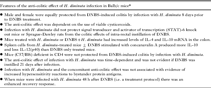
Dinitrobenzene sulphonic acid is a haptenizing agent and when instilled directly into the colon of rodents results in an acute, self-limiting colitis that shares some similarities with Crohn's disease, one of the major categories of IBD in humans. Balb/c mice infected with H. diminuta (confirmed by blood eosinophilia or goblet cell hyperplasisa in the small intestine) were significantly protected from colitis induced by intra-rectal instillation of DNBS (3 mg) (Table 2) (Hunter et al. Reference Hunter, Wang, Hirota and McKay2005). Infection was accompanied by an increased propensity of spleen cells and mesenteric lymph node cells to produce IL-10 upon mitogenic stimulation. In addition, RT-PCR analyses revealed increased IL-10 mRNA in colonic segments excised from DNBS+H. diminuta-treated mice compared to those obtained from DNBS only or naïve control mice. Based on the known anti-inflammatory effects of IL-10, the hypothesis that infection with H. diminuta inhibited DNBS-induced colitis via IL-10 was tested by treating mice with an anti-IL-10 neutralizing antibody: administration of 200 μg of anti-IL-10 antibody ablated the ability of H. diminuta to reduce DNBS-induced colitis. These data add to the growing awareness that infection with helminth parasites has the ability to ameliorate concomitant disease conditions and indicate that IL-10 can be an important component of the anti-inflammatory effect. Similar findings have been presented for mice infected with Heligosomoides polygyrus given trinitrobenzene sulphonic acid (TNBS) (Setiawan et al. Reference Setiawan, Metwali, Blum, Ince, Urban, Elliott and Weinstock2007); however, the same investigators reported that this nematode also reduces the inflammation that spontaneously develops in mice genetically deficient in IL-10 (Elliott et al. Reference Elliott, Setiawan, Metwali, Blum, Urban and Weinstock2004).
Infection with Hymenolepis diminuta inhibits colitis induced in mice by direct instillation of dinitrobenzene sulphonic acid into the colon
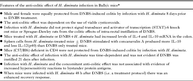
* , data summarized from Hunter et al. (Reference Hunter, Wang, Hirota and McKay2005) and Persaud et al. (Reference Persaud, Wang, Reardon and McKay2007).
Given the complexity of host-parasite interactions it would be naïve to suggest that any concomitant health or anti-inflammatory benefit of infection with a helminth parasite would be dependent on a single molecule. Infection with H. diminuta results in time-dependent increases in mRNA for FOXP3, and arginase-1 and found in inflammatory zone (FIZZ)-1 (Persaud et al. Reference Persaud, Wang, Reardon and McKay2007) that are indicative of the presence of regulatory T cells (Tregs) and alternatively activated macrophages (AAMs), respectively. Both of these cell types have been associated with helminth infections and the anti-inflammatory effects of infection with helminths (Wilson and Maizels, Reference Wilson and Maizels2006; Reyes and Terrazas, Reference Reyes and Terrazas2007). The putative role of these cells in the anti-colitic effect of infection with H. diminuta needs to be determined. Also, as noted above, mice lacking CD4 are capable of expelling H. diminuta but these mice were not protected from DNBS-induced colitis by infection with H. diminuta. Thus, much remains to be done to define the roles of, and interactions (if any) between FOXP3+ Tregs, AAMs, CD4+ cells and IL-10 in: (1) the immunologically-mediated expulsion of H. diminuta from the mouse small intestine, and (2) in the inhibition of DNBS-induced colitis.
In addition, one must question the role of neural factors, enteroendocrine-derived mediators (e.g. serotonin) and alterations in the composition of the normal microflora following infection with H. diminuta in the protection against DNBS-colitis. For instance, the microflora is recognized as a trigger for the development of colitis, and thus at least a portion of the anti-colitic effect of infection with H. diminuta could be via modulation or maintenance of the normal gut flora.
While there are many examples of helminth amelioration of concomitant disease in murine model systems (McKay, Reference McKay2009), and to a lesser extent human disease (Summers et al. Reference Summers, Elliott, Urban, (Jr.), Thompson and Weinstock2005), the investigator should be mindful to two issues: (1) helminths may not be beneficial in all situations, and (2) there is the omnipresent possibility for deleterious side-effects of any novel treatment and the spectre of iatrogenic disease. In the case of the former, oxazolone-induced colitis is driven by IL-13 and is characterized by increased numbers of eosinophils in the colon. Mice infected with H. diminuta and administered oxazolone had significantly greater colonic inflammation than oxazolone-only treated mice (Hunter et al. Reference Hunter, Wang and McKay2007). The enhanced colitis was associated with increases in eosinophils (and eosinophil peroxidase), but whether or not these cells, that are mobilised in response to infection with helminth parasites, are responsible for the increased inflammatory disease remains to be determined. In the context of potential side-effects of either infection with parasitic helminths or a therapy based on rodent-helminth model systems, data from long-term follow-up studies that are needed to address this have not been reported.
Finally, there will be circumstances in which the effect of infection with H. diminuta is neutral, as recently described for the development of gastric ulceration following acetic acid treatment (McKay and Wallace, Reference McKay and Wallace2009). The caveat is added that this study does not negate the possibility that infection with H. diminuta could affect gastritis in mice or upper gastro-intestinal ulceration induced by other agents such as non-steroidal anti-inflammatory drugs or infection with Helicobacter pylori.
CONCLUDING COMMENTS
Hymenolepis diminuta is in many ways a model parasite: its life-cycle is readily maintained in the laboratory, permissive and non-permissive hosts are known and in the case of the latter, the kinetics of the spontaneous expulsion are defined, although the intricacies of the immunology underlining this are not fully known. Use of this model has confirmed and extended findings from other helminth-rodent model systems, underscoring the universality of a host TH2 response to infection with parasitic helminths, and identifying STAT6 as a crucial requirement for successful expulsion from the murine gut. Arguably of more importance is the acquisition of knowledge that can shape the development of novel therapies to treat not only parasitic diseases but also autoimmune and inflammatory diseases, the incidence of which is rapidly increasing in westernised societies. There is little doubt that H. diminuta can be a source of novel immunoregulatory drugs and the application of new technologies that allow the characterization and purification of molecules from small amounts of material (Johnston et al. Reference Johnston, MacDonald and McKay2009) make this a realistic possibility should the challenge be taken up. Similarly, while one can speculate on the use of a ‘therapeutic helminth’ (McKay, Reference McKay2009) there is clearly an aspect of palatability here and the spectre of unforeseen consequences when introducing an organism into a novel environment. However, defining how the immune response mobilised to combat infection with a parasitic helminth can inhibit or block other diseases can reveal new ways to treat these diseases in the absence of helminth infection. Parasitism is a hetero-specific interaction that evolves over millennia and by using the H. diminuta-rodent model system, and others, we have gained insight to the workings of mammalian immune system and can develop new approaches to combat omnipresent diseases (including parasitic ones) and emergent disorders such as idiopathic inflammation, diabetes and cardiovascular disease that take a huge toll on human health and well-being.
ACKNOWLEDGEMENTS
Research conducted in the authors' laboratory was funded by operating grants from the Canadian Institutes for Health Research (CIHR), the Crohn's and Colitis foundation of Canada (CCFC) and the Natural Science and Engineering Research Council of Canada (NSERC).