A key and highly successful intervention in intramammary infection and mastitis control in the dairy cow has been managing the presence of pathogenic organisms on the teat (Dodd and Bramley, Reference Dodd and Bramley1985). This includes reduction in the prevalence of infected lesions on or near the teat orifice mostly by good milking machine operation and by adroit application of disinfectant immediately post milking. This has been most successful in the drastic reduction of intramammary infections and subsequent disease (clinical mastitis) by the colonising/contagious bacterial pathogens Streptococcus agalactiae, Streptococcus dysgalactiae and Staphylococcus aureus, which can be practically eliminated from a herd (Hillerton et al., Reference Hillerton, Bramley, Staker and McKinnon1995). Environmentally-originating bacteria are less well controlled, but by preventing exposure of the teat in the period after milking before teat canal closure is complete and by using an effective disinfectant, the rate of these new infections can be minimised.
Colonising bacteria penetrate the teat mostly either via growth in the teat canal (Lacy-Hulbert and Hillerton, Reference Lacy-Hulbert and Hillerton1995) or directly during milking by a physical ‘ímpact’ force (Thiel et al., Reference Thiel, Cousins, Westgarth and Neave1973). The means of entry of Streptococcus uberis, the commonest bacterial pathogen in herds pastured or bedded with plant-based materials, appears to be only directly but unlikely to be aided by direct force (Lacy-Hulbert and Hillerton, Reference Lacy-Hulbert and Hillerton1995). The timing of such entry, whether through an opaque teat canal, or a completely closed canal (as such infections are the commonest in the dry period: Neave et al., Reference Neave, Dodd and Henriques1950), is unclear.
Bramley (Reference Bramley1976) showed different outcomes from the infusion of Escherichia coli directly into the mammary gland between before (no infection) and immediately after milking (infection common). The fate of Str. uberis, according to time of introduction is unknown. Given that milk, and probably the interior of the mammary gland, does not in the first instance provide all of the nutritional requirements of Str. uberis (Kitt and Leigh, Reference Kitt, Leigh, Horaud, Bouvet, Leclercq, de Montclos and Sicard1997) and that in vitro growth in milk has a lag phase up to seven hours (Kliem and Hillerton, Reference Kliem and Hillerton2002), the timing of introduction of these bacteria may affect the establishment of infection and subsequent disease. This has been examined by experimental infection of an infectious dose of Str. uberis directly into the udder through the teat canal at various times in the inter-milking interval including immediately after complete machine milking, both when the following milking occurs after an 8- and a 16-h interval, mimicking twice-daily milking.
Materials and methods
This study was carried out compliant with the UK Animals (Scientific Procedures) Act 1986 (IAH request 3000). A condition of ethical approval was that diagnosis of any moderate clinical mastitis (a level when a commercial farmer would withhold milk and initiate antimicrobial therapy), using a quarter and systemic score system, must result in the animals being withdrawn from the study and being treated appropriately with antibiotic products. Use of the terms mild, moderate and severe clinical mastitis follows the terminology of IDF (2011).
Twelve multiparous, Holstein-Friesian dairy cows, approximately 100 d post partum and free of intramammary infection in the current lactation, were used. They were milked at 07.30 and 15.30 daily in an experimental parlour equipped to record quarter milk yields.
Foremilks from each quarter were examined visually for colour, flecks and clots, a sample used to measure milk electrical conductivity (MEC) and a 20 ml sample collected aseptically for bacteriological examination and somatic cell count (Hillerton et al., Reference Hillerton, Bramley, Staker and McKinnon1995). Rectal temperatures were also measured for each cow. These observations were made from 4 d pre infusion until 7 d post infusion or until antimicrobial treatment was initiated. Milk samples were cultured on aesculin blood agar for 48 h at 37°C and any bacterial colonies counted and identified and cell counts determined (International Dairy Federation, 1981).
Selected quarters, two diametrically opposed per cow, of two cows per treatment (control and 5 MI) were infused either with 1 ml sterile skim (SK) milk solution or 500 cfu initial titration of Str. uberis 0140J in the SK solution at each time point detailed in Fig. 1. Infusion was directly into the teat sinus, bypassing the teat canal. The SK solution was used, as a prior test showed 500 cfu/ml in pyrogen free phosphate buffered saline decayed to 16 cfu/ml in the two-hour period between preparation and completion of infusion. Post infusion recovery using the SK solution was 340 cfu/ml.
Timeline to show approximate time of infusions relative to milkings.

Infusions were timed to give intervals of 1 (MI1), 4 (MI4), 8 (MI8), 12 (MI12) and 16 (MI16) hours before the next milking thus at 1 h before afternoon milking, 4 h before afternoon milking, immediately after morning milking, 4 h after afternoon milking and immediately after afternoon milking (Fig. 1).
All quarters infused with Str. uberis, whether developing clinical mastitis or not, were treated on diagnosis of moderate clinical mastitis or at the conclusion of the observation period, using the normal farm protocol of intramammary penethamate hydriodide.
Results
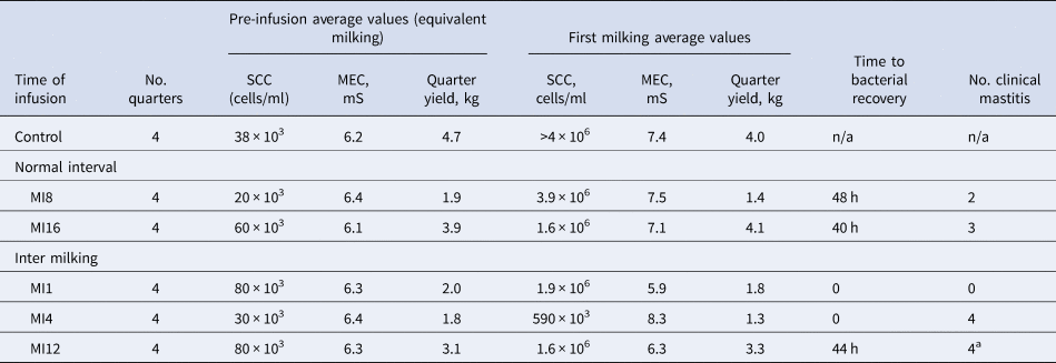
The changes in milk parameters and their timing relative to the time of infusion are shown in Table 1.
Changes in milk parameters in response to infusion of Str. uberis into mammary gland quarters at various milking intervals, hours prior to next milking (MI)

n/a – not applicable.
a Bacteria recovered from only 3 quarters.
The quarters infused with SK solution developed a transitory mild mastitis (rise in average cell count from 38 × 103 to >4.0 × 10⁶ cells/ml, increase in MEC, decrease in yield and a few small clots in foremilk) at the next milking (16 h) after infusion. Body temperature did not rise. These changes decayed and had resolved by 72 h post infusion.
When bacteria were infused into the mammary gland immediately after milking, followed by an 8 h interval until the next milking, a mild mastitis (rise in average cell count from <20 × 103 to 3.9 × 10⁶ cells/ml, average rise in body temperature of 1°C, rise of MEC of 1.0 mS and yield decrease of 30%, and the quarters had some local heat and firmness but no clots in foremilk) occurred in all quarters at the next milking. Str. uberis were first recovered, but only from two of the four quarters, 48 h (4 milkings) later. These two quarters reached the threshold for clinical mastitis 56 h after infusion, they were treated with antibiotics and withdrawn from the study as required in the ethical approval. The other two quarters had resolved all indicators of mastitis by 96 h post infusion.
When infusion was followed by a 16 h interval a mild mastitis (rise in average cell count from <60 × 103 to 1.6 × 10⁶ cells/ml, yield decrease of 20% but no rise in body temperature or MEC, and quarters and foremilk appeared normal) occurred in all quarters at the next milking. Up to 106 cfu/ml Str. uberis were recovered from three of the four quarters 40 h (4 milkings) later, coincident with three of these quarters meeting the threshold for moderate clinical mastitis. These three quarters were treated with antibiotics and withdrawn from the study. Despite the timing of milkings between the MI8 and MI16 groups resulting in a longer inter milking interval allowing more time for the bacteria to establish in MI16, the first bacterial recoveries were after essentially the same length of time (milkings) post infusion.
When bacteria were infused one hour before milking (MI1) no effect was found at that milking. However, at the subsequent milking (after a further 8 h) the cell count had risen from an average 80 × 103 to 1.9 × 10⁶ cells/ml but there was no effect on body temperature, milk yield, MEC, foremilk appearance or the udder. No bacteria were recovered from any of the four infused quarters.
When bacteria were infused 4 h before milking, at that next milking the foremilk from all four quarters was significantly clotted, all quarters were hot and hard, average MEC had increased and yield had decreased 35%. All quarters met the criteria to be diagnosed as moderate clinical mastitis and so were treated with antibiotics and withdrawn from the study. Subsequently the cell count at treatment was found to have increased from an average of 30 × 103 to only 590 × 103 cells/ml.
When bacteria were infused 12 h before milking, at that next milking all quarters appeared normal, neither MEC nor yield had changed. Subsequently average cell count was shown to have increased from 80 × 103 to 1.6 × 10⁶ cells/ml. All infused quarters met the criteria for moderate clinical mastitis 44 h after infusion with severe clotting in foremilk and an average cell count of 67 × 106 cells/ml. They were treated with antibiotics and withdrawn from the study. Str. uberis were recovered from three of the four quarters (>106 cfu/ml).
All quarters that received intramammary antibiotic treatment recovered as determined by visual normality of milk and the udder, and recovery of quarter milk yield.
Discussion
The control infusion of SK solution induced a sterile mastitis. This is significant in that any bacterial invasion of the mammary gland is likely to be in an extraneous medium that will induce a foreign material response. The inflammation lasted up to 72 h suggesting that the neutrophil diapedesis was continuous, although transitory, and the SK had stimulated the cellular defence system. Thus, the response to SK mimics any natural invasion of the mammary gland of the lactating dairy cow but is not protective. The SK induced mastitis did not protect against the bacterial mastitis (see Anderson, Reference Anderson1985).
Infusion of Str. uberis post milking (both 8 and 16 h before the next milking) induced both a moderate clinical mastitis and a bacterial infection in 5 of 8 quarters. This probably mimics a natural bacterial invasion and mastitic event with the gland responding mildly, measurable by cell count, MEC and yield but not easily observable at the next milking by anyone milking the cows in a commercial operation. The measured and observed signs developed to reach the clinical threshold over 2 to 3 d.
The MI1 group showed no change at the first milking after infusion. Eight hours later cell count had risen, and this persisted for up to 72 h but no other parameters changed at any time. It appears that if Str. uberis invades the mammary gland shortly before milking it may have no udder health significance. Probably the bacteria were milked out of the gland before growth occurred. Kliem and Hillerton (Reference Kliem and Hillerton2002) showed a lag phase before growth occurred in milk. Further, the temporary persistence of a higher cell count would provide cellular defences.
The most immediate and severe response at the first milking after infusion occurred in the MI4 group with an immediate clinical mastitis in all quarters. The MI12 group also developed a clinical mastitis in all quarters but only 2 d after infusion. Bacteria were not recovered from the MI4 group, probably because of insufficient time for their growth. Hence the severity of mastitis, beyond that of SK only infusion, was due to the response of the challenged mammary gland epithelium at a time when alveolar milk was being produced, and less than one quarter of the daily yield, probably insufficient to dilute the bacteria (Elliott et al., Reference Elliott, Dodd and Brumby1960). Whereas, at MI12 up to half the daily milk production had occurred and the alveoli would be releasing milk into the udder cistern. These two groups demonstrate that any infection by Str. uberis in the intermilking interval, sufficiently long before the next milking, is most likely to result in a bacterial infection and clinical mastitis. The mastitis resulting from the SK solution is not protective when the bacteria are introduced in the middle of the intermilking interval.
Protection of the lactating mammary gland from Str. uberis at all times, possibly except soon before milking, appears necessary to prevent a mastitis and an infection. This is consistent with the relative lack of effect of conventional mastitis control on new infections by aesculin-positive streptococci (Hillerton et al., Reference Hillerton, Bramley, Staker and McKinnon1995), with the need to ensure closure of the teat canal as soon as possible after milking and the need to use effective teat disinfection with a product of some persistence (Williams and Mein, Reference Williams and Mein1985).
Acknowledgement
This work was undertaken with the superb technical support of the late Andy Walton.