INTRODUCTION
Species of Hematodinium are parasitic dinoflagellates that infect a wide taxonomic range of crustacean hosts throughout the world (Shields, Reference Shields1994; Stentiford and Shields, Reference Stentiford and Shields2005). These parasites have been reported in the blue crab Callinectes sapidus along the East and Gulf coasts of the USA since the 1970s (Newman and Johnson, Reference Newman and Johnson1975; Messick, Reference Messick1994; Shields, Reference Shields1994; Messick et al. Reference Messick, Jordan and Van Heukelem1999; Messick and Shields, Reference Messick and Shields2000; Shields and Squyars, Reference Shields and Squyars2000). The prevalence of the disease has been reported as high as 100% in focal outbreaks in blue crabs along the East Coast (Messick, Reference Messick1994; Messick and Shields, Reference Messick and Shields2000). Hematodinium can cause severe pathology to their blue crab hosts (Newman and Johnson, Reference Newman and Johnson1975; Messick and Shields, Reference Messick and Shields2000; Shields et al. Reference Shields2003). Hosts infected with a related species of parasite eventually die from malfunction of the hepatopancreas, degradation of muscle tissue, and respiratory dysfunction (Taylor et al. Reference Taylor, Field and Parslow-Williams1996; Stentiford et al. Reference Stentiford, Neil and Coombs1999, Reference Stentiford, Neil and Coombs2000). Naturally infected blue crabs often die over a period of 14 to 35 days depending on the intensity of the infections (Messick and Shields, Reference Messick and Shields2000). Experimentally infected blue crabs start dying 14 to 17 days post-inoculation (p.i.), with cumulative mortalities of 86% by 40 days p.i. (Shields and Squyars, Reference Shields and Squyars2000) and 100% by 55 days p.i. (Messick and Shields, Reference Messick and Shields2000). Hematodinium infections in blue crabs may potentially contribute to the recent population decline of this species in Chesapeake Bay (Messick and Shields, Reference Messick and Shields2000; Shields, Reference Shields2003; Stentiford and Shields, Reference Stentiford and Shields2005).
Hematodinium spp. are members of the Syndiniales, an order of parasitic dinoflagellates that infect a variety of protistan, invertebrate and fish hosts. Two species of Hematodinium have been described, the type species, H. perezi (Chatton and Poisson, Reference Chatton and Poisson1931) and H. australis (Hudson and Shields, Reference Hudson and Shields1994). Recent work suggests that the species infecting blue crabs (and other Western Atlantic portunids) is different from the species infecting cold-water hosts in boreal climates (Jensen et al. Reference Jensen, Califf, Lowe, Hauser and Morado2010; Small et al. Reference Small, Shields, Hudson and Reece2007a). Little is known about the transmission of Hematodinium spp. and their development within the crustacean hosts. This is largely due to their poorly understood life cycles. The species of Hematodinium from the blue crab shares similar morphological characteristics with H. perezi, and was identified as H. perezi in previous studies (Newman and Johnson, Reference Newman and Johnson1975; Messick, Reference Messick1994; Shields and Squyars, Reference Shields and Squyars2000). However, recent DNA analyses suggest that it is different from the Hematodinium species isolated from one of the type hosts (Liocarcinus depurator, Small et al. Reference Small, Shields, Hudson and Reece2007a); we therefore refer to the parasite as Hematodinium from C. sapidus.
Like most other species of Hematodinium, the life cycle of Hematodinium from the blue crab is unknown, although several morphologically distinct stages have been reported from the haemolymph and tissues (Newman and Johnson, Reference Newman and Johnson1975; Shields, Reference Shields1994; Messick et al. Reference Messick, Jordan and Van Heukelem1999; Messick and Shields, Reference Messick and Shields2000; Shields and Squyars, Reference Shields and Squyars2000) and from short-term cultures (7–14 days) of the parasite (Small, Reference Small2004; Small et al. Reference Small, Shields, Neil, Taylor and Coombs2007b). However, the Hematodinium-like species from the Norway lobster, Nephrops norvegicus, has been established in vitro (Appleton and Vickerman, Reference Appleton and Vickerman1998). In cultures of the parasite from the Norway lobster, macro- and micro-dinospores give rise to the filamentous trophonts, which develop into ‘gorgonlocks’ trophonts. The latter divide further into either clump colonies that develop into more filamentous trophonts, or they become arachnoid trophonts. Over time, the arachnoid trophonts become arachnoid sporonts that undergo sporogony to produce sporoblasts, which then develop into dinospores (Appleton and Vickerman, Reference Appleton and Vickerman1998). In the blue crab host, the vermiform plasmodium (plasmodes of Chatton and Poisson (Reference Chatton and Poisson1931), or filamentous trophonts of Appleton and Vickerman (Reference Appleton and Vickerman1998)), ameboid trophonts, clump colonies, and dinospores can be observed in the haemolymph of infected hosts (Newman and Johnson, Reference Newman and Johnson1975; Shields, Reference Shields1994; Messick et al. Reference Messick, Jordan and Van Heukelem1999). It is unclear how those stages develop within the blue crab host or what their role is in the life history of the parasite. However, the filamentous trophont is always found in early infections (Messick and Shields, Reference Messick and Shields2000; Shields and Squyars, Reference Shields and Squyars2000).
We undertook culture studies with Hematodinium from the blue crab, C. sapidus, to characterize the developmental stages of the parasite in vitro. Once established in culture, we explored the life cycle, infectivity and effect of temperature and light on the growth of the parasite in vitro.
MATERIALS AND METHODS
Host collection
Blue crabs, Callinectes sapidus, were collected from Delmarva Peninsula, Virginia, USA during our routine field surveys in 2009–2010, using commercial traps baited with menhaden. Crabs were placed under wet towels in plastic bushel baskets or coolers, and transported to the Eastern Shore Laboratory (ESL), Wachapreague, Virginia. The diagnosis and status of Hematodinium infections in these crabs were determined using the haemolymph smear assay described by Stentiford and Shields (Reference Stentiford and Shields2005). Crabs were housed temporarily in flow-through troughs in ESL and brought to the Virginia Institute of Marine Science using aerated coolers. Infected crabs were held individually in 38L aquaria (salinity=22±1 psu, temperature=23±2°C) equipped with biological filters (Whisper) and filled with crushed coral as a substrate. Healthy crabs were held together in re-circulating troughs or individually in aquaria during experimental trials at approximately the same salinity and temperature as the infected crabs above. Crabs were fed portions of squid 3 times per week. Water quality was monitored periodically and water changes made when necessary to ensure that water quality parameters remained within acceptable limits: ammonia (0–0 3 ppm), nitrite (0–0 6 ppm), pH (7 4–8 2).
Culture medium
The culture medium was modified from that used by Appleton and Vickerman (Reference Appleton and Vickerman1998), with the addition of blue crab serum to boost the growth of Hematodinium parasites. Blue crab serum was prepared from haemolymph collected from uninfected adult crabs and allowed to clot. The clot was subsequently homogenized at ∼4°C (on ice) with a PowerGen 125 electronic homogenizer (Fisher Scientific). The resulting supernatant was then carefully removed with a pipette and centrifuged at 1300 g for 30 min at 7°C to remove cellular debris. Crab serum was then filtered through Nalgene® disposable filters (Pore size=0 45 μm, Nalgene Labware), aliquoted and stored at −80°C until use. To make the culture medium, 5% (v /v) of blue crab serum and 10% (v /v) heat-inactivated fetal bovine serum (Sigma Aldrich, F4135) were added to balanced Nephrops saline (NaCl 27 99 g/l; KCl 0 95 g/l; CaCl2 2 014 g/l; MgSO4 2 465 g/l; Na2SO4 0 554 g/l; HEPES 1 92 g/l; pH 7 8 as described by Appleton and Vickerman, Reference Appleton and Vickerman1998). Penicillin (100 IU ml−1) and streptomycin (100 μg ml−1) were initially added to minimize contamination. The culture medium was then sterilized by filtering through Nalgene Filters (0 45 μm).
Hematodinium isolation and culture
Hematodinium cells were isolated from the haemolymph of infected blue crabs using the method described by Small et al. (Reference Small, Shields, Neil, Taylor and Coombs2007b). The juncture between the basis and ischium of the fifth walking leg of each infected crabs was wiped with 70% ethanol, then approximately 0 5–1 ml (depending on the intensity of Hematodinium infection) of haemolymph was drawn into a 1-ml syringe equipped with 27-ga needle. The extracted haemolymph was added to 10 ml of culture medium, gently mixed in a sterile 25 cm2 tissue-culture flask, and incubated for 20 min at room temperature (23°C). The suspension was then transferred into a new sterile culture flask and incubated for another 20 min. After 2 incubation steps, the majority of crab haemocytes adhered to the plastic surfaces of culture flasks, thus enriching the number of parasite cells suspended in the medium. If, after this step, there were still noticeable numbers of haemocytes remaining in the suspension, an additional incubation step was performed to further enrich the parasite suspension. The Hematodinium suspensions were then transferred into a new culture flask pre-loaded with medium to make a final volume of 12 ml. After this initial isolation, approximately 50% of the media in the resulting cultures were refreshed weekly. To establish subcultures, original cultures with suspended cells and clumps of parasites were agitated gently to mix the cell suspensions, and 3–5 ml of material were carefully transferred into sterile culture flasks pre-loaded with medium. Unless specifically stated, all cultures were maintained in a NUAIRE biosafety II cabinet at room temperature (23°C) or in an incubator (VWR) held at 23°C. Cultures were examined twice a week with an Olympus IX50 inverted microscope equipped with Hoffman modulation contrast filters. Images were captured using a Nikon DXM 1200 digital camera.
Anecdotally, we maintained a small number of initial cultures in the dark and found further progression and development of the parasite. Thereafter, all cultures were maintained in the dark or shielded against ambient light. The effects of light and temperature on development of Hematodinium were tested in subsequent experiments.
Effects of temperature and light on in vitro development of Hematodinium cells
Hematodinium cells were isolated separately from 3 crabs with heavy amoeboid trophont infections and cultured at 10, 15 and 23°C, respectively, to assess the effect of temperature on the in vitro development of the parasite. The density of the trophont cells was quantified using a haemacytometer (Hausser Scientific, Horsham, PA, USA), and adjusted to a final density of approximately 106 cells/ml with culture medium in sterile 25 cm2 culture flasks. Aliquots of cell suspensions (1 ml per well, 4 replicates per isolate) were seeded into 12-well culture plates (Nalge Nunc, Denmark) pre-loaded with 1 ml of the culture medium in each well. Culture plates were sealed with Parafilm and kept in the dark in a NUAIRE biosafety II cabinet (23°C), a VWR incubator (15°C) or a Thermo Scientific incubator (10°C).
To test the effect of light on in vitro development of the parasite, 2 isolates of amoeboid trophonts were obtained from infected crabs as described above, and seeded into 25 cm2 tissue culture flasks. To test the effect of light on in vitro development of Hematodinium, 6 cultures from the above 2 isolates were maintained at 23°C under 3 different light conditions (2 replicates for each setting): (1) covered against light in a NUAIRE biosafety II cabinet – light intensity was 0 00 μmol s−1 m−2, (2) kept in the same biosafety cabinet but exposed to a normal day/night light cycle in the laboratory – light intensity ranged from 0 5 to 1 0 μmol s−1 m−2, and (3) incubated in a VWR incubator with a continuous exposure of light at approximately 2 0 μmol s−1 m−2.
Upon establishing the cultures, the culture media were refreshed (50%) once a week thereafter. The cultures were examined weekly with an Olympus IX50 inverted microscope and images were captured to evaluate the development of Hematodinium cells. The growth and development of the cells were evaluated qualitatively due to difficulty of counting attached or suspended cells, particularly when the cells formed large arachnoid syncytia adhering to the flask surface.
Infectivity of in vitro cultured Hematodinium cells
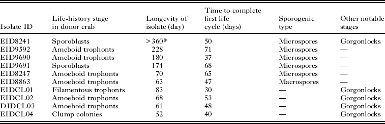
To test the infectivity of the culture isolates, suspended cells (arachnoid trophonts and sporonts) were collected from one isolate (EID8241, Table 1) that had been maintained in the laboratory continuously after 3, 9 and 12 months post-isolation, and used as inocula for experimental infection of adult crabs. Parasite cells in the inocula were enumerated and adjusted to a final density of approximately 106 cells/ml. Naïve adult blue crabs (110–150 mm carapace length) collected from the York River (a non-endemic area) were tested for the presence of infections by examining haemolymph smears stained with 0 3% neutral red. They were then injected with 100 μl of the inocula (approximately 105 parasite cells per crab) using disposable sterile 1-ml syringes with 27-ga needles, as described by Shields and Squyars (Reference Shields and Squyars2000). Inoculated crabs were housed together in a re-circulating 760 L trough (salinity=24±2 psu, temperature=19±2°C), equipped with a pre-conditioned biological filter. Crabs were fed semi-weekly with squid portions and were monitored daily for mortalities. Dead or moribund crabs were assessed for Hematodinium infections using the haemolymph smear assay described above. At various times after inoculation crabs were dissected and samples of gill, muscle, epidermis, heart, hepatopancreas and foregut were collected and preserved in Bouin's fixative for subsequent histological diagnosis as reported by Wheeler et al. (2007). After 3 weeks post-inoculation, all of the remaining crabs were dissected and processed for histology to assess the status of Hematodinium infection.
Summary of the isolates of Hematodinium sp. from the blue crab, C. sapidus, which completed development in vitro

* Those isolates are still alive and maintained in the laboratory.
RESULTS
Over 25 isolates of Hematodinium obtained from infected blue crabs were cultured during this study. Ten of the isolates (Table 1) completed the entire developmental cycle in vitro. One of these isolates (EID8241) has been maintained in our laboratory for more than 12 months through serial subcultivation with fresh media. The remaining 15 isolates developed partially through several life-history stages, then gradually died out 4–6 weeks after isolation. Numerous life-history stages, including, filamentous trophonts, amoeboid trophonts, arachnoid trophonts, arachnoid sporonts, sporoblasts, prespores and dinospores were routinely observed in these cultures. The transition and development of the observed life-history stages is presented below. For consistency, we follow the nomenclature of the life-history stages presented by Appleton and Vickerman (Reference Appleton and Vickerman1998) for the Hematodinium-like species from the Norway lobster when describing the various stages and development of the Hematodinium sp. from C. sapidus.
Filamentous trophonts
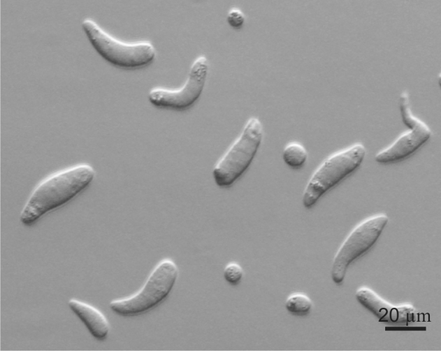
Filamentous trophonts, or vermiform plasmodia, were observed in circulating haemolymph or tissues of blue crabs with very light Hematodinium infections. This stage is easily distinguishable from host haemocytes as the trophont is motile, vermiform, and up to 100 μm in length in the haemolymph. In haemolymph and in culture the filamentous trophonts displayed slow motility such as lateral stretching, flexing and writhing movements, with cytoplasmic streaming inside the cell. Neutral red uptake occurred in the lysosomes of viable filamentous trophonts, making them distinctly visible compared to the host haemocytes that only had limited uptake of the dye. The filamentous trophonts were uninucleate, binucleate or multinucleate. The size of the filamentous trophonts varied markedly, ranging from 13 to 58 μm in length (mean=31 3±11 9 μm S.D., n=25) (Fig. 1). The filamentous trophonts multiplied by what appeared to be budding wherein they developed constriction points and separated at the constriction. In cultures, this stage was very brief, usually only occurring over 1–3 days. They also underwent a division process resembling merogony (cf. segmentation) with rapid asexual division that led to the next stage, the amoeboid trophonts.
In vitro culture of filamentous trophonts of Hematodinium sp. from Callinectes sapidus. Hoffman modulation contrast.

Amoeboid trophonts
Amoeboid trophonts were the most common stage observed in haemolymph or tissues of crabs with light to moderate infections. The amoeboid trophonts were oval or spherical and ranged in size from 9 to 17 μm in diameter (12 0±1 9 μm, n=25), and were similar in size and appearance to the semi-granulocytes of blue crabs (Fig. 2). However, the amoeboid trophonts displayed a vivid uptake of Neutral red stain in their lysosomes, which become bright red upon uptake of the vital dye. Unlike filamentous trophonts, amoeboid trophonts displayed little to no motility in crab haemolymph or in vitro culture. In haemolymph this stage often possessed a single short lobopod or irregular margin. In culture the cells were often more rounded and globular, less amoeboid in shape. Uninucleate, binucleate or multinucleate cells of this stage have been observed in both diseased crabs and in in vitro cultures. Amoeboid trophonts were observed to undergo binary fission to produce more trophonts. In culture, amoeboid trophonts maintained their oval or spherical shape for 1–3 days, and then adhered to the bottom of the culture flasks or plates, and developed into arachnoid trophonts.
In vitro culture of amoeboid trophonts of Hematodinium sp. from Callinectes sapidus. The cells often have large numbers of refractile granules. Hoffman modulation contrast.

Arachnoid trophonts and sporonts
The arachonoid trophont stage was the primary stage for rapid amplification of the parasite in culture. This stage was not observed circulating in the haemolymph of infected crabs as it is apparently a tissue phase of the parasite. Shortly after the amoeboid trophonts adhered to the culture flasks or plates, they began developing pseudopodial branches with anastomosis of the pseudopods into an elaborate syncytial network fringed by adherent reticulopodia-like pseudopods (Fig. 3A). The centroids of these trophonts were clearly undergoing rapid division to produce an apparent multicellular stage. Through continuous asexual division, these arachnoid forms expanded progressively outward over the substrate of the culture vessels (Fig. 3B, C). Surprisingly, isolated syncytial networks that came into contact with each other merged to form a larger syncytial network. Approximately 4–6 weeks after isolation, arachnoid trophonts developed into arachnoid sporonts.
In vitro development of arachnoid trophonts and sporonts. (A) Initial development of arachnoid trophonts 1 day after establishment of amoeboid trophonts in culture. Note the formation of the adherent reticulopodial-like extensions (arrow). (B) An arachnoid trophont 7 days after initiation of a culture of amoeboid trophonts. Note the syncytial network (arrow) with the mass of cells developing medially within. (C) Arachnoid trophont 3 weeks after isolation, with additional proliferation of sporonts within the mass of cells. The central area is packed with differentiating cells and the syncytial network has retracted. (D) Arachnoid trophont 6 weeks after isolation showing the development of the arachnoid sporont as a cellular mass, with developing schizonts (arrows). Hoffman modulation contrast.

Light had a profound effect upon the survival and development of the arachnoid stages in culture. Under an ambient light cycle in the laboratory, most of our cultures developed into the arachnoid trophont stage within 4–6 weeks; however, they would then cease growing and eventually die out. However, cultures maintained in the dark progressed to form arachnoid sporonts (see below) (Fig. 3D). The arachnoid trophonts in these cultures often possessed enlarged centroid cells, reminiscent of germinative cells that were undergoing apparent budding on the periphery of the cell (Fig. 4). Asexual division in these centroid cells rapidly led to a large mass of cells, the whole complex termed an arachnoid sporont. The arachnoid sporont was characterized by the central area becoming a cellular mass, with the syncytial network gradually contracting and vanishing within the expansion of the central mass of cells. This cellular mass continued to grow, eventually progressing to release compact, spherical sporoblasts from within the centre or edges of the mass. The arachnoid syncytia often fragmented with agitation or suction from a sterilized pipette, and could be subcultured when transferred to a new vessel containing fresh medium.
In vitro development of the arachnoid trophont showing a group of enlarged, centroid cells within the syncytial network. Hoffman modulation contrast.

Sporoblasts
Sporoblasts that detached from the arachnoid sporonts formed cellular aggregates or eventually dispersed into single cells free in the culture (Fig. 5). Sporoblasts accumulated to high densities in cultures, eventually giving rise to prespores, and then developing further into dinospores. Sporoblasts were morphologically similar to amoeboid trophonts, but perhaps more rounded than the amoeboid trophonts. When transferred into new medium, the sporoblasts were able to establish and initiate the development of new arachnoid trophonts rather than develop into dinospores.
(A) In vitro culture of sporoblasts (individuals and aggregates of cells) shown detached from the substrate. (B) Prespores (arrows) developing shortly after release of sporoblasts from the arachnoid trophont. (C) Micro-dinospores from in vitro culture. (D) Macro-dinospores from culture fixed with 2 5% glutaraldehyde. Hoffman modulation contrast.

In 4 of the 10 culture isolates that completed the entire developmental cycle in vitro, a different type of sporoblast developed and it developed synchronously with the previously described sporoblasts (Fig. 6A). To avoid confusion in terms, we refer to the latter type of sporoblast as a schizont. Schizonts were embedded among the normal sporoblasts; however, they were much larger, being at least 3 times larger than the sporoblasts (Fig. 3D). As they progressed in their development, multiple cellular bodies formed within the schizonts. After a few weeks, the contents became motile and began writhing within the schizonts. When sporoblasts became detached from the arachnoid syncytium, the schizonts were also released into suspension. The motile cellular bodies within eventually broke through the schizont membrane and developed into aggregates of filamentous trophonts (Fig. 6B), which were morphologically similar to the gorgonlocks stage observed in cultures of Hematodinium-like parasites from the Norway lobster, N. norvegicus (Appleton and Vickerman, Reference Appleton and Vickerman1998). The gorgonlocks gradually separated into distinct filamentous trophonts, then underwent merogony (segmentation) to develop into aggregates of cellular clumps (Fig. 6C), morphologically similar to the ‘clump colonies’ described by Appleton and Vickerman (Reference Appleton and Vickerman1998). When transferred into fresh medium, these clump colonies started forming syncytial networks, developing into arachnoid trophonts and then arachnoid sporonts (Fig. 6D). This apparently represents an alternative pathway for the multiplicative proliferation of Hematodinium cells in the blue crab host as it was not observed in every culture, nor has it been reported from the parasite in N. norvegicus. However, during the course of our studies clump colonies were occasionally observed in haemolymph of infected crabs, indicating that this stage is not likely a culture artifact.
(A) In vitro culture of schizonts (arrows) arising from arachnoid sporonts. (B) Gorgonlocks (clumps of filamentous trophonts) (arrows) protruding through the wall of the schizont. (C) Clump colonies (arrows) arising from gorgonlocks in vitro. (D) Establishment of arachnoid trophonts from clump colonies transferred into new medium. Hoffman modulation contrast.

Prespores
In most successful cultures, the sporoblast stage developed further into a prespore – an apparent transition stage before developing into dinospores (Fig. 5B). This stage was occasionally observed in the circulating haemolymph of heavily infected crabs collected in the field. However, heavy infections with prespores exhibited limited uptake of neutral red. Prespores were not motile in haemolymph and cultures. The prespores were circular and ranged in size from 8 to 16 μm in diameter (12 6±2 3 μm, n=25).
Dinospores
After approximately 6–9 weeks of culture in the dark, 6 isolates successfully completed development of the life cycle and generated large numbers of dinospores. Two types of dinospores, micro-dinospores or macro-dinospores, were observed in individual isolates, but they were never observed together in a single culture (Fig. 5 C, D). Of the 6 isolates that completed sporogenesis, 5 produced micro-dinospores and 1 produced macro-dinospores. The macro-dinospores ranged in size from 11 to 17 μm (14 3±1 6 μm, n=25) excluding measurement of the flagella. The micro-dinospores ranged in size from 6 to 12 μm (9 2±2 4 μm, n=25). Both spore types were actively motile in culture suspensions after the development of their flagella. Macro-dinospores were sluggish, typically moving along the substratum, while micro-dinospores were vigorous swimmers, easily moving throughout the culture medium. Only a small portion of the dinospores developed into filamentous trophonts in culture. When moved into separate cultures, most dinospores survived from to 3–7 days.
Effects of temperature and light on in vitro development of Hematodinium sp. cells
Temperature affected cell growth and development of Hematodinium cells in vitro. Amoeboid trophonts cultured at 10°C did not fully attach to the substrate of the culture plates, as did those cultured at room temperature (23°C), and their development stagnated at the early formation of arachnoid trophonts after approximately 14 days, dying out 40 days after the initiation of the cultures. Cultures maintained at 15°C developed into arachnoid trophonts similar to those grown at room temperature, and then progressed slowly until dying out after 54 days. The cultures grown at 23°C developed similarly to other cultures maintained at room temperature and eventually developed into arachnoid trophonts and arachnoid sporonts. No dinospores were produced upon termination of the experiment (72 days after initiation of cultures) even though some sporoblasts or schizonts were occasionally observed at the end of the culture period.
Light intensity had a profound effect on cell growth and development of Hematodinium in culture. The 2 isolates (2 replicates of each) exposed to light died out weeks after the initial formation of arachnoid trophonts. The 2 cultures (2 replicates of each) not exposed to light (dark treatment) completed development of the life cycle in vitro, and produced dinospores 7 weeks after the initiation of the cultures. The cultures (2 replicates of each) maintained on an ambient light/dark schedule survived and eventually developed into arachnoid trophonts and arachnoid sporonts. However, these cultures did not produce dinospores and were terminated 8 weeks after initiation of cultures.
Infectivity of culture isolates
At least 1 culture of Hematodinium from the blue crab remained infectious after being cultured for several months. Hematodinium isolate #8241 (Table 1) was infectious after 3, 9, and 12 months in culture. Of 12 adult blue crabs inoculated with 3-month-old cultured parasites, 3 developed light infections with either amoeboid or filamentous trophonts. The other crabs died within 1 week due to handling stress or introduced bacterial infections during inoculation. Of 15 adult crabs inoculated with 9-month-old cultured parasites, 7 developed light infections with either amoeboid or filamentous trophonts. Of the 12 crabs inoculated with 12-month-old cultured parasites, 8 developed light to moderate infections. The remaining crabs had not developed infections when they died or were assessed at the end of the experiments.
DISCUSSION
This is the first report of the in vitro life cycle of the Hematodinium sp. from Callinectes sapidus. We successfully established several continuous in vitro cultures of the parasite using a lobster saline augmented with fetal bovine serum and blue crab serum in cultures grown in the dark at room temperature. One of those isolates has been maintained in our laboratory for more than 12 months through serial subculture with fresh media. After 3, 9 and 12 months, it was still capable of initiating new infections when inoculated into crabs. Hematodinium cells underwent continuous developmental changes in vitro, which appeared consistent with the in vivo development and life cycle of the parasite within the blue crab host, as many but not all of the in vitro stages were also observed in infected crabs (Fig. 7). During in vitro culture, Hematodinium sp. was observed progressing through various developmental stages that included filamentous trophonts, amoeboid trophonts, arachnoid trophonts and sporonts, sporoblasts, prespores and dinospores over a 2 to 3-month period. Similar developmental stages have been observed in cultures of the boreal species of Hematodinium from the Norway lobster, N. norvegicus (Appleton and Vickerman, Reference Appleton and Vickerman1998).
Schematic diagram of the in vitro life cycle of Hematodinium sp. from Callinectes sapidus. (1) Filamentous trophonts represent the initial stage of infection in a blue crab host. (2) Amoeboid trophonts arise from merogony of the filamentous trophont. This stage is often found in the circulating haemolymph of infected crabs. (3) Arachnoid trophonts arise from the syncytial network laid down by the amoeboid trophonts. They were the main proliferative stage of the parasite in vitro. (4) Arachnoid sporonts develop as a mass within the arachnoid trophont. They give rise to sporoblasts (5) which occur as single cells or aggregates of cells, which are released from the arachnoid sporonts when fully developed. (6) Prespores arise as the transition between sporoblasts and dinospores. (7) Dinospores, micro-dinospores or macro-dinospores, arise from the prespores and represent the highly motile stage in the life cycle of the parasite. (8) Schizonts also developed from within arachnoid sporonts; they apparently represent an alternate pathway for proliferation of parasites in crab hosts (dashed arrows). The schizonts develop into gorgonlocks (9) that undergo schizogony (cf. segmentation) and develop into clump colonies (10). The clump colonies develop into arachnoid trophonts when subcultured in vitro.

There are several differences in the in vitro life cycle of the Hematodinium sp. from C. sapidus and that in N. norvegicus. Notably, the gorgonlocks did not arise directly from filamentous trophonts in the parasite from the blue crab; rather it was an ephemeral stage arising from the presumptive ‘schizont’. The gorgonlocks gave rise to filamentous trophonts, which developed into clump colonies. In the parasite from the Norway lobster, the gorgonlocks develops from the filamentous trophont, perhaps arising from a delay in cytokinesis during budding by the filamentous form (Appleton and Vickerman, Reference Appleton and Vickerman1998). In addition, the schizont stage has not been reported in the parasite from the Norway lobster. In the parasite from the blue crab, schizonts were usually observed late in the development of arachnoid sporonts, embedded in the cellular mass of developing sporoblasts. The development of the schizont appears to be a shunt in the life cycle that can lead to sustained infection as well as the rapid amplification of the parasite in the crab host. The mechanism that triggers arachnoid sporonts to develop schizonts instead of sporoblasts is unknown, but the sporoblasts are no doubt tied to the production of dinospores. Lastly, the centroid ‘germinative’ cells in the arachnoid trophont have not been described previously. They appear to be zones for budding and rapid asexual division, perhaps giving rise to the developing sporont. While not attempted in this study, further comparisons of these stages using transmission electron microscopy should be very revealing.
The arachnoid trophont and sporont stages are the primary stages for asexual multiplication of Hematodinium sp. in vitro. Moreover, these stages likely account for the rapid proliferation of parasites in internal organs of diseased hosts. Unlike filamentous trophonts, amoeboid trophonts and prespores, the arachnoid trophonts and sporonts have not been observed in circulating haemolymph of diseased crabs. These stages occur intertwined in the hepatopancreas, and possibly the epidermal tissues of infected hosts, but they have not been previously described from the blue crab host. The delicate syncytial network of the arachnoid trophonts or sporonts are difficult if impossible to identify in histological sections from infected blue crabs (Shields, unpublished data); however, these stages have been observed in the hepatopancreas of Norway lobsters infected with Hematodinium using an indirect fluorescent antibody technique (Field and Appleton, Reference Field and Appleton1996), and within abdominal skeletal muscle and myocardial muscle (Field and Appleton, Reference Field and Appleton1995).
In culture, single or aggregated sporoblasts detach from the arachnoid sporont and occur free within the culture vessel. Within the crab host, we speculate that sporoblasts disseminate throughout the body via the haemolymph, settling in new sites to initiate additional foci within the infected host. These sporoblasts look morphologically similar to amoeboid trophonts in early infections, but they are at higher densities in the haemolymph, more rounded or globular in nature, and give rise to dinospores. These observations are supported by studies of Hematodinium in other crustaceans. Meyers et al. (Reference Meyers, Koeneman, Botelho and Short1987) and Appleton and Vickerman (Reference Appleton and Vickerman1998) speculated that amoeboid trophonts were present in circulating haemolymph in early infections, whereas the high-density subspherical parasites in haemolymph of chronically infected hosts probably represented sporoblasts. Our culture findings support their observations that high-density infections are likely comprised primarily of sporoblasts and prespores.
Two types of dinospores, macro-dinospores and micro-dinospores, were observed in vitro in this study and in vivo in other studies. These dinospores can survive up to 7 days in 20–35 psu (Li et al. 2010), whereas amoeboid trophonts are only viable for 24 h in seawater (Li et al. 2011). The apparent behavioural differences between macro-dinospores and micro-dinospores require further study. Waterborne transmission via dinospores is a likely mode of transmission for Hematodinium sp. in the blue crab (Frischer et al. Reference Frischer, Lee, Sheppard, Mauer, Rambow, Neumann, Brofft, Wizenmann and Danforth2006; Shields and Overstreet, Reference Shields, Overstreet, Kennedy and Cronin2007). There is also evidence that transmission of other Hematodinium spp. infections occurs in association with host moulting (Meyers et al. Reference Meyers, Botelho, Koeneman, Short and Imamura1990; Eaton et al. Reference Eaton, Love, Botelho, Meyers, Imamura and Koeneman1991; Stentiford et al. Reference Stentiford, Neil and Atkinson2001; Shields et al. Reference Shields, Taylor, Sutton, O'Keefe, Collins, Ings and Pardy2005; Frischer et al. Reference Frischer, Lee, Sheppard, Mauer, Rambow, Neumann, Brofft, Wizenmann and Danforth2006; Shields et al. Reference Shields, Taylor, O'Keefe, Colbourne and Hynick2007). However, until transmission experiments are done with moulting hosts, this mode of transmission remains undetermined. Understanding the cues for dinospore production in vitro would facilitate transmission studies, and lead to a clearer picture of the processes that leads to infection by these important pathogens.
ACKNOWLEDGEMENTS
We thank Kersten N. Wheeler for her help with this project. John Tiggelaar helped with the infection trial at 12 months. This research was supported by the National Science Foundation's Ecology of Infectious Diseases (EID) programme (OCE BE-UF0723662). This is VIMS contribution 3171.