The Holt–Oram syndrome is an autosomal dominant condition with variable clinical expression. Mutations in the TBX5 gene located on chromosome 12q24.1 constitute an underlying cause.Reference Faria, Rabenhorst, Pereira Ada and Krieger1 Here, we report on a Holt–Oram syndrome case with associated anomalous right coronary artery originating from the left Valsalva sinus, crossing between the aorta and the pulmonary trunk (the interarterial subtype) and known to be associated with sudden death.Reference Davis, Cecchin, Jones and Portman2–Reference von Kodolitsch, Franzen, Lund, Koschyk, Ito and Meinertz4
Case report
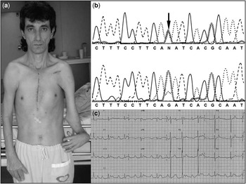
A 36-year-old man presented with skeletal characteristics compatible with the Holt–Oram syndrome (Fig 1a). He was a wall painter, asymptomatic, even during strenuous physical activities. Physical examination showed a palpable right ventricular impulse, midsystolic pulmonary ejection murmur, and a fixed splitting second heart sound. The diagnosis was confirmed by identification of a novel sequence variant in the TBX5 gene (Fig 1b), characterised by a G–A substitution at nucleotide-1 of intron 7 (exon 8 acceptor splicing site, c.663-1G greater than A).
(a) Frontal authorised picture. See skeletal malformations: left and right hypoplastic thumbs with low implantation, left hand with radial deviation, left hypoplastic radius, mild hemithorax hypoplasia, hypoplastic clavicles, and narrowed shoulders. (b) Genetic analysis disclosed a c.663-1G greater than A substitution at the exon 8 acceptor splicing site. (c) Electrocardiography showing bradycardia due to a high degree of atrioventricular block, enlargement of both atria, and diffuse T-wave alterations. (A = adenosine, C = cytosine, G = guanine, N = heterozygous nucleotide, T = thiamine).

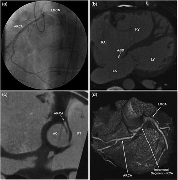
Electrocardiography showed bradycardia due to a high degree of atrioventricular block with normal QRS duration (nodal block pattern; Fig 1c). Echocardiography showed a large secundum atrial septal defect (18 millimetres) and mild right ventricular hypokinesis. Cineangiography registered 40 millimetres of mercury of systolic pulmonary artery pressure, confirmed the atrial septal defect, and showed that an anomalous right coronary artery originated from the left Valsalva sinus (Fig 2a). Contrast-enhanced multi-detector computed tomography also confirmed the atrial septal defect (Fig 2b), and clearly demonstrated the anomalous vessel coursing between the aorta and the pulmonary trunk (Fig 2c, d). Tests of myocardial ischaemia were normal. The patient required a permanent atrioventricular pacemaker and underwent surgery.
(a) Coronary angiography. The catheter was on the ostium of the anomalous right coronary artery (ARCA) in the left Valsalva sinus. The left main coronary artery (LMCA) was also contrasted by a very close ostium. (b) Contrast-enhanced multi-detector computed tomography. The arrow points to a large secundum atrial septal defect (SASD). (c) Moreover, the exam showed clearly the interarterial crossing of the ARCA (arrow). (d) A three-dimensional surface reconstruction with the pulmonary trunk removed. See the origin and trajectory of LMCA and ARCA. The initial segment of the ARCA had an intramural trajectory into the aortic wall (AO). LA = left atrium, LV = left ventricle, PT = pulmonary trunk, RA = right atrium, RV = right ventricle.

Surgery corrected the atrial septal defect with a polytetrafluoroethylene patch. The anomalous right coronary artery arose side-by-side to the left main coronary ostium, above the intercoronary commissure, with a very acute angled takeoff. This initial segment had a deep intramural trajectory into the aortic wall. Its initial portion was unroofed and incised (enlarged and repaired with a saphenous vein patch). The intercoronary commissure was not manipulated and neo-ostia were not created. He was discharged on only acetylsalicylic acid, and 2 years later, he remains asymptomatic, working without limitations.
Discussion
Surprisingly, this patient performed routinely exhaustive physical activities being always asymptomatic, despite the large atrial septal defect. He had a high degree of atrioventricular block. Atrioventricular and bundle branch conduction systems disorders occur commonly in the Holt–Oram syndrome. The genetic variant in the TBX5 gene of this patient (Fig 1b) was not previously reported. It is located at the exon 8 acceptor splicing site, a highly conserved region among species. This variant was not observed in 100 ethnic-matched control chromosomes, and nor is it described by any group that sequences TBX5 for molecular diagnosis of the Holt–Oram syndrome.
The interarterial subtype of anomalous coronary arteries (right or left arteries) is the second most common cause of sudden death in athletes (following hypertrophic cardiomyopathy).Reference Davis, Cecchin, Jones and Portman2–Reference von Kodolitsch, Franzen, Lund, Koschyk, Ito and Meinertz4 Ischaemia is considered to be a mechanism of arrhythmias and sudden death. It is believed that the anomalous vessels do not suffer extrinsic compression by the aorta or pulmonary trunk due to the physiological levels of pressure in these vessels during a normal cardiac cycle. However, in the presence of an acute angled takeoff, a slit-like opening may occur in these vessels. In addition, the initial segment generally has an intramural course into the aortic wall, and the aortic medial layer can promote compression. Several stress tests, such as inotropic stimulus, high heart rate, intravenous fluid infusion, and increased coronary flow, can worsen the slit-like origin.Reference Davis, Cecchin, Jones and Portman2–Reference von Kodolitsch, Franzen, Lund, Koschyk, Ito and Meinertz4 In addition, in course of time, chronically modified coronary flow can cause endothelial dysfunction resulting in inadequate vasodilatation, vasospasms, or collapses.Reference Davis, Cecchin, Jones and Portman2–Reference von Kodolitsch, Franzen, Lund, Koschyk, Ito and Meinertz4
Anomalous coronary arteries were not previously described in the Holt–Oram syndrome patients. Causality may be difficult to establish in these cases. Indeed, the association described may be casual since coronary abnormalities are not rare (present in 0.2–1.2% of the general population).Reference Davis, Cecchin, Jones and Portman2, Reference von Kodolitsch, Franzen, Lund, Koschyk, Ito and Meinertz4 In spite of this fact, the anomalous coronary artery should be listed as a possible development anomaly associated with the Holt–Oram syndrome.
Surgical management of the interarterial subtype of coronary anomaly is indicated when there is proved ischaemia, ventricular arrhythmias, or worrisome symptoms such as chest pain, palpitations, syncope, or dyspnoea.Reference Vianna, Gonzalez and Dallan3–Reference Romp, Herlong and Landolfo6 Surgical correction of asymptomatic paediatric or young patients is recommended, but it is a debatable issue.Reference Mirchandani and Phoon5, Reference Romp, Herlong and Landolfo6 In this case, unroofing was decided during the operation mainly because the takeoff angle and the intramural trajectory were very accentuated.
A widely used operation in this scenario is coronary artery bypass grafting, in spite of graft thrombosis being frequent and secondary to competitive flow. Other techniques are vessel reimplantation or creation of neo-ostia and have been scarcely performed, mainly because of the not so rare incidence of valvar aortic insufficiency and emergency bypass grafting with these techniques.Reference Vianna, Gonzalez and Dallan3, Reference Mirchandani and Phoon5 The procedure used in this case seems to be promising. Unroofing the anomalous vessel from its intramural course allows expressive enlargement of the slit-like ostium, with or without incision of the initial segment. Nevertheless, it should be noted that the long-term outcomes of all these surgical methods are still poorly unknown.Reference Vianna, Gonzalez and Dallan3–Reference Romp, Herlong and Landolfo6