INTRODUCTION
Abnormalities in elasmobranchs have not been commonly reported. Many of these records are focused on albinism, as Schwartz (Reference Schwartz1959) reported albinism in a white adult male cow-nose ray, Rhinoptera bonasus caught in Maryland. Joseph (Reference Joseph1961) reported an albino female cow-nose ray taken in Chesapeake Bay. McKenzie (Reference McKenzie1970) found it in a juvenile scalloped hammerhead shark Sphyrna lewini caught in Georgia, and Talent (Reference Talent1973) in an embryo grey smoothhound shark. Also in Mustelus californicus, the bat ray Myliobatis californica (De Jesus-Roldan, Reference De Jesus-Roldan1990), tiger shark Galeocerdo cuvier, and the giant electric ray Narcine entemedor (Sandoval-Castillo et al., Reference Sandoval-Castillo, Mariano-Meléndez and Villavicencio-Garayzar2006).
Other abnormalities in elasmobranchs include the malformation in the cephalic region, mainly in some rays when pectoral fins are not merged with the head, as in the case of the blonde ray Raja brachyura, the starry ray Amblyraja radiata and the freshwater stingray Potamotrygon motoro (Templeman, Reference Templeman1965; Rosa et al., Reference Rosa, Gomes and Gadig1996). Escobar–Sánchez et al. (Reference Escobar-Sanchez, Galvan-Magaña, Downton-Hoffman, Carrera-Fernandez and Alatorre-Ramirez2008) also reported abnormalities in the longtail stingray Dasyatis longa in the Gulf of California, Mexico. Some skeletal deformities have been reported in a school shark Galeorhinus galeus (Officer et al., Reference Officer, Clement and Rowler1995) and in other elasmobranchs (Triaenodon obesus, Hemiscyllium ocellatum, Mustelus antarticus and Furgaleus macki) (Heupel et al., Reference Heupel, Simpfendorfer and Bennett1999) with caudal deformities and severe spinal curvatures.
The bicephaly has been reported in other elasmobranchs. Castro-Aguirre & Torres-Villegas (Reference Castro-Aguirre and Torres-Villegas1979) described bicephaly in the cow-nose ray Rhinoptera steindachneri on the eastern coast of Baja California Sur, Mexico. In sharks, the bicephaly cases have been in the reef shark Carcharhinus perezi (Bonfil, Reference Bonfil1989), and in the blue shark, Prionace glauca (Linnaeus, 1758). Goto et al. (Reference Goto, Taninuchi, Kuga and Iwata1981) described the deformity in four specimens caught in Japanese waters. Ferreira et al. (Reference Ferreira, Ferreira and Amorim2002) found embryos with two heads in Brazil, and Mancini et al. (Reference Mancini, Casas and Amorim2006) records the occurrence of a blue shark with morphological abnormalities in the trunk and cephalic region from a female caught in Brazil. In the Pacific Ocean, Galván-Magaña et al. (Reference Galván-Magaña, Escobar-Sánchez and Carrera-Fernández2011) reported a case of bicephaly in embryos of the blue shark caught in the Gulf of California and in the western coast of Baja California Sur. Here we describe cephalic abnormalities of blue shark foetuses in Mexican waters.
MATERIALS AND METHODS
Two pregnant blue shark females were caught in October 2008 in the San Lazaro fishing camp in Baja California Sur, Mexico (24°45′N 112°8′W). The reproductive tracts of two gravid females were removed and preserved in 10% formalin for posterior analysis. We examined the uterus of both females and abnormal foetuses were found inside them. The embryos with bicephaly were dissected to observe the internal organs to describe external and internal characteristics. The other abnormal embryos were also reviewed and measured. Embryos were deposited in the Morphophysiology Laboratory from the Centro Interdisciplinario de Ciencias Marinas—IPN in La Paz, Baja California Sur, Mexico.
RESULTS
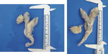
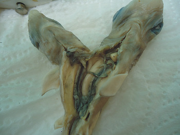
The first female examined (236 cm total length (TL)) caught on 2 October 2008 had 46 embryos, one of which had bicephaly. This deformed embryo was the smallest (8 cm TL) compared with the other embryos (15 cm TL) that were normal. The embryo was a female, with a body with two heads clearly separated. The right cephalic region was significantly smaller than the left head. Each head had nostrils, eyes and mouths and gill slits well-developed. The shark body had normal first and second dorsal, anal, pectoral, pelvic and caudal fins and umbilical cord. The left cephalic structure was bigger and well-formed (Figure 1). Internally, we observed that their organs were well-formed, and that the only organ shared was the liver. Each head and part of its body had heart, stomach and reproductive tract (Figure 2).
Prionace glauca foetus with bicephalia showing dorsal and ventral view (E, eyes; Ys, yolk sac; Df, dorsal fin; n, nostrils; m, mouth; gs, gills slits; Uc, umbilical cord; pf, pectoral fins).

Blue shark foetus showing internal organs (rt, reproductive tract; h, heart).

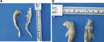
The second gravid female (222 cm TL) caught on 3 October 2008 had 40 embryos, two of which (one male and one female) presented deformed heads. Both embryos were small (8.5 cm TL) in relation to the other normal embryos (15 cm TL). They did not have eyes and part of their snout was not well-formed (Figure 3). The male embryo presented only the cranial cartilage; the gill slits were present but not fully open, whereas the female embryo had the eye arches, an incomplete snout, but mouth and gill slits were complete (Figure 3). In both embryos, the body was complete with pectoral, pelvic, anal and caudal fins. Internally their organs had developed normally.
Prionace glauca foetuses: (A) Dorsal view of female and male; (B) Ventral view of female and male showing the cephalic abnormalities (F, female; M, male; s, snout; m, mouth; gs, gill slits; cc, cranial cartilage).

DISCUSSION
A possible explanation for these deformities in blue shark is the high number of embryos that they produce, as it is considered the most fecund shark species. Castro et al. (1999) indicated variable fecundity in the blue shark, ranging from 28 to 54 embryos; however, the maximum fecundity reported was up to 135 embryos (Compagno, Reference Compagno1984). These kind of abnormalities have been observed during embryonic development in which external factors such as pollution were attributed to these malformations (Goto et al., Reference Goto, Taninuchi, Kuga and Iwata1981; Ferreira et al., Reference Ferreira, Ferreira and Amorim2002; Mancini et al., Reference Mancini, Casas and Amorim2006). However, the fishing area close to Baja California Sur is clear of pollution and we cannot consider it a cause for abnormalities in blue shark, as considered by Galvan-Magaña et al. (2011). Heupel et al. (Reference Heupel, Simpfendorfer and Bennett1999) attributed skeletal deformities to parasitic infection, injury, bad nutrition or congenital abnormality. We consider that the abnormalities found in these blue shark embryos is because of the large number of embryos produced by the mother and their uteri are too small to permit normal development of all embryos.
ACKNOWLEDGEMENTS
We would like to thank the fishermen of San Lázaro (BCS, Mexico) for helping in the sampling. We would also like to thank the Morphophysiology Laboratory at CICIMAR-IPN. The first author thanks CONACYT and PIFI. F.G.M. and R.I.O.B. thank the Instituto Politécnico Nacional for fellowships (COFAA and EDI). We would also like to thank Angela M. Cheek for editing the English-language text.