There has been a decrease in mortality rates during the perioperative period amongst infants with CHD. Therefore, more studies are evaluating neurodevelopmental outcomes after surgery.Reference Picciolini, Squarza and Fontana1 Occurrence rates of patients undergoing congenital heart surgery have been reported at 2–25%.Reference Furnish, Mueller, Kiser, Dufficy, Sullivan and Beyer2,Reference Andropoulos, Stayer, Diaz and Ramamoorthy3 Children exhibit less tolerance for alterations in cerebral hyperemia or ischaemia and are at a higher risk for sustaining secondary brain injuries. In addition, children under 4 years are more likely to develop neurological dysfunctions.Reference Hayashida, Kin and Tomioka4 Low regional cerebral oxygen saturation levels have been shown to play a key role in the pathogenesis of neurological dysfunctions.Reference Schober, Feiner, Bickler and Rollins5 However, evaluation of cerebral haemodynamics and regional cerebral oxygen saturation is not routine clinical practices for infants during CHD surgery in developing countries.
Mechanical ventilation affects cerebral oxygen saturation and cerebral blood flow in infants. Hypercapnia and hypocapnia are associated with hypoventilation and/or hyperventilation. They have been associated with the imbalance between brain oxygen supply and demand.Reference Ricci, Garisto and Favia6–Reference Zhang, Xie, Han, Huang, Ou-Yang and Lu8 Studies have highlighted the influence of modified ultrafiltration on cerebral oxygen saturation. The findings from these studies were, however, not conclusive.Reference Medlin and Sistino9 Due to the incomplete development in brain circulation and poor cerebral autoregulation of infants, the effects of carbon dioxide on cerebral oxygen saturation have not been determined in these paediatric patients under the age of Reference Picciolini, Squarza and Fontana1,Reference Vutskits10,Reference Rhondali, Mahr and Simonin-Lansiaux11 especially with modified ultrafiltration.Reference Medlin and Sistino9
This study aims to evaluate the effects of different ventilation levels on regional cerebral oxygen saturation and cerebral blood flow before and after modified ultrafiltration, during surgery in infants with ventricular septal defects. The flow velocity of middle cerebral artery was measured by transcranial Doppler (TCD) sonography while regional cerebral oxygen saturation was measured by near-infrared spectroscopy.
Materials and methods
Study design and patients
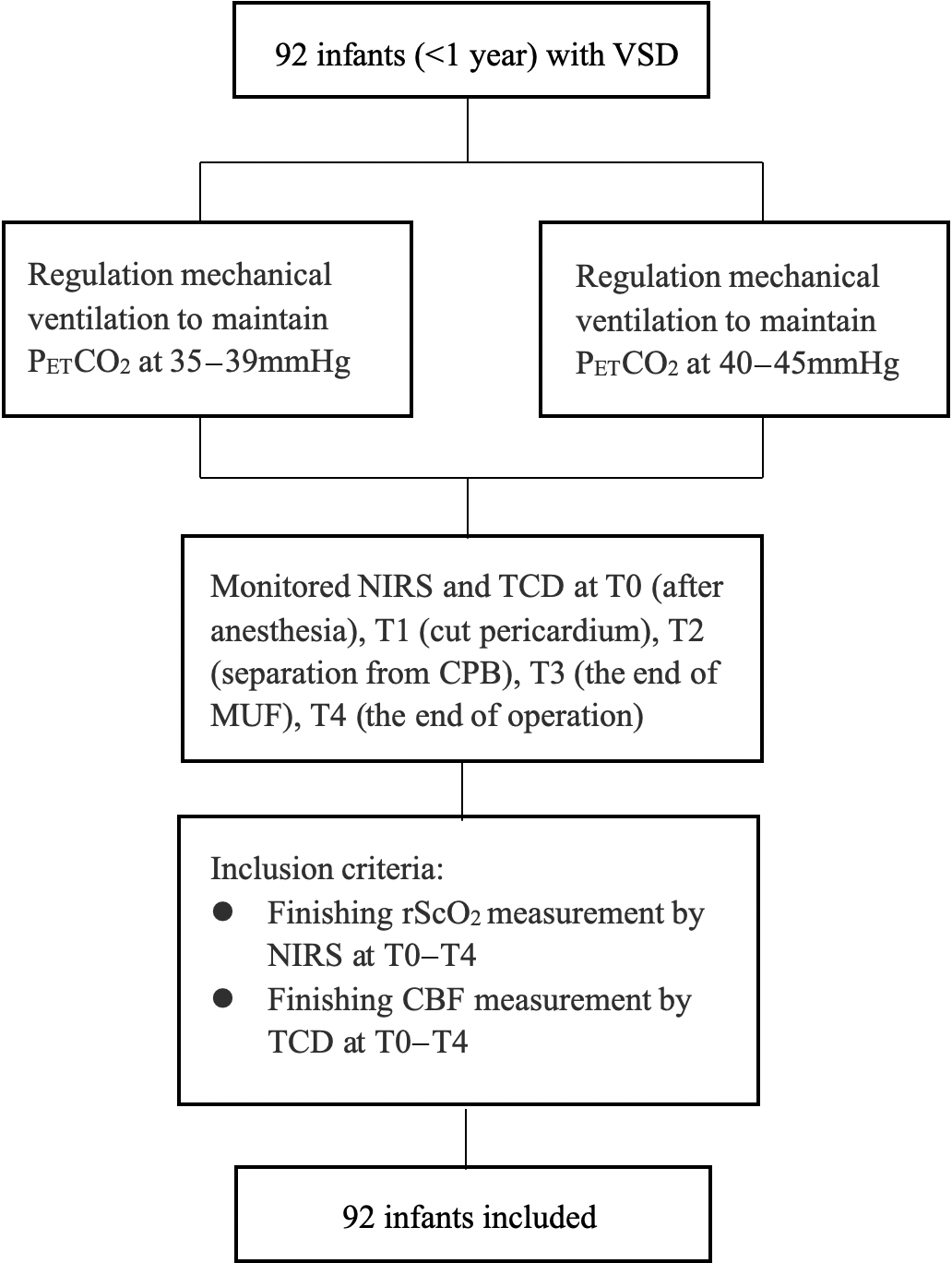
This was an interventional study performed at Capital Medical University affiliated Beijing An Zhen Hospital. This study was carried out from July, 2017 to April, 2018. Ethical approval was obtained from the Institutional Review Board (IRB 2017030X). Verbal and written informed consents were obtained from the infants’ parents or guardians before surgery. Infants undergoing cardiopulmonary bypass for the complete repair of ventricular septal defects were enrolled. Exclusion criteria included emergent or urgent procedure, and pre-existing congenital abnormality of any other organs, especially neurological disorders. The study process was show in Figure 1.
Study flowchart. CBF = cerebral blood flow; CPB = cardiopulmonary bypass; MUF = modified ultrafiltration; NIRS = near-infrared spectroscopy; PETCO2 = end-expiratory tidal pressure of carbon dioxide; rScO2 = regional cerebral oxygen saturation; TCD = transcranial Doppler; VSD = ventricular septal defect.

Anaesthesia and mechanical ventilation strategies
After placement of the routine monitors (electrocardiogram, blood pressure, and pulse oximetry), a standardised anaesthetic technique was used. Anaesthesia was performed with sevoflurane 1.0–1.5 minimum alveolar concentration (MAC), and a peripheral intravenous line was inserted. Then, sufentanil 0.5 μg·kg−1, and pipecuronium 0.1 mg kg−1 were inducted. Followed by cuffed endotracheal tube was inserted successfully, volume-controlled ventilation was implemented. By adjusting the tidal volume (6–10 ml·kg−1) and respiratory frequency, the end-expiratory tidal pressure of carbon dioxide was maintained at 35–39 mmHg (relative high ventilation group) or 40–45 mmHg (relative low ventilation group) before and after cardiopulmonary bypass, respectively. The fraction of inspired oxygen was set at 50%, the inspiratory to expiratory ratio was set at 1:1.5 in both groups. The end-expiratory tidal pressure of carbon dioxide was continuously monitored by a CO2 analyzer (Capnomac Ultima, Datex, Tewksburg, Massachusetts, United States of America). Before cardiopulmonary bypass was initiated, all the infants received the same anaesthetic maintained with sevoflurane 0.5–1.0 MAC, sufentanil (2–4 µg kg−1·h−1), and pipecuronium (0.08–0.16 mg·kg−1·h−1). Depending on the age, weight, and haematocrit of the infants, the cardiopulmonary bypass procedures were performed with standard techniques. Priming volume was approximately 250 ml and contained crystalloid, albumin, and preserved red blood cells. The amount of preserved red blood cells in the priming was calculated to achieve a haematocrit >28% during cardiopulmonary bypass. The prime was always completed with 0.5 g·kg−1 body weight mannitol 200 g·L−1 and 0.5 g·kg−1 body weight human albumin 200 g·L−1. Perfusion flow was maintained between 100 and 120 ml·kg−1·min−1 while haematocrit was maintained between 28 and 30%. After cardiopulmonary bypass, modified ultrafiltration was used in all infants. A blood cell saver device was used for blood protection during operation.
Haemodynamic monitoring
All infants were instrumented with radial arterial and internal jugular venous catheters to allow routine arterial pressure monitoring and advanced haemodynamic monitoring by pressure recording analytical method (MostCare, Vygon, Vytech, Padova, Italy). Fluctuations of the heart rate, cardiac index, and mean artery pressure, within 20% of the base value, were maintained by using fluid boluses or vasodilatory/inotropic (dopamine 2–5 µg·kg−1·min−1).
Blood samples for arterial blood gas analysis were obtained at three time points and are as follows: after induction of anaesthesia (T0), during separation from cardiopulmonary bypass (T2), and at the end of modified ultrafiltration (T3). Arterial carbon dioxide pressures and haematocrit were also measured as well at these time points. Arterial blood sample collection at the three time points was chosen because of the low blood volume in infants.
Cerebral oxygen saturation and cerebral blood flow monitoring
A near-infrared spectroscopy sensor was placed centrally on the forehead at 1 cm above the eyebrow. It was used to monitor the regional cerebral tissue oxygen saturation (neonatal or infant sensors; INVOS 5100 C, Somanetics, Troy, MI, USA) of each frontal cortex. A 2-MHz pulsed-wave transcranial Doppler sonographic probe (DWL Elektronische Systeme, Sipplingen, Germany), which was placed in the proximal segment of the middle cerebral artery, and is used to measure the middle cerebral artery blood flow velocity.
Data collection
We studied patients during the period between endotracheal intubation and the end of surgery. Two repeated measurements of middle cerebral artery flow velocity, resistance index, and pulsation index were made at 1-minute intervals after induction of anaesthesia (T0), on opening the pericardium (T1), separation from cardiopulmonary bypass (T2), the end of modified ultrafiltration (T3), and at the end of operation (T4). Regional cerebral oxygen saturation, mean artery pressure, heart rate, cardiac index data, haematocrit, and temperature were continuously recorded. Surgical information that was recorded was the time of cardiopulmonary bypass and detailed operational procedure. The use of inotropes was also recorded.
Statistical analysis
Statistical analysis was performed using SPSS 22.0 (IBM Corp, Armonk, New York, United States of America). Measurement data were reported as mean ± standard deviation. Statistical differences in regional cerebral oxygen saturation, middle cerebral artery flow velocity, resistance index, pulsation index, mean artery pressure, heart rate, and cardiac index amongst the different time points in each group were determined by repeated-measures analysis of variance. The Student’s t-test was performed to analyse statistical differences between the two groups. The proportions of the binary variables were compared using the χ2 test. A p-value of <0.05 was considered to be significant.
Results
Patients
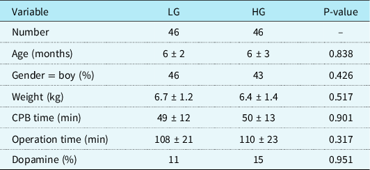
A total of 92 infants with ventricular septal defects were enrolled in this study. In accordance with an end-expiratory tidal pressure of carbon dioxide, 46 patients were enrolled in the relative high ventilation group while 46 patients were enrolled in the relative low ventilation group. There were no complications observed during transcranial Doppler measurements and regional cerebral oxygen saturation monitoring.
There were no significant statistical differences in gender, mean age, weight of infants, inotropic use, blood products use, cardiopulmonary bypass time, and time of operation between the two groups (summered in Table 1). There were also no significant statistical differences in all kinds of blood products during CPB.
Demographics and perioperative clinical data

Data are in mean ± SD for continuous variables and in percentage (%) for binary variables
CPB = cardiao pulmonary bypass; HG = relative high ventilation group; LG = relative low ventilation group.
Regional cerebral oxygen saturation and cerebral blood flow values
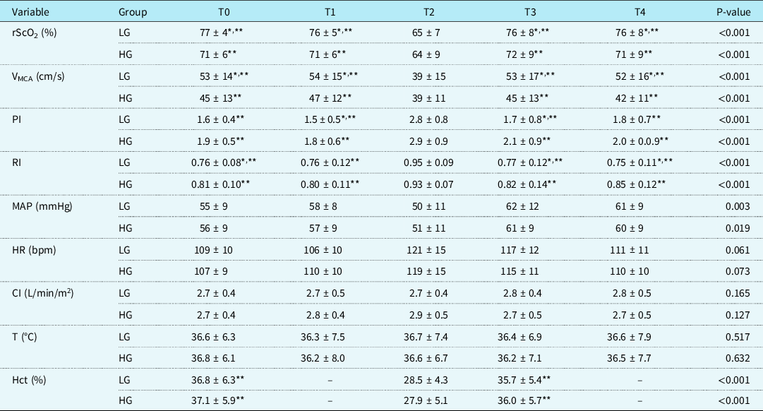
Cerebral haemodynamic parameters of the two groups at each time point during the operation were presented in Table 2. The relative low ventilation group exhibited a significantly higher regional cerebral oxygen saturation at each time point, except at the T2 time point (77 ± 4, 76 ± 5, 76 ± 8, 76 ± 8, p < 0.001, respectively). The regional cerebral oxygen saturation for all infants was the lowest at T2. In addition, the middle cerebral artery flow velocity value was high in the relative low ventilation group compared to the relative high ventilation group at each time point, except for T2 (53 ± 14, 54 ± 15, 53 ± 17, 52 ± 16, p < 0.001, respectively). The T2 time point exhibited the lowest regional cerebral oxygen saturation and middle cerebral artery flow velocity between the two groups (p < 0.001). On the contrary, pulsation index and resistance index showed the opposite trend.
rScO2, VMCA, and haemodynamic parameters (
 $$\overline {\rm{X}}$$
± s)

CI = cardiac index; Hct = haematocrit; HR = heart rate; MAP = mean artery pressure; PI = pulsation index; RI = resistance index; rScO2 = regional cerebral oxygen saturation; T = temperature; VMCA = flow velocity of the middle cerebral artery
*p < 0.05 when LG compared to HG
**p < 0.05 compared with T2
Haemodynamic variables
The haematocrit was significantly elevated from 28.5 ± 4.3 at T2 to 35.7 ± 5.4 at T3 (p < 0.001) in the relative low ventilation group, and was elevated from 27.9 ± 5.1 at T2 to 36.0 ± 5.7 at T3 (p < 0.001) in the relative high ventilation group. In addition, the mean artery pressure significantly increased from 50 ± 11 at T2 to 62 ± 11 at T3 (p = 0.003) in the relative low ventilation group, and increased from 51 ± 11 at T2 to 61 ± 9 at T3 (p = 0.019) in the relative high ventilation group. Other haemodynamic data (heart rate and cardiac index) did not show any significant differences between the two groups at each time point. There was no significant difference in temperature between the two groups.
Respiratory parameters
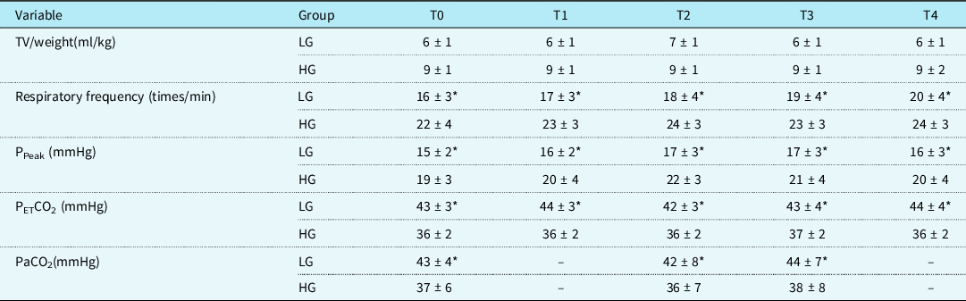
Compared to the relative high ventilation group, the relative low ventilation group showed a lower respiratory frequency, tidal volume, and peak airway pressure and a higher end-expiratory tidal pressure of carbon dioxide (p < 0.001). These results are shown in Table 3.
Respiratory parameters of two groups during anaesthesia (
 $$\overline {\rm{X}}$$
± s)

PETCO2 = end-expiratory tidal pressure of carbon dioxide; PPeak = peak airway pressure; TV = tidal volume
*p < 0.05 when LG compared to HG
Discussions
Few studies have documented the impact of carbon dioxide at a range of 35–45 mmHg and modified ultrafiltration on regional cerebral haemodynamics in infants. In this study, the regional cerebral oxygen saturation and middle cerebral artery flow velocity were impacted by modified ultrafiltration, significantly low upon separation from cardiopulmonary bypass when compared to other time points, and significantly high in the 40–45 mmHg end-expiratory tidal pressure of carbon dioxide range than in the 35–39 mmHg range.
Oxygen supply to the brain relies on tight regulation of cerebral blood flow.Reference Vutskits10,Reference Rhondali, Mahr and Simonin-Lansiaux11 Therefore, a decrease in regional cerebral oxygen saturation could signal a decrease in cerebral blood flow and/or inadequate oxygen supplyReference Schober, Feiner, Bickler and Rollins5 and vice versa. Adjustment of the mean artery pressure and end-expiratory tidal pressure of carbon dioxide increases cerebral blood flow and improves the balance between oxygen supply and demand in the brain during paediatric cardiac surgery. However, changes in mean artery pressure could exhibit a negative impact on infants with concomitant CHD.Reference Grüne, Kazmaier, Stolker, Visser and Weyland12 Due to these outcomes, it’s important to determine the influence of different end-expiratory tidal pressures of carbon dioxide on regional cerebral oxygen saturation and cerebral blood flow in those infants.
Existing scientific evidence suggests that carbon dioxide is a powerful modulator of cerebral vasomotor tone, which can dilate the cerebral arteriole, facilitate oxygen transport and cerebral perfusion.Reference Zhang, Xie, Han, Huang, Ou-Yang and Lu8 However, these studies seldom researched the effect of normocapnia, which was mostly adopted for cardiac surgery patients, and mostly investigated the active role of carbon dioxide in adults or children but not infants. In this study, we found that the middle cerebral artery flow velocity and regional cerebral oxygen saturation in the 40–45 mmHg end-expiratory tidal pressure of carbon dioxide range were improved. Studies have established a 30% increase in cerebral blood flow for every 7.5 mmHg increase in the end-expiratory tidal pressure of carbon dioxide in healthy volunteers.Reference Vutskits10 In this study, we observed a 24% increase in cerebral blood flow for infants younger than 1 year, a finding that is not in concordance with that found by. Thus, our results imply that small changes in end-expiratory tidal pressure of carbon dioxide might have a clinically significant impact on cerebral haemodynamics during CHD surgery in infants younger than 1 year.
The early post-bypass phases do not supply adequate cerebral oxygenation because the blood pressure is unstable at a time before the brain is fully protected by hypothermia. Studies have shown that cardiopulmonary bypass is prone to brain damage and continuous regional cerebral oxygen saturation. Transcranial Doppler monitor inhibits these neurological complications.Reference Abu-Sultaneh, Hehir and Murkowski13 In this study, regional cerebral oxygen saturation and middle cerebral artery flow velocity decreased while pulsation index and resistance index increased when after separation from cardiopulmonary bypass. Some studies demonstrated that a gradual decrease in regional cerebral oxygen saturation did have close influences on coronary reperfusion and rewarming.Reference Polito, Ricci and Di Chiara14 During rewarming, cerebral metabolism and oxygen consumption would increase beyond the increase in cerebral blood flow. With transcranial Doppler, it has been shown that after rewarming, the cerebral blood flow velocity increases 65% while the regional cerebral oxygen saturation decreases by 25%.Reference Kotlinska-Hasiec, Czajkowski and Rzecki15 Pulsation index is an important index for measuring cerebrovascular elasticity. Intracranial hypertension or focal oedema increases pulsation index. Resistance index indicates the blood flow resistance, and is high when there is a buildup of acidic metabolites, ischaemia hypoxic stage, etc. We demonstrated that ischaemia–reperfusion injury, hypothermia, inflammation, and hemodilution that were caused by cardiopulmonary bypass increased cerebrovascular resistance, decreased elasticity of cerebral vessels, reduced carbon dioxide reactivity, impaired cerebrovascular autoregulation, and decreased cerebral blood flow. These effects triggered an imbalance in cerebral perfusion and cerebral oxygen supply and demand.Reference Abu-Sultaneh, Hehir and Murkowski13,Reference Fogel, Li and Elci16 Cerebral blood flow and cerebral oxygen saturation were highly dependent on the end-expiratory tidal pressure of carbon dioxide. These factors did not vary after cardiopulmonary bypass. It might be caused by the impaired carbon dioxide activity and poor cerebral autoregulation.
After CPB, modified ultrafiltration was used to reduce hemodilution and its potential adverse effects.Reference Medlin and Sistino9 The concept of modified ultrafiltration was first proposed by Naik et alReference Naik, Knight and Elliott17 and is now routinely used in cardiac surgery for infants. In this study, there was an increase in cerebral blood flow, cerebral oxygen saturation, mean artery pressure, and haematocrit after modified ultrafiltration. A short duration ultrafiltration circuit leads to a severe left-to-right shunt (steal blood phenomenon), no matter the modified ultrafiltration flow rate.Reference Rodriguez, Ruel, Broecker and Cornel18 An increase in cardiac function could supplement for this “steal” and causes a lower oxygen extraction ratio.Reference Medlin and Sistino9,Reference Aeba, Katogi, Omoto, Kashima and Kawada19,Reference Kotani, Honjo and Osaki20 Moreover, the increased cerebral oxygen saturation might be associated with an increased mean artery pressure. Autoregulatory studies in neonates have reported a linear relationship between regional cerebral oxygen saturation and mean artery pressure.Reference Vutskits10,Reference Wyatt and Meek21 Some studies have, however, documented that no correlation exists between regional cerebral oxygen saturation and mean artery pressure in humans.Reference Lucas, Tzeng, Galvin, Thomas, Ogoh and Ainslie22 There is no conclusive evidence on the correlations between mean artery pressure and regional cerebral oxygen saturation. Recent studies determining the coherence between continuous bedside near-infrared spectroscopy and arterial pressure have suggested that cerebral autoregulation can transiently be impaired in critically ill infants.Reference Rhondali, Mahr and Simonin-Lansiaux11,Reference Wong, Leung and Austin23 The frequency of impaired cerebral autoregulation is associated with low regional cerebral oxygen saturation and systemic hypotension.Reference Wong, Leung and Austin23,Reference Tsuji, Saul and du Plessis24 Furthermore, increased cerebral oxygen saturation could be attributed to increased haematocritReference Medlin and Sistino9 that promotes arterial oxygen content and lowers the oxygen extraction ratio. In this study, modified ultrafiltration gradually decreased the resistance index and pulsation index. This could be attributed to the fact that increased haematocrit, reduced inflammatory mediators and relieved cell oedema, reduce cerebrovascular resistance, increase elasticity of cerebral vessel, improve carbon dioxide reactivity, recover cerebrovascular autoregulation,Reference Shen, Wang, Zhang, Jiang and Yang25 and therefore, improve cerebral perfusion and cerebral oxygen metabolism. These factors elevate the regional cerebral oxygen saturation and middle cerebral artery flow velocity. However, these conclusions need to be verified by more studies.
For infants with ventricular septal defects, the lower the obstructive pulmonary hypertension, the better the recovery of cardiopulmonary vascular function after surgery. However, studies have suggested that these children exhibit difficulties in cerebral oxygenation maintenance during surgery, and therefore, the incidences of long-term cognitive decline may be higher.Reference Robertson, Justo, Burke, Pohlner, Graham and Colditz26 Hyperventilation reduces pulmonary vascular resistance and increases cerebrovascular resistance. These outcomes decrease cerebral blood flow.Reference Ricci, Garisto and Favia6,Reference Li, Zhang and Holtby7,Reference Dodds27 It has been demonstrated that hyperventilation might result in the poor cerebral autoregulation and low cerebral oxygen saturation.Reference Redlin, Koster and Huebler28 Maintaining an end-expiratory tidal pressure of carbon dioxide at 40–45 mmHg improved cerebral perfusion and cerebral oxygen saturation, avoided pulmonary vascular bed over-exploitation, and limited the left-to-right shunt. We recommend that in infants younger than 1 year who undergo VSD repair, attention must be paid to adequate cerebral blood flow and balance of cerebral oxygen supply and demand to avoid hyperventilation.
There are several limitations in our study. Although, middle cerebral artery flow velocity by transcranial Doppler ultrasonography, rather than cerebral blood flow, most researches validated that the middle cerebral artery flow velocity is a reliability index of cerebral blood flow (measured using intravenous Xenon-133).Reference Giller, Bowman, Dyer, Mootz and Krippner29,Reference Valdueza, Balzer, Villringer, Vogl, Kutter and Einhäupl30 End-expiratory tidal pressure of carbon dioxide is significantly correlated with arterial carbon dioxide pressure amongst infants, however, in this study, no arterial carbon dioxide pressure data were obtained.Reference Karsli, Luginbuehl, Farrar and Bissonnette31 Besides technical factors, several physiological factors have been found to have an influence on regional cerebral oxygen saturation and middle cerebral artery flow velocity. These physiological factors include cardiac output and body temperature. All measurements were done before surgery to eliminate the impact of surgical stimulation on cerebral haemodynamics changes. Body temperature remained unchanged throughout the study. In addition, it is important to evaluate the impact of near-infrared spectroscopy and transcranial Doppler monitoring on the incidence of perioperative complications in infants. It is also important to determine how near-infrared spectroscopy and transcranial Doppler provides continuous regional cerebral oxygen saturation. Outcomes from these studies may be a suitable target for cerebral directed therapy.
Conclusion
Modified ultrafiltration improved cerebral oxygen haemodynamics. The regional cerebral oxygen saturation and middle cerebral artery flow velocity performed better when the end-expiratory tidal pressure of carbon dioxide was 40–45 mmHg during relative low ventilation in infants. It is important to regulate ventilation to precisely achieve cerebral oxygen balance in infants undergoing ventricular septal defect surgery.
Acknowledgements
We thank Dongni Zhang and Nan Wang for their assistance in data collection.
Financial support
The work was supported by grants from the National Natural Science Foundation of China (No. 81471902 and No. 81871592), Beijing Municipal Administration of Hospitals Clinical Medicine Development of Special Funding Support (ZYLX 201810), and Beijing Hospitals Authority Youth Program (QML20180602).
Conflict of interests
The authors declare no conflict of interest.
Ethical standards
Ethics approval for this trial was obtained from the Institutional Ethics Committee of Capital Medical University affiliated Beijing Anzhen Hospital (IRB: 2017030X).