Introduction
Anisakis simplex is a parasitic gastrointestinal nematode with a complex life cycle; the definitive hosts are marine mammals (Smith & Wootten, Reference Smith and Wootten1978) in which the parasite undergoes the third and fourth moults preceding maturity. Humans may become accidental hosts by consuming raw, lightly salted, smoked or marinated marine fishes infested by third stage (L3) A. simplex larvae (Oshima, 1987; Takabe et al., Reference Takabe, Ohki, Kunihiro, Sakashita, Endo, Ichikawa, Sekido, Amano, Nakatani, Suzuki and Shimanda1998; Molina-Garcia & Sanz, Reference Molina-Garcia and Sanz2002). As anisakiasis is a serious disease that can be fatal and cause an allergic reaction in patients sensitive to A. simplex allergens (Nakatani et al., Reference Nakatani, Suzuki and Shimanda1998; Valls et al., Reference Valls, Pascual and Esteban2005), it is necessary to intensify research on the biochemistry and physiology of the parasite's larvae. Current knowledge is still fragmentary including information on the amino acid, protein profile of the L3 and their protease activity (Viglierchio & Görtz, Reference Viglierchio and Görtz1972; Sakanari & McKerrow, Reference Sakanari and McKerrow1990; Morris & Sakanari, Reference Morris and Sakanari1994; Perteguer et al., Reference Perteguer, Raposo and Cuellar1996; Nguyen et al., 1999; Dziekońska-Rynko et al., Reference Dziekońska-Rynko, Rokicki and Jabłonowski1997a, Reference Dziekońska-Rynko, Rokicki and Jabłonowskib, Reference Dziekońska-Rynko, Rokicki and Jabłonowski2002). Recently we have published data on saccharide types and their contents in A. simplex L3 and L4 (Łopieńska-Biernat et al., Reference Łopieńska-Biernat, Żółtowska and Rokicki2006). Trehalose metabolism enzymes of the larvae have also been elucidated (E. Łopieńska-Biernat et al., unpublished data). The present paper focuses on enzymes involved in glycogen catabolism.
In nematodes, glycogen may be broken down by hydrolysis, amylases (α-amylase and glucoamylase), or phosphorolysis, the process involving glycogen phosphorylase (Żółtowska et al., Reference Żółtowska, Łopieńska, Rokicki and Dmitryjuk2001, Reference Żółtowska, Łopieńska, Rokicki and Dmitryjuk2002). The hydrolytic glycogen degradation pathway in nematodes is controlled by α-amylase (Żółtowska, Reference Żółtowska2001a; Mohamed, Reference Mohamed2004), an enzyme found in A. simplex. A preliminary characterization of α-amylase isolated from A. simplex larvae was published by Łopieńska et al. (Reference Łopieńska, Żółtowska and Rokicki2001).
The literature lacks information on A. simplex glucoamylase (exo-1,4-α-d-glucan-glucohydrolase EC 3.2.1.3), the other hydrolytic enzyme involved in the glycogen hydrolysis pathway, as well as on glycogen phosphorylase, involved in the phosphorolytic pathway.
Our earlier paper (Łopieńska-Biernat et al., Reference Łopieńska-Biernat, Żółtowska and Rokicki2006) described significant differences in the content of endogenous storage carbohydrates, including glycogen, in A. simplex L3 and L4 larvae. We hypothesize that the differences are accompanied by alterations in activities and properties of the glycogen catabolism enzymes. The present work tested this hypothesis.
Materials and methods
The study involved third (L3) and fourth (L4) larval stages of A. simplex. The L3 were isolated from fresh Baltic herring (Clupea harengus). The nematodes, cleaned of host tissue remains and rinsed several times in 0.65% NaCl, were kept in 0.65% NaCl at 4°C until analysed. The L4 were obtained from in vitro cultures, run according to the modified method of Grabda (Reference Grabda1976). The cultures were kept in an atmosphere containing 5% CO2 at 37°C.
Preparation of A. simplex extracts
Samples (100 mg) of larvae (L3 or L4) were homogenized in a glass blender with 1 ml 0.65% NaCl. The homogenate was centrifuged for 15 min at 800 × g. The supernatant was used to determine enzymatic activities and protein content.
Enzymatic activity assays
The activity of α-amylase was determined using the technique of Caraway (Reference Caraway1959), and expressed in activity units (u). One unit is equivalent to the amount of the enzyme that would break down 10 mg of starch until the iodine–starch reaction disappeared during 2 h incubation at 37°C and pH 6.0 for L3 or pH 6.5 for L4. The glucoamylase activity was measured using the method of Dahlqvist (Reference Dahlqvist1968), and expressed in enzymatic units (u). One unit is equivalent to the amount of the enzyme that would release 1 μmol glucose during 1 h at 37°C pH 6.5. The activity of glycogen phosphorylase was determined from the enzyme's ability to synthesise glycogen, as described by Cori et al. (Reference Cori, Illingworth, Keller, Colowick and Kaplan1955). Inorganic phosphate formed during the reaction was assayed by the microtechnique of Chen et al. (Reference Chen, Toribara and Warner1974). Phosphorylase activity was also determined from the enzyme's ability to break down glycogen. Glucose-1-phosphate, released during the reaction, was measured using the technique of Michal (Reference Michal and Bergmeyer1981). The enzymatic unit (u) is equivalent to the amount of the enzyme that would release 1 μmol glucose-1-phosphate (G-1-P) during 1 h at 37°C. Enzyme activities were referred to 1 mg protein determined by Bradford (Reference Bradford1976).
Enzyme characterization
The effect of keeping the larvae at a low temperature on their enzymatic activities was determined in the following way. Samples (100 mg) of fresh L3 and L4 larvae were placed at − 100°C and enzymatic activities in the larvae were assayed at one-week intervals. The intracellular distribution of the enzymes was determined by differential centrifugation described by Van den Bossche & Borgers (Reference Van den Bossche and Borgers1973). Their optimum pH activity was determined by incubating larval extracts in 70 mm venoral/sodium acetate buffer of pH 3.20–8.0. The optimal temperature of the enzymatic activities was determined by incubating the larval extracts for 1 h at 25–85°C, at optimal pH. In each subsequent measurement the temperature was raised by 5°C. The thermal stability of the enzymes was determined in the following way: buffered enzymatic extracts without substrates were preincubated for 15 min at 25–95°C and cooled down; the preincubation being followed by incubation, which was carried out with an appropriate substrate for 1 h at 37°C. The effects of various chemical compounds on the amylases were determined by adding 10 mmol aqueous solutions of MgCl2, CaCl2, CH2ICOOH, EDTA, and Tris as well as 1 mmol ZnCl2. The effects of 10 mmol ATP, CH2ICOOH, ZnCl2, caffeine, and pyridoxal phosphate were tested on glycogen phosphorylase.
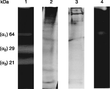
Molecular weights of protein fractions in the extracts were determined from mass standards (Sigma Wide Molecular Range markers) using sodium dodecyl sulphate–polyacrylamide gel electrophoresis (SDS–PAGE) (Weber & Osborn, Reference Weber and Osborn1969) and PAGE (Davis, Reference Davis1964) techniques. The analysis performed with the Scan Gel 1.45 software (Kucharczyk Electrophoretic Technique, Warsaw, Poland) allowed the comparison of percent contributions of individual fractions to the total protein pool. α-Amylase in extracts from the two stages of A. simplex larvae were identified after PAGE on polyacrylamide gel prepared with 1% soluble starch (fig. 1), according to the procedure described by Żółtowska (Reference Żółtowska2001a).
Separation of Anisakis simplex proteins and amylases by PAGE. Lane 1, the activity of L3 α-amylase; lane 2, proteins in L3; lane 3, proteins in L4; lane 4, the activity of L4 α-amylase. α1,α2 and α3 isoenzymes of α-amylase. Protein was detected by staining with Coomassie Brilliant Blue R 250; the activity was located by iodine staining after PAGE.

The results presented are arithmetic means of five independent analyses. The data obtained were subjected to statistical treatment using Student's t-test.
Results
The two larval stages were found to differ in their respective protein contents. The difference in mean protein contents between L3 and L4 was highly significant (P < 0.01). The L4 contained 2.5 times more protein than the L3. The mean protein contents in L3 and L4 extracts were 13.12 ± 7.55 and 31.11 ± 20.09 mg g− 1 wet weight, respectively.
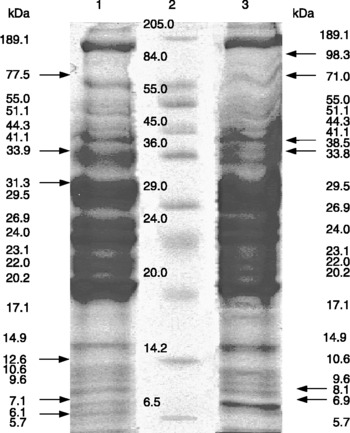
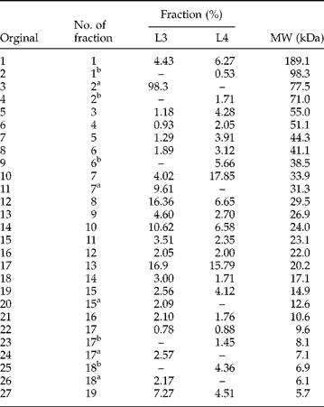
PAGE analysis of the L3 extracts revealed 12 protein fractions, and as few as seven in L4 (fig. 1). The denaturing electrophoresis (SDS–PAGE) produced similar, but not identical, patterns in the two larval stages (fig. 2). The extracts analysed were found to contain 22 protein fractions in each larval stage, 17 fractions showing identical mobility and molecular weight in both stages (table 1). The remaining five fractions (denoted in table 1 as a and b in L3 and L4 respectively) were stage-specific proteins. The contributions of proteins with a molecular weight higher than 30 kDa (table 1, fractions 1–7) were 2–4 times higher in the L4 extracts than in the L3. In contrast, the contributions of proteins with a lower molecular weight, except for those of fractions 15 and 19, were higher in L3 than in L4 (table 1, fractions 8–19).
Separation of protein of third (L3) and fourth stage (L4) larvae of Anisakis simplex by SDS–PAGE. Lane 1, proteins in L3; lane 2, molecular mass standards; lane 3, proteins in L4.

Molecular weights (MW) and per cent contribution of individual protein fractions in extracts from third (L3) and fourth stage (L4) larvae of Anisakis simplex after SDS–PAGE.
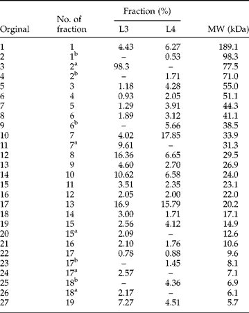
Contribution of individual fractions in total pool of protein. The fractions specific for larval stages: afor L3; bfor L4.
Amylolytic activity in the L3 extracts was displayed by three protein fractions. They were called α1, α2, and α3 isoenzymes, their molecular weights were 64, 29 and 21 kDa, respectively. On the other hand, the L4 extracts produced only one active fraction, corresponding to α1 isoenzyme of the L3 (fig. 1).
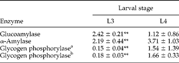
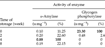
The L3 and L4 of A. simplex possess enzymes of both glycogen catabolism pathways. The activity of the hydrolytic glycogen degradation enzymes, α-amylase and glucoamylase, was very high in L3, and activity of glycogen phosphorylase was low (table 2). In contrast, phosphorylase activity in L4 was ten times higher than in L3. Differences in mean activities of analogous enzymes between L3 and L4 were highly significant in all cases. Keeping the larvae at − 10°C led to a gradual increase in α-amylase activity in the extracts (table 3). In contrast, activity of glycogen phosphorylase decreased relatively rapidly (table 3). Moreover, 95% of the total enzyme activity in the fresh larvae was due to form a of glycogen phosphorylase, whereas the trace activity detected after two weeks was ascribed exclusively to form b of the enzyme.
Activities of carbohydrate catabolism enzymes in third (L3) and fourth stage (L4) larvae of Anisakis simplex (u mg−1 of protein).
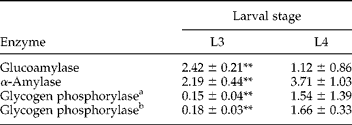
** P < 0.01.
Activity of glycogen phosphorylase determined: ain the direction of breakdown of glycogen, bsynthesis of glycogen.
Changes in enzymatic activities during storage of Anisakis simplex third stage larvae at −10°C.
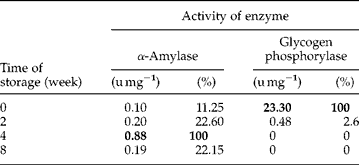
Values in bold are maximal values, assumed to be 100%.
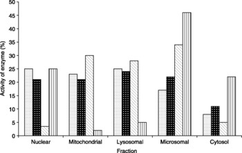
The pH optimum for α-amylase was 6.5 in L4; 6.0 and 8.0 in L3. The pH optimum for glucoamylase was 6.5 in both stages, whereas that for glycogen phosphorylase was 6.8 in L4 and 7.0 in L3. The highest glucoamylase activity, both in L3 and in L4, was observed in the microsomal fraction (fig. 3). The very low activity of the enzyme was in the nuclear and cytosol fractions of L3, those fractions showing relatively high activity in L4. In contrast, intracellular distribution of glycogen phosphorylase was similar in the two larval stages (fig. 3).
Intracellular distribution of glycogen phosphorylase and glucoamylase in third (L3) and fourth stage (L4) larvae of Anisakis simplex. ( ) glycogen phosphorylase L3; (
 ) glycogen phosphorylase L4; (▧) glucoamylase L3; (
 ) glucoamylase L4.

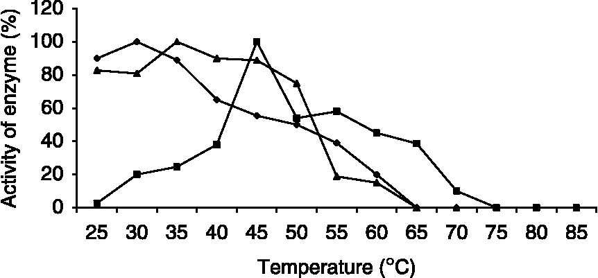
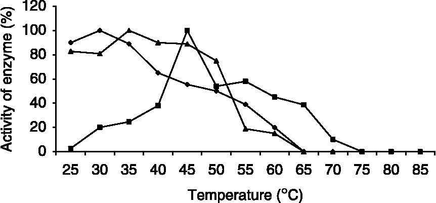
The limited amount of material necessitated following the temperature dependence of enzymes activities only in L3 (fig. 4). The activities of α-amylase and glycogen phosphorylase were high (>80%) at a relatively low temperature of 25°C. α-Amylase showed optimal activity at 30°C and glycogen phosphorylase at 35°C. The activities of both enzymes decreased rapidly at a temperature higher than 50°C. In contrast, glucoamylase became more active when the temperature rose above 35°C, its peak of activity being recorded at 45°C (fig. 4).
Dependence of activities of amylolytic enzymes and glycogen phosphorylase from Anisakis simplex third stage larvae on temperature. For the conditions of analysis see Material and methods section. The relative activities were expressed as percentages of maximum activity; 2.19 ± 0.44 (u mg− 1) for α-amylase; 2.42 ± 0.21 (u mg− 1) for glucoamylase and 0.15 ± 0.04 (u mg− 1) for glycogen phosphorylase. ( ) alpha amylase; (
 ) glucoamylase; (
 ) glycogen phosphorylase.
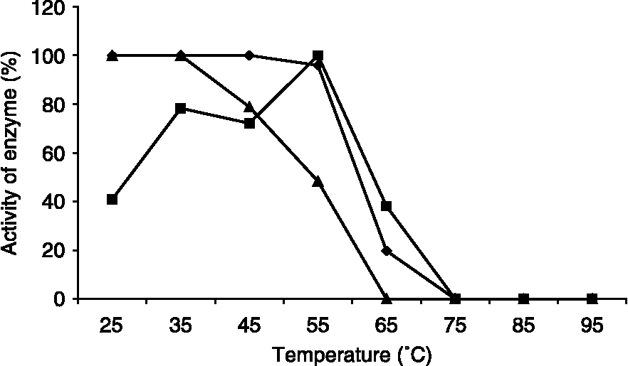
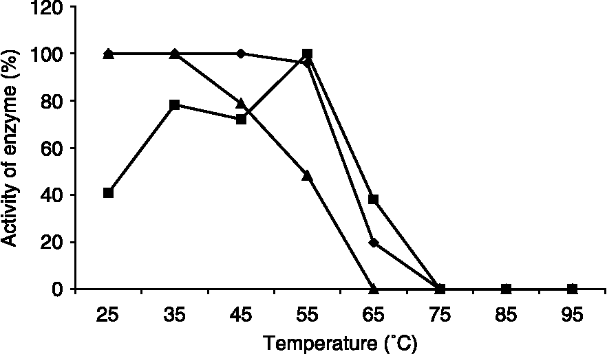
The activity of α-amylase was found to be unaffected by the 15-min preincubation (without substrate) at temperatures up to 55°C (fig. 5). The thermal stability curve of glucoamylase was different. An increase in preincubation temperature from 30 to 55°C was accompanied by increased activity of this enzyme. Temperatures higher than 65°C resulted in inactivation of both amylases. Glycogen phosphorylase proved to be very thermolabile (fig. 5).
Thermal stability of amylases and glycogen phosphorylase from Anisakis simplex third stage larvae. For the conditions of analysis see Material and methods section. The relative activities were expressed as percentages of maximum activity; 1.89 ± 0.20 (u mg− 1) for α-amylase; 2.04 ± 0.21 (u mg− 1) for glucoamylase and 0.09 ± 0.02 (u mg− 1) for glycogen phosphorylase. () alpha amylase; () glucoamylase; () glycogen phosphorylase.
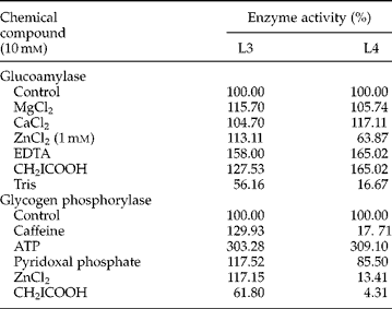
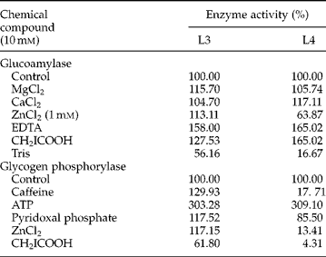
The same enzymes isolated from the two larval stages often responded differently to salts added to their respective media. For example, zinc ions increased glucoamylase in L3 by about 13% and inhibited it in L4; caffeine increased glycogen phosphorylase activity by 30% in L3 and produced a decrease of about 80% in L4 (table 4). Even when the changes in the enzyme from the two stages proceeded in the same direction, the intensity of the enzymatic response differed, e.g. Tris inhibited glucoamylase activity by about 40% in L3 and by 83% in L4. The activity of glycogen phosphorylase in both stages of A. simplex larvae was distinctly reduced by iodoacetic acid and strongly stimulated by ATP (table 4).
Effects of chemical compounds on the activities of glucoamylase and glycogen phosphorylase in third (L3) and fourth stage (L4) larvae of Anisakis simplex.
Discussion
Under natural conditions, the larval stages of A. simplex studied in this work live in different hosts: L3 occur in fish and marine invertebrates, while L4 dwell in the intestinal tract of marine mammals (Smith & Wootten, Reference Smith and Wootten1978). Thus, the native habitats of the two stages differ in terms of both their physical and chemical parameters. We assumed that the differences should be evident also at the level of proteins, including enzymatic proteins.
The metabolic differences were confirmed by the data of protein analysis in extracts of both larval stages of A. simplex. The total protein pool showed both quantitative and qualitative differences reflected in the presence of five stage-specific fractions (table 1, fig. 2). Similar results were reported by Frank & Grieve (Reference Frank and Grieve1991) for secretion–excretion proteins of L3 and L4 of Dirofilaria immitis.
The levels of activity and properties of analogous glycogen catabolism enzymes in the two larval forms confirm the suggestion that the third moult involves changes which, perhaps, proceed at the expression level of the genes coding for the enzymes.
Glycogen, the major storage carbohydrate of Anisakis (Łopieńska-Biernat et al., Reference Łopieńska-Biernat, Żółtowska and Rokicki2006), was catabolized in L3 dominated by hydrolysis, whereas activities of hydrolytic and phosphorolytic enzymes in L4 were similar. Results of this study are in agreement with data reported for Hysterothylacium aduncum and Cystidicola farionis (Żółtowska et al., Reference Żółtowska, Łopieńska, Rokicki and Dmitryjuk2001, Reference Żółtowska, Łopieńska, Rokicki and Dmitryjuk2002). Those fish-parasitizing nematodes showed an ontogenetic increase in phosphorolytic enzyme activity. It can be concluded that the two larval stages of A. simplex differ primarily in that L4 are more able to phosphorolyse glycogen (table 2).
The present study is the first to demonstrate the activity of glycogen phosphorylase in A. simplex larvae. Glycogen phosphorylase present in L3 of A. simplex showed properties typical of enzymes described from other species (Cori et al., Reference Cori, Illingworth, Keller, Colowick and Kaplan1955; Bhuiyan et al., Reference Bhuiyan, Rus'd, Kitaoka and Hayashi2003). Active in a near-neutral medium, it was very sensitive to thermal effects and protected by the substrate (table 4, figs 4 and 5). Similarly to phosphorylases in other nematodes, Ascaris suum and Litomosoides carinii, it was activated by ATP and pyridoxal phosphate (Donahue et al., Reference Donahue, Yacoub, Kaeini, Masaracchia and Harris1981; Yacoub et al., Reference Yacoub, Allen, Payne, Masaracchia and Harris1983; Beg et al., Reference Beg, Fistein, Ingram and Storey1996). Caffeine activation of the enzyme (table 4) suggests that glycogen phosphorylase activity in L3 is controlled in a typical way, by the cAMP level, whereas, some properties of the enzyme from L4 were not typical for glycogen phosphorylases, e.g. the inhibition by caffeine and ZnCl2.
The activities of glycogen catabolism enzymes correlate positively with the glycogen content in L4, four times higher than that in L3 as demonstrated in our earlier study (Łopieńska-Biernat et al., Reference Łopieńska-Biernat, Żółtowska and Rokicki2006). In conclusion, glycogen is not only the major energy supplier for L4 of A. simplex but also a substance that is intensively metabolized by those larvae.
We suggest that the increase in α-amylase activity in L4 (table 2) may, at least in part, be explained by adaptive changes with respect to the type of food the larvae find in their host. The major carbohydrate source for the L4 present in the digestive system of predatory marine mammals is glycogen and its breakdown products. This may explain the relatively high activity of hydrolases in glycogen catabolism in L4 of A. simplex. The activity of α-amylase was similar in larval and adult nematodes H. aduncum isolated from fish; but that nematode species completes its life cycle without changing its host (Żółtowska et al., Reference Żółtowska, Łopieńska, Rokicki and Dmitryjuk2002).
The present results obtained allowed us to conclude that the two larval stages differ significantly not only in the level of activity of the enzymes studied (table 1), but that there are more profound differences. They involve properties of individual, seemingly identical enzymes present in the two stages. This is shown particularly in α-amylase. The enzymes of the two stages differ in their intracellular distribution and pH optima as well as in their responses to some chemical compounds (Łopieńska et al., Reference Łopieńska, Żółtowska and Rokicki2001). Due to organ-specific differences, α-amylases of parasitic nematodes show isoenzymes. For example, A. suum isolates contained muscle-, intestine-, and haemolymph-specific α-amylases. Moreover, each was represented by at least two isoenzymes (Ženka & Prokopič, Reference Ženka and Prokopič1984; Żółtowska, Reference Żółtowska2001b). Mohamed (Reference Mohamed2004) reported the presence of three isoenzymes in extracts of the invasive L3 of the entomopathogenic nematode Heterorhabditis bacteriophora. The presence of α-amylase isoenzymes in L3 of A. simplex, first described by Taniguchi in 1967 (reported by Smith & Wootten, 1978), were confirmed in the present study, although the larvae showed as few as three active forms instead of six reported by Taniguchi. It is not clear why L3 have so many α-amylase isoenzymes, while L4 contain only one of them. A reverse situation and a higher diversity of amylase pattern had been anticipated in the older developmental stage, similarly to that found in the shrimp Palaemon elegans (Van Wormhoudt & Farvel, Reference Van Wormhoudt and Farvel1988).
The conclusion that, following the third moult, the larvae of A. simplex synthesize enzymes with different properties is strongly supported by the comparison of properties of glucoamylase and glycogen phosphorylase in the present work and α-amylase studied earlier (Łopieńska et al., Reference Łopieńska, Żółtowska and Rokicki2001). In the present study, we do not answer all the questions of glycogen catabolism in larval A. simplex, but we have succeeded in providing a basis for further detailed studies on glycogen metabolism in this interesting parasite.
Acknowledgements
The research was supported by the Ministry of Education and Science grant no. PO4C02428.