I. INTRODUCTION
Many commercial processes for pharmaceutical, chemical, and biomedical applications require immobilization or encapsulation of biomolecules on/in a solid host material. Similarly, environmental, industrial, and clinical sensors, detectors or diagnostic devices, and biological assay platforms all depend on the immobilization/encapsulation of biomolecules on/in a solid material. For immobilization of biomolecules, one important challenge is in preserving the native biological activity and biochemical functionality of the biomolecule after immobilization. Generally, nonspecific interactions of the biomolecule with the host material and steric or orientation-dependent hindrances can limit the biochemical function and activity in the immobilized state. An emerging method for bioimmobilization is entrapment of biomolecules in sol–gel-derived silica. Sol–gel processing offers potential in integrating delicate biomolecules within solid hosts due to its aqueous-based nature requiring low temperature and soft reaction conditions.Reference Braun, Rappoport, Zusman, Avnir and Ottolenghi1–Reference Lin and Brown9 The resulting porous silica host may restrict the global movements of the biomolecules that may lead to denaturation, while maintaining its segmental activities where reagent binding and catalysis can occur. Another key property of sol–gel-derived materials is the freedom in tuning the chemical and structural properties of the hosting environment, where the organic or biological molecule is immobilized; the scale of porosity can be tailored from the nano- to the microscale for specific applications. Sol–gel-derived silica matrices also contain molecular water, resembling an aqueous environment, which contributes to retention of the native structure and reactivity of the encapsulated biomolecules.
Many combinations of the inorganic components with organic molecules or biomolecules can be prepared by modifications of generic sol–gel approaches. For example, organic template-assisted techniques utilize hybrid sol–gels to stimulate the ordered growth processes, which can be related to biomineralization.Reference Mann, Burkett, Davis, Fowler, Mendelson, Sims, Walsh and Whilton10–Reference Weiner and Addadi12 Another general class of hybrid sol–gels is those systems where biomolecules are physically entrapped/hosted in the inorganic matrix. Protein-, enzyme-,Reference Braun, Rappoport, Zusman, Avnir and Ottolenghi1–Reference Gill and Ballesteros4 and even living cell-encapsulatedReference Carturan, Campostrini, Dire, Scardi and Dealteriis5, Reference Pope6 sol–gel-derived matrices have already exhibited their diversity and potential in applications including chemical or biosensors and bioreactors.Reference Dave, Dunn, Valentine and Zink7–Reference Lin and Brown9 Sol–gel-derived hybrids can also be used for controlled release of bioactive compounds for drug delivery.Reference Böttcher, Slowik and Suss13 An overview of bioapplications of sol–gel material can be found in a comprehensive review article.Reference Avnir, Coradin, Lev and Livage14
Similarly, sol–gel-derived silica matrices can be synthesized with specific chemical and structural functionalities for immobilization or encapsulation of DNA. Such systems can be useful in numerous applications. For example, passive attachment of DNA or oligonucleotide strands on glass surface is the basic principle for hybridization-based bioassay platforms and lab-on-a-chip applications. DNA immobilization on solid supports, or encapsulation in microspheres and in liposomes, is potentially one of the most important tool for gene delivery systems. In this respect, there is a great potential for using sol–gels to extend the application and use of genetically engineered materials in practical applications in gene therapy, pharmaceutical, molecular biology, and food and biotechnology. Innovative uses of such gene delivery systems include modifying and repairing cells functions for therapeutic healing.Reference Ledley15 A number of methods including viral-,Reference Flotte and Carter16, Reference Mah, Byrne and Flotte17 nonviral-,Reference Schatzlein18, Reference Rettig and Rice19 and physical stimuli-mediatedReference Hosseinkhani, Aoyama, Ogawa and Tabata20 transfection approaches have been investigated to improve the gene transfection for selective delivery and successful gene expression. However, designing new processes, allowing material improvements, and enhanced directed evolution of reagent genes still remain as challenging tasks for expanding technological use of gene delivery systems for therapeutic or diagnostic applications.
Despite the studies in adapting the sol–gel approaches for designing such DNA-based biochips,Reference Fidanza, Glazer, Mutnick, McGall and Frank21–Reference Durucan, Pantano, Sakka, Almeida and Kozuka26 there are only limited number of studies reporting direct sol–gel encapsulation of DNA in silica matrices and these are restricted to the understanding of molecular-level DNA–silica interactionsReference Pierre, Bonnet, Vekris and Portier27 and to the use of DNA as a templating agent for sol–gel polycondensation.Reference Numata, Sugiyasu, Hasegawa and Shinkai28, Reference Shinkai, Takeuchi and Bae29 The first study investigating the feasibility of sol–gel method for DNA encapsulation was by Pierre et al.,Reference Pierre, Bonnet, Vekris and Portier27 showing that low molecular weight small DNA molecules can be encapsulated within tetramethylorthosilicate-based pure and in hybrid polyvinyl alcohol–silica gels. However, only limited amount of DNA could be reextracted from these silica hosts, suggesting a molecular complexation between the silica and DNA after encapsulation. The diversity of DNA encapsulation has recently become more evident with emerging applications and practical uses. For instance, sol–gel approaches have been amenable for entrapment of a variety of DNA-based biorecognition elements such as DNA enzymesReference Shen, Mackey, Rupcich, Gloster, Chiuman, Li and Brennan30 and DNA aptamersReference Rupcich, Nutiu, Yu and Brennan31 for developing optical sensors and diagnostic systems. More recently, ormosil silica gels of tetraethylorthosilicate (TEOS) and aminopropytriethoxysilane were used to entrap natural DNA molecules in an attempt to obtain adsorbents for environmentally harmful and toxic pollutants.Reference Satoh, Fugetsu, Nomizu and Nishi32 Additionally, some investigators tried to expand DNA encapsulation by modified sol–gel approaches to achieve different physical forms of silica host.Reference Fujiwara, Shiokawa, Hayashi, Morigaki and Nakahara33 However, there is no systematic study reporting the effect of presence and amount of DNA on gelation behavior of alkoxide precursors. There is also limited analytical information on the microstructural and chemical properties of the resultant silica hosts and on correlation of these factors with DNA encapsulation efficiency.
In this study, we prepared DNA-encapsulated silica under mild chemical conditions by a two-step acid/base catalysis method. This was achieved by mixing partially hydrolyzed TEOS solution and ionic buffer solution that was used to dissolve low molecular weight natural double-stranded DNA. 29Si nuclear magnetic resonance (NMR) spectroscopy and gas adsorption studies were performed to characterize the silica gels. Sol–gel processing parameters such as gelation pH and DNA concentration greatly influenced the gelation kinetics and microstructure of the silica. The DNA encapsulation efficiency in these gels was dependent on the scale of porosity, mainly controlled by the gelation pH. Furthermore, DNA–silica hybrids can accumulate DNA-intercalating molecules from aqueous solutions, indicating the conservation of the double-stranded native form of the DNA during sol–gel encapsulation. As a result, a modellistic DNA encapsulation mechanism correlating the silica microstructure and DNA holding efficiency has been proposed.
II. MATERIALS AND METHODS
A. Preparation of DNA–silica gels
The encapsulation has been achieved by introduction of DNA-containing buffer solution into a partially hydrolyzed TEOS [Si(OC2H5)4; Sigma-Aldrich Chemie GmbH, Steinheim, Germany] solution before completion of hydrolysis and condensation reactions. First, TEOS was dissolved in anhydrous ethanol (EtOH, C2H5OH, 200 proof; Merck KGaA, Darmstadt, Germany) at 1:1 volume ratio in a borosilicate glass beaker at room temperature; the pH of TEOS:EtOH mixture was around 6. After obtaining a homogeneous solution by mixing for 10 min, 0.1 M HCl solution was added dropwise, and the mixture was further stirred for 5 min. The solution pH decreased to 4.2. Later on, DNA (double-stranded low molecular weight salmon sperm DNA, <5% protein impurity, 50–5000 base pairs; Fluka, Sigma-Aldrich Chemie GmbH, Steinheim, Germany) molecules dissolved in Tris-EDTA [TE, 10x concentrate, 1 M Tris-0.01 M ethylenediamine tetraacetic acid (C10H16O8N2; EDTA); Sigma] buffer were added into the sol. This was again achieved under continuous stirring by dropwise addition. The concentration of TE buffer solution was 40 μg DNA/mL and had a pH value of 8.4. Then, DNA-incorporated mixture was further stirred for 5 min.
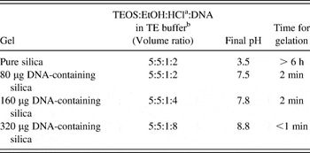
In this way, three DNA-containing silica gels were prepared, which were different in terms of the total amount of DNA-containing buffer solution that was added into HCl-catalyzed TEOS–EtOH sol. Sol–gel formulations are given in Table I. The TEOS:EtOH:HCl (volumetric) ratio was 5:5:1 and identical for all cases. The total amount of buffer addition was 2, 4, and 8 mL, corresponding to a total DNA amount of 80, 160, and 320 μg, respectively. In addition, a plain silica gel was also prepared in the same way as a control sample for which 2 mL TE buffer solution was added to the TEOS-based sol mixture instead of DNA-containing TE buffer solution. This sample is referred as pure silica hereafter.
Sol–gel formulations for pure silica and for DNA–silica gels.

a 0.1 M aqueous solution.
b Concenration was 40 μg DNA/mL in the case of DNA-containing formulations.
Before gelation, the sols were poured into polystyrene petri dishes or test tubes for aging. Aging was performed in ambient conditions, while the solution containers were kept sealed with Parafilm™ (Bemis Flexible Packaging, Oshkosh, WI). Typically, gels were transparent at this stage. The covers were removed when the gels reached the syneresis stage, where the by-product alcohol and/or water expel out from the gel network and accumulate on top of the gels. This typically occurred in 3 days. The gelation of the sols was monitored by visual examination. The complete gelation was considered as the physical state, where the gelling product reached an observed viscosity preserving its shape and remaining intact without any distortion when tilted 45° angle. The gels in the test tubes practically enabled such examination. Following gelation, bulk gel products were transparent or opaque controlled by the size and level of their porosity. Finally, chunk gels were ground using alumina mortar and pestle and were sieved to obtain −200 mesh powders.
B. Characterization of DNA-containing silica powders
The size and level of porosity of the gels were examined by nitrogen gas adsorption tests according to Branuer–Emmett–Teller (BET) method. Quantachrome Autosorb-1-C/Ms analyzer (Quantachrome Corp, Boynton Beach, FL) was used in gas adsorption analyses. Powdered samples, which were analyzed, were approximately 0.2 g, and all the samples were degassed in high purity nitrogen gas for 16 h before measurement. The pore size and distribution were calculated by using BJH (Barrett-Joyner-Halenda) method according to systems software.
In addition, molecular-level chemical structure of the gels was investigated by 29Si NMR spectroscopy. All NMR analyses were performed in solid state. Bruker Superconducting FT-NMR spectrometer (Avance 300 MHz WB, Bruker BioSpin, Rheinstetten, Germany) was used for the NMR measurements. The relative percentages of the resultant chemical species corresponding to the different chemical bonding of Si were evaluated by comparing the relative intensities of the NMR peaks.
An UV–Vis spectroscopy-based optical testing method was used for assessment of DNA encapsulation efficiency. This method provides a semiquantitative measure for DNA amount present in sol–gel-derived silica and was based on monitoring the extent of intercalation of a reagent molecule into the DNA-containing silica gels. This technique is referred as “ethidium bromide intercalation test” hereafter. Ethidium bromide (EtBr, C21H20BrN3; Sigma, Sigma-Aldrich Chemie GmbH, Steinheim, Germany) is a well-known reagent molecule used as a DNA indicator and can bind to DNA through intercalation into the strands.Reference Satoh, Fugetsu, Nomizu and Nishi32, Reference Nafisi, Saboury, Keramat, Neault and Tajmir-Riahi34 This interaction can be followed by optical measurement. In a typical EtBr intercalation test, first, an EtBr solution was prepared by dissolving EtBr powders in de-ionized (DI) water (10 μg/mL). Then, pure or DNA-containing silica powders were immersed into EtBr solution for incubation for 3 h. Same amount of powder was used in each measurement. Typically, 35 mg sample was suspended in 3.5 mL of EtBr solution. After 3 h, the solid powders were separated from the EtBr solution by centrifugation at 10,000 rpm for 5 min, and the supernatants were analyzed by UV–Vis spectrophotometer. By comparing the changes in absorption maxima at 285 nm (A 285), which is specific to EtBr molecules, extent of EtBr intercalation into DNA molecules was determined. A Varian Cary 100 Bio UV–Vis spectrophotometer (Agilent Technologies Pty Ltd., Mulgrave, VIC, Australia) was used in all optical absorption measurements utilizing standard quartz cuvettes with a path length of 1 cm. The scan rate for each measurement was 600 nm/min, and DI water was used for the baseline correction.
III. RESULTS
A. Gelation kinetics
The final solution pH values and gelation times for pure and DNA-containing silica formulations are listed Table I. Pure silica had a pH value of 3.5 before gelation, and the gelation time was around 6 h. The final pH of the solution increased, and the gelation occurred in shorter times in the presence of DNA. Upon addition of 80 μg DNA, the final pH increased up to 7.5, and gelation time was determined to be around 2 min. There was not much difference in gelation time and final pH value when the DNA amount was further increased to 160 μg. The final pH increased to 8.8 for the silica gel with highest DNA incorporation (320 μg DNA) and gelation time was shorter than a minute. These findings imply that in the presence of DNA, hydrolysis and condensation reactions progressed extensively faster compared with gelation of pure silica.
B. Chemical characterization (NMR analyses)
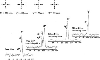
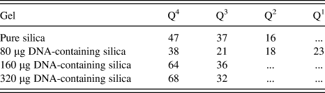
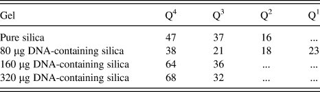
In Fig. 1, 29Si NMR spectra are shown for pure silica and DNA-containing silica gels. The silica networks for all gels were different, and the presence of DNA affected the Si–O–Si network formation efficiency. The molecular structures of Qn species and associated chemical shifts (δ, in ppm) are also shown in Fig. 1, and Table II summarizes the relative percentages of the Q4, Q3, Q2, and Q1 species for each sample. The sum of Q1 (−84 ppm), Q2 (−90 ppm), and Q3 (−100 ppm) species designates the amount of Si–O–X groups (X: hydrogen or alkyl), whereas Q4 species designates Si–O–Si bonding.Reference Glaser, Wilkes and Bronnimann35 In other words, it shows the degree of completion in sol–gel reactions. In pure silica, the level of Si–O–Si network formation was around 47%, and Si atoms are either in Q4, Q3, or Q2 coordination. Upon addition of 80 μg DNA, the amount of Q4 species in the resultant silica decreased to 38%, and there was remarkable amount of Si atoms in Q3, Q2, and Q1 coordination. Mostly, Q4 together with some Q3 coordinations were present upon further increase in the DNA amount in the sol. The Si–O–Si network formation was also more complete for more DNA-containing gels; Q4species increased to 64% in the presence of 160 μg of DNA and to 68% with 320 μg DNA addition. It is also noteworthy to point out that no additional NMR peak besides Si–O and Si–O–X bonding was observed for the DNA-containing silica gels, revealing that the DNA molecules were not chemically associated with the Si–O–Si network.
29Si NMR spectra of pure and DNA-containing silica gels and the molecular structures of expected chemical species for sol–gel-derived silica and associated chemical shifts (δ, in ppm) for different Si coordinations.

Relative percentages of Qn species as determined from the 29Si NMR data for pure silica and DNA-silica gels.
C. Microstructural characterization (BET analyses)
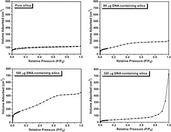
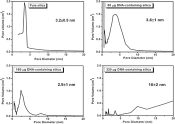
Figure 2 shows the nitrogen gas adsorption isotherms for pure silica and DNA-containing silica gels. The average pore size and pore distribution are presented in Fig. 3. The adsorption isotherm data for pure silica revealed a type I isotherm typical for microporous adsorbents.Reference Brunauer, Deming, Deming and Teller36, Reference Storck, Bretinger and Maier37 For pure silica gel, the average pore size was determined to be 3.2 ± 0.5 nm. The isotherm of 80 μg DNA-containing silica resembles that for pure silica. However, there is a slight increase in the total pore volume and average pore size. Further increase in DNA amount leads to a mesoporous microstructure as indicated by the characteristic type IV adsorption isotherm of 160 μg DNA-containing silica. This change is also evident from the increase in adsorbate volume in high pressure region (P/P 0 is higher than 0.3 P/P 0) of the representative isotherm. The average pore size was determined as 3.6 ± 1 nm and 2.9 ± 1 nm for 80 and 160 μg DNA-containing silica gels, respectively. However, within the limitations of the BET technique, these differences in the absolute average pore sizes can be considered trivial. This is because the pore size determinations according to BJH model are not reliable when the pore size reaches to limiting value at around 2.5 nm.Reference Groena, Peffera Louk and Pérez-Ramı́rez38 However, both adsorption isotherms and average pore size distribution graphs suggest that the scale of porosity is changing from microporous to a mixed character of micro- and mesoporous upon increasing amount of DNA. The increase in the scale of pores is more remarkable upon further increase in the DNA reaching to 320 μg. The average pore size was determined to be around 10 ± 2 nm. The adsorption isotherm for 320 μg DNA-containing silica is distinctly different than the rest of the isotherms. There is an abrupt increase in the gas adsorbate amount at P/P 0 around 0.9 indicating presence of bigger pores. The pore size distribution curve also implies the presence of pores bigger than 20 nm. To sum up, DNA addition changes the physical properties—here, porosity—of the resulting silica gels, where the average pore size of the silica increases with DNA content.
Gas adsorption isotherms for pure silica and DNA-containing silica gels.

Pore size distributions and average pores sizes for pure silica and DNA-containing silica gels.

D. DNA encapsulation efficiency
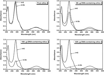
DNA encapsulation efficiency of the silica matrices is illustrated by the UV–Vis spectra data obtained from EtBr intercalation tests shown in Fig. 4. The encapsulation efficiency can also be considered as DNA holding capacity, as it will be further explained in Sec. IV. In Fig. 4, UV–Vis absorption spectra of supernatant EtBr solutions, which were exposed to pure or DNA-containing silica powders for 3 h, are shown. To comment on the extent of EtBr adsorption to the silica powders, the initial (t= 0) absorption spectra of EtBr solutions (at a concentrations of 10 μg/mL) are also shown. This specific concentration corresponds to initial composition of the EtBr solution, i.e., before adding the silica powders.
UV–Vis absorption spectra of supernatant EtBr solutions exposed to pure silica or to silica gels containing different amounts of DNA.

The UV–Vis spectra of EtBr solution in contact with pure silica for 3 h are almost the same as the original spectra of EtBr solution at t = 0. This implies that no EtBr uptake from the initial solution took place, as there is no DNA present in this sample. This finding also excludes any nonspecific adsorption of EtBr to silica powders, which can be considered as a concern for the validity of the EtBr intercalation tests. For the DNA-containing silica powders, on the other hand, a decrease in absorption intensities was observed, suggesting a certain uptake of EtBr molecules by the powders. EtBr permeation into gel was also obvious from the visual evidence as the initial orange color of the EtBr solution faded and became lighter after 3 h of incubation. Meanwhile, white silica powders immersed in the EtBr solution turned to orange. In fact, this color change for EtBr solution was quantified by UV–Vis measurements and was related to the DNA amount present in the gel. As shown in Fig. 5, in the cases of 80 and 160 μg DNA-containing silica gels, after 3 h immersion, intensities of absorption at 285 nm (A 285) decreased to approximately 2.20 and 1.00 from an initial value of 3.40. The amount of encapsulated DNA present in the gel affected the adsorption of EtBr. These decreases correlate with the quantities of DNA present in the silica gels. The A 285 absorption value dropped to half, when the DNA amount was doubled, indicating a 2-fold increase in EtBr adsorbtion by the DNA–silica gel, as it would be expected. However, when DNA incorporation was further increased to 320 μg, the A 285 decreased to 0.22. When 80 and 320 μg DNA-containing gels were compared, there is almost a 10-fold increase in the EtBr intercalation efficiency even though there is only a 4-fold increase in the DNA amount. This finding suggests that there is no absolute correlation between the amount of DNA initially present in sol formulation and DNA that had been encapsulated. The point will be further clarified and reason for such a difference will be postulated in the Sec. IV.
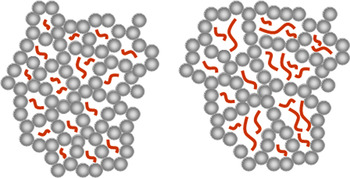
Representative schematics for encapsulation of DNA in microporous (left) and mesoporous (right) silica host, illustrating the differences in DNA holding capacity due to microstructural variations.

IV. DISCUSSION
The presence of DNA greatly influences the gelation chemistry and the Si–O–Si network formation extent in the sol–gel-derived silica gels. This chemistry-dependent factor in turn determines the size and distribution of the porosity and DNA encapsulation efficiency.
One obvious effect of DNA presence was on gelation time, and the gelation of silica occurred relatively faster in DNA-containing sol formulations as compared to that of pure silica. This is because of the changes in the solution pH due to presence of DNA in buffer solution altering the catalysis conditions. In this study, the silica gels were formed from aqueous solutions of TEOS:EtOH for which the hydrolysis was initiated by an acid catalyst (HCl). So, for the pure silica formulation hydrolysis and subsequent condensation, reactions were extensively occurred in the acidic pH conditions. The final pH value measured before gelation (after 6 h) for pure silica was around 3.5. On the other hand, for the silica gels with DNA, where the DNA-containing basic buffer solutions were added subsequently into the partially hydrolyzed gels, the final pH values were in the alkaline range, in the pH range approximately 7–8.8. Additionally, the final pH value was higher when starting formulation contained more DNA-containing buffer solution. The gelation times for the silica gels with DNA were only a couple of minutes, remarkably shorter than those for pure silica. In this respect, gelation of DNA-containing gels occurred by a two-step acid/base catalysis mechanism. The pH dependence of gelation is well known for the silica sols, resulting from the difference in relative hydrolysis rates and condensation reactions at different pH values.Reference Brinker, Keefer, Schaefer and Ashley39–Reference Jones and Fischbach41 During gelation at acidic pH values, achieved during initial times of gelation upon addition of HCl solution, the hydrolysis reaction rate is relatively high. However, at acidic pH values, the condensation reaction is extremely sluggish, leading to long gelation times (6 h) for acid-catalyzed silica formulation. The relative rates of hydrolysis and condensation reactions are also different for alkaline pH values in the range of pH = 7–10 but in an opposite manner.Reference Groena, Peffera Louk and Pérez-Ramı́rez38–Reference Brinker, Keefer, Schaefer, Assink, Kay and Ashley40 At basic pH values, the condensation reaction is fast, but as the hydrolysis rates are slow, complete gelation would still occur at much longer times. For a two-step acid-/base-catalyzed sol as achieved for DNA-containing sol, therefore, an optimum overall gelation rate can be attained. Initially, when the solution pH is reduced to acidic range by addition of the HCl solution, hydrolysis reaction starts immediately and efficiently, where condensation reaction still proceeds slowly. Upon subsequent addition of DNA-containing buffer solution, the pH values increase toward pH > 7, where condensation is also favored. In this way, much shorter gelation times can be achieved for DNA–silica sols. This is due to basic nature of DNA molecules containing hydroxyl (–OH) binding active sites on their sugar–phosphate backbone and due to presence of nitrogenous base pairs between the strands forming the typical helix configuration of DNA molecules.
Accompanying NMR, analyses also support the idea that the presence of DNA promotes gelation and enhances Si–O–Si network completion. This effect is more clear at higher DNA concentrations as shown by the NMR data in Fig. 1. NMR results are somewhat in agreement with the observations on gelation time. Overall, DNA addition drives the gelation conditions to the alkaline pH regions leading to enhanced condensation and gelation. This two-step catalysis approach may also bring about some additional advantages toward effective DNA encapsulation beyond attaining faster gelation. Most biological molecules function in solutions that are at around physiological pH (7.4) values and their stabilities are sensitive to ionic conditions of their environment. During encapsulation by a two-step acid/base catalysis, the pH values of the gel do not reach to extremities of very low or high pH, and gelation takes place at moderate pH values for most of the time. A final sol pH value of 7.5–8.8 for DNA-containing formulations implies the same, the gelation occurring and finalizing in a moderate pH range, potentially complementary for encapsulation of DNA without any structural degradation or functional lost.
The gas adsorption tests results revealed that pH-controlled gelation mechanism also affected the size and scale of pores of the silica gels. Acid-catalyzed pure silica was microporous. The DNA-containing silica showed a gradual translation from microporosity to mesoporosity and further to macroporosity with increasing DNA amount, as it would be typically expected for silica networks formed by base catalysis. Therefore, the gels catalyzed with low DNA concentration were condensed similar to acid-catalyzed gels, and they exhibited a condensed network with much smaller pores. The small porosity indicates that condensation of silica network occurred by entanglement of linear or random branched sols, which is characteristic during gelation at low acidic conditions.Reference Brinker and Scherer42, Reference Ying and Benzinger43 At high amount of DNA presence, condensation is similar to those for base-catalyzed gels where three-dimensional network condensation occurs by integration of colloidal silicates leading to a more open, i.e., a mesoporous structure.
NMR studies also give some insights about the DNA encapsulation mechanism. According to the 29Si NMR data, the silica network for pure gel and DNA-containing gels was similar and was mostly composed of Q4 and Q3 sites. The absence of any different configurations for Si besides Q4 and Q3coordination for DNA-encapsulated gels excludes the possibility of attachment of DNA and Si–O–Si matrix. For DNA, the absence of any chemical interaction with the surrounding medium is important in maintaining the stability and reactivity. DNA encapsulation occurs by physical entrapment/confinement within the pores of silica.
Some critical findings in regard to DNA encapsulation mechanism were obtained through the EtBr adsorption test. First of all, this test works as a semiquantitative analytical tool to assess presence and encapsulation of DNA in silica matrices. Additionally, it also shows that the encapsulated DNA molecules were preserved in their native state, as the EtBr intercalation requires double-stranded DNA molecules. This means that the chemical and the structural changes in hosting environment during formation of silica were not detrimental on the form and state of the DNA molecules. Finally, EtBr absorption test also shows that there is no direct absolute correlation between the amount of DNA in the initial sol formulation and DNA that had been eventually encapsulated. The encapsulation efficiency (or DNA holding capacity) for the sol–gel-derived silica strongly depends on the scale and the size of the porosity attained. In other words, the limit for total DNA that can be encapsulated is determined by the structural characteristic, mainly by the average pore size governed by the gelation chemistry. The pore size-dependent encapsulation mechanism can be explained with the schematic in Fig. 5. This figure shows the difference in DNA holding capability of microporous and mesoporous silica hosts. An efficient encapsulation requires a size match between the average pore size and the length of the DNA fragments. The DNA used in this study was commercially supplied salmon sperm DNA comprising DNA molecules of variable lengths smaller than 5000 base pairs. The spacing between neighboring base pairs in a DNA helix is around 0.35 nm. It is useful to recall that the typical pore size for the sol–gel-derived silica matrices are on the order of 2–20 nm. This means that these gels can accommodate the small DNA fragments with 6–60 base pairs. At such “pore size match” requirement, if the average pore size is relatively big, then all the DNA fragments with a size similar or smaller than average pore size will be effectively encapsulated. This will correspond to encapsulation of DNA fragments of relatively broader size distribution. The net result is that more DNA molecules will remain trapped in such open silica with larger pores. If the pore size is too small, then DNA molecules will still encapsulated selectively according to their sizes. However, this time there would be limited amount of DNA fragments with appropriate sizes matching with the pore size. This proposed description also explains the anomalous EtBr uptake by the macroporous gel prepared with highest DNA amount, i.e., for the gel formulation with 320 μg of DNA.
Besides the physical confinement of DNA molecules within the pores of sol–gel-derived silica, another critical factor for DNA–silica hybrids is the charge interactions with the silica host and DNA. In fact, such interactions are the basis for design strategies for synthetic gene delivery systems, and polycations enabling gene complexation via electrostatic interaction with DNA have been extensively investigated as carriers in gene delivery for achieving effective gene transfection. In the case of sol–gel-derived DNA–silica hybrids, however, considering the isoelectric point of silica (pH = 2), association or complexation of negatively charged DNA molecules with silica surfaces can be excluded. The condensation and silica network development at around neutral conditions mediate accumulation of anionic charges on the silica network and on the pore surfaces.
In this regard, the pathway for encapsulation of DNA is somewhat analogous to encapsulation of proteins in sol–gel-derived silicates. Encapsulated proteins typically reside in the pores that are of a similar size to the proteins, resulting from the templating of silica around protein. Such a size match is important to retain native stability and reactivity of the protein upon encapsulation. Similarly, DNA itself directs the formation and evolution of the silica network, and size of the DNA molecules determines the pore size and the encapsulation efficiency. These DNA-doped silica hosts may provide promising matrices for the development of biosensors, bioreactors, and bioassay platforms. Such solid biocomposites encapsulating DNA in a chemically inert and durable host can be also useful for environmental cleanup applicationsReference Satoh, Fugetsu, Nomizu and Nishi32, Reference Yamada and Aono44, Reference Yamada, Kato, Nomizu, Sakairi, Ohkawa, Yamamoto and Nishi45 for scavenging small harmful organic pollutants or ionic species, which exhibit an intrinsic and highly selective affinity to entrapped DNA.
V. CONCLUSIONS
In this study, the natural double-stranded DNA molecules were encapsulated in silica at ambient temperature and mild chemical conditions. During this encapsulation, the DNA molecules maintained their native form without degradation and did not form a chemical bond with the porous silica host. The DNA encapsulation efficiency was mainly controlled by the size and scale of the porosity of the silica, which was directed by the solution pH during gelation. The gelation pH was regulated by the DNA amount, leading to an alkaline condensation condition and inducing formation of meso-/macroporous silica networks for the gels with higher DNA concentrations. It was found that an effective encapsulation requires a size match between the DNA molecules and hosting pores, and only the small DNA strands with comparable length with the pore size were amenable for encapsulation.
ACKNOWLEDGMENTS
The authors (D.K. and C.D.) acknowledge ÖYP Program of Middle East Technical University and METU-BAP (Grant No. 2007-03-08-05) for the financial support. We would like to thank Meral Yücel and Ayşe Eda Aksoy for valuable advices and discussions.