Case report
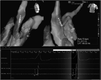
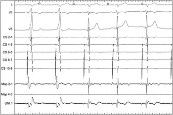
A 17-year-old boy was referred because of recurrent episodes of paroxysmal tachycardia, despite antiarrhythmic drug treatment. He had a structurally normal heart. The electrocardiogram showed ventricular pre-excitation, with positive delta waves in leads II, III, and aVF, negative delta wave in leads aVR and aVL, and positive delta waves from V1 to V6, suggesting an accessory pathway location in the left anterolateral region. An electrophysiological study was conducted after written informed consent. Two quadripolar catheters were placed at the right ventricular apex and His bundle via the femoral vein, and a decapolar catheter was inserted in the coronary sinus via the right jugular vein. The electrophysiological study revealed a retrograde concentric conduction during right ventricular pacing, without decremental property, with a ventriculo-atrial refractory period of 300 ms. After intravenous 12-mg bolus injection of adenosine, ventriculo-atrial conduction was blocked for few seconds, and then remained unchanged, with the earliest atrial activation recorded at the His-bundle region. In addition, antegrade conduction over a left anterolateral accessory pathway was noted during atrial pacing manoeuvres. The atrial pacing protocol did not induce any tachyarrhythmia, with or without isoproterenol. A standard (4 mm) 7-Fr radiofrequency catheter (Therapy; St Jude Medical, Sylmar, California, United States of America) was used to map the tricuspid annulus, through a femoral access, and the mitral annulus, via a transseptal approach, during sinus rhythm, by using a three-dimensional mapping system (Ensite NavX; St Jude Medical). The electrophysiological study and the three-dimensional propagation map showed the shortest atrioventricular interval with a QS pattern in the unipolar electrograms recorded on a left anterior position. However, radiofrequency applications at this location failed to eliminate the ventricular pre-excitation. Then, the ablating catheter was inserted through the right femoral artery and advanced retrogradely into the left ventricle. Electrophysiological and three-dimensional mapping was performed at the mitral annulus, to explore the ventricular insertion site, subvalvular mitroaortic curtain, and sinuses of Valsalva (above the cusps), respectively. An aortic root angiogram and coronarography were obtained to delineate the aortic root anatomy and origin of the coronary arteries. Heparin was administered to maintain activated clotting time above 250 seconds. The earliest site of ventricular activation during normal sinus rhythm was in the left sinus of Valsalva at the junction of the left coronary cusp with the non-coronary cusp (20 ms before the onset of delta wave), where a small sharp spike preceded ventricular electrogram (Figs 1 and 2). Ablation at this site using 15 W, increasing to 3 W for 60 seconds, with a tip temperature <50°C, eliminated the action potential conduction, with immediate normalisation of electrocardiogram within 10 seconds of radiofrequency application. Subsequent programmed atrial and ventricular stimulation demonstrated only atrioventricular nodal conduction, without induction of tachycardia, even during infusion of isoproterenol. Post-procedure coronary angiography showed normal coronary arteries. At 6-month follow-up, he is asymptomatic with no therapy and his electrocardiogram continues to show no pre-excitation.
Three-dimensional propagation map (Ensite Navx; St Jude Medical) with shortest atrioventricular interval at the junction of the left coronary cusp with the non-coronary cusp.

Ablation site electrogram and normalisation of electrocardiogram after radiofrequency application.

Discussion
A very limited number of cases of successful ablation of an accessory pathway localised to the left anterior region from the non-coronary sinus of Valsalva have been reported.Reference Tada, Naito, Taniguchi and Nogami 1 , Reference Calo, Sciarra, De Ruvo, Franceschetti, Pellegrini and Lioy 2 The structural relationships between the right and left ventricular outflow tracts and the aortic root are crucial for a safe and effective ablation in this location. Because the aortic valve is “wedged” between the mitral and tricuspid valves, it is possible to ablate the left anterior and anteroseptal pathways from the Valsalva sinuses.Reference Tada, Naito, Taniguchi and Nogami 1 – Reference Suleiman, Powell, Munger and Asirvatham 3 The mitroaortic junction is not simply formed by two fibrous annuli, but shares a common structure, the subaortic curtain, that simultaneously supports two of the aortic cusps (left coronary and non-coronary) and the anterior leaflet of the mitral valve. In this site, the muscle fibres of the left atrium are continuous with those of the anterior mitral valve leaflet. Therefore, if an accessory pathway connecting the left atrial and ventricular myocardium is present in this region it can be successfully ablated between the aortic non-coronary and left coronary cusps. However, the aortic cusp ablation includes the risk of aortic cusp rupture, aortic valve damage, and coronary artery occlusion and complete heart block.Reference Park, Wi and Joung 4 Coronary angiography of the aortic root should be performed before energy application to visualise the anatomy of the aortic root, to optimise the distance of the ablation catheter from the coronary ostia, and to rule out coronary anomalies. It has been suggested that coronary ostia should be at least 1 cm away from the ablation site during aortic cusp ablation with a temperature <55°C.Reference Kilicaslan, Uz, Isilak and Tokatli 5 In this report, the accessory pathway was successfully eliminated at the junction of the left coronary cusp with the non-coronary cusp and might have been localised on the left side anterosuperior to the membranous septum within the aortomitral continuity. In conclusion, a careful radiofrequency catheter ablation approach through the cusps of the aortic valve appears to be an effective alternative technique to treat left anterior accessory pathways, after failure of traditional retrograde or transseptal approaches.
Acknowledgement
The authors would like to acknowledge the support of CPL Ricardo Pimenta during the electrophysiology procedure.
Financial Support
This research received no specific grant from any funding agency, commercial, or not-for-profit sectors.
Conflicts of Interest
None.
Ethical Standards
The authors assert that all procedures contributing to this work comply with the ethical standards of the relevant national guidelines on human experimentation and with the Helsinki Declaration of 1975, as revised in 2008, and has been approved by the institutional ethical committee.