Prostate cancer is a significant health problem worldwide. It is the most common visceral cancer among men in North America, and second only to lung cancer as a cause of cancer-related death in men. The development of prostate cancer in humans represents a multistep process, thought to arise from an accumulation of mutations in tumour-related genes that lead from transformation of benign prostatic epithelium to prostatic intraepithelial neoplasia (PIN), with progression to locally invasive disease and ultimately metastasis (Ref. Reference Abate-Shen and Shen1).
As our understanding of the molecular pathways involved in prostate carcinogenesis has grown, novel models have been developed to satisfy our desire for increased understanding, to explore treatment regimes and ultimately, for transfer to improvements in patient care. In the past, we have achieved this with xenograft techniques involving primary or genetically altered cell lines derived from localised and metastatic diseased tissue. Although mice do not spontaneously develop prostate cancer, the murine models of human cancer have provided new tools for the investigation of prostate carcinogenesis. In many other diseases, murine models have provided invaluable information regarding pathogenesis and novel disease approaches (Ref. Reference Guilbault2). In cystic fibrosis for example, the variety of models reflect different aspects of the heterogenicity of phenotypes observed in humans: intestinal, pancreatic, hepatobilary and lung disease, as well as those reflecting the increased susceptibility to respiratory infections.
Developing transgenic prostate cancer models has proved similarly challenging, because no one model appears to represent the entire continuum of the disease. As the second generation of these models is further developed, the expectation has risen that they will continue to be refined in the future to recreate the molecular, physiological and clinicopathological features of human prostate cancer.
Animal models of prostate cancer
Rats were utilised as one of the first models of prostate cancer, being one of the few species that develop this disease spontaneously. The best defined model is the Dunning rat model, which shows well-differentiated, nonmetastatic and slow-growing tumours (Ref. Reference Dunning3). It mimics human disease in that it is initially androgen dependent, eventually becoming androgen independent. Refinements to this model have produced rats that develop highly metastatic tumours, which spread to lymph nodes and lung (Ref. Reference Isaacs4).
Prostate cancer also occurs spontaneously in canines. The tumours are different from those in humans in that they lack a functional androgen receptor and thus are androgen independent. Dogs develop bone metastasis, which mimics advanced human disease and thus allows trials of bone-directed regimes (Ref. Reference Aquilina5). However, their disadvantages are numerous and primarily include difficulties with cost and care (Ref. Reference Navone6).
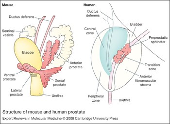
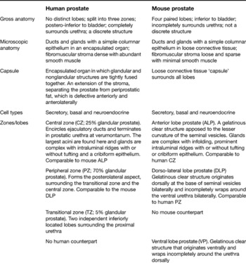
The transgenic mouse model has provided an attractive model for researchers. However, the prostates of the human and mouse have key similarities as well as marked differences, which must be considered when one attempts to make conclusions across species (Table 1 and Fig. 1). The peripheral zone is where 70% of human prostate cancer arises and in the mouse, the comparable area is the dorsolateral prostate. However, this identification is based on descriptive information rather than molecular characteristics (Ref. Reference Shappell7).
Structure of mouse and human prostate. The peripheral zone is where 70% of human prostate cells arise. In the mouse the comparable area is the dorsolateral prostate (see also Table 1).

Anatomical and structural comparison of human and mouse prostate

Transgenic murine models do have their flaws. A lot of time and resources must be invested and generation and maintenance of the mice is often expensive. When investigating new pathways or genetic defects, mice may die before prostate cancer actually develops, if indeed it does develop. The genetic background is known to have a modifying influence on the formation and/or progression of prostate cancer (Ref. Reference Kim8). Meticulous care must therefore be taken when initially describing the genetic background and breeding regimes involved in creating new transgenic models.
First-generation mouse models
The first murine models utilised prostate-specific expression of viral oncogenes. They have been useful in analysing disease progression and resistance to traditional androgen-deprivation therapy. However, the viral antigens used are not usually associated with cancer and their effects are pleiotrophic. Also, the prostates developed extensive neuroendocrine differentiation, which is relatively uncommon in humans (Refs Reference Garabedian, Humphrey and Gordon9, Reference Masumori10, Reference Abate-Shen and Shen11, Reference Powell12). The transgenic adenocarcinoma of the mouse prostate (TRAMP) model was one of the first effective models developed. This model uses the short probasin (−426/+ 28 bp PB) promoter to express the SV40 early genes (T/t antigen) specifically targeted to the terminally differentiated tall columnar epithelial cells of the prostate (Refs Reference Greenberg13, Reference Gingrich14). The males develop locally invasive disease that becomes metastatic. High-grade PIN is evident by 12 weeks, with metastasis (predominantly to lung and lymph nodes) by 30 weeks.
A 12 kb fragment of the prostate-specific probasin promoter was isolated [long probasin (LPB)] and it elicited a higher level of expression in the mouse prostate (luminal epithelial cells), which was developmentally and differentially regulated by androgen. This promoter was used to create the LPB-Tag (LADY) model when linked to the large-T antigen of SV40 (with deletion of small-t antigen expression) (Ref. Reference Kasper15). The major advantage of this model is that expression is high with a less-aggressive disease pathology, progressing to invasive carcinoma with neuroendocrine differentiation, but with no metastasis. Metastatic potential can be conferred on nonmetastatic LADY tumours by expression of the LPB-hepsin transgene (Ref. Reference Klezovitch16). Hepsin is a cell-surface serine protease that is markedly upregulated in human prostate cancer.
A transgenic mouse model was developed that utilised nucleotides –6500 to +35 of the mouse gene encoding cryptdin-2 (Defcr2) to direct expression of SV40 large-T antigen to a subset of neuroendocrine cells in all lobes of the murine prostate (Ref. Reference Garabedian, Humphrey and Gordon9). These mice develop PIN and progress to invasive carcinoma with extensive neuroendocrine differentiation, which finally results in metastasis (to lymph nodes, liver, lung and bone) by 6 months of age.
Modelling tumourigenesis in mice
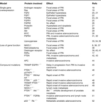
Given the inherent flaws with the first-generation models, researchers were motivated to create a model that recapitulated better the genetic events that occur in human prostate cancer. Strong prostate-specific promoters such as probasin and PSA were developed to drive target gene expression, which led to the so-called ‘second-generation’ murine models (Ref. Reference Roy-Burman17). Many target genes have been manipulated in either gain- or loss-of-function strategies, including cell-cycle regulators, steroid hormone receptors, oncogenes, tumour-suppressor genes, homeobox genes, growth factors and their receptors and pro- and anti-apoptotic genes (Table 2).
Summary of mouse models of prostate cancer

Abbreviations: APC, adenomatous polyposis coli; EGFR, epithelial growth factor receptor; FGF, fibroblast growth factor; FGFR, FGF receptor; KIP1, kinase inhibitor protein 1; PIN, prostate intraepithelial neoplasia; PSP94, prostate secretory protein 94; PTEN, phosphatase and tensin homologue deleted on chromosome 10; Rb, retinoblastoma; RXR, retinoid X receptor; SKP, S-phase kinase-associated protein; TRAMP, transgenic mouse model of prostate cancer.
Promoter selection
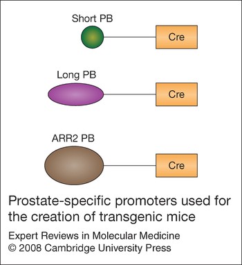
Promoter selection is critical for targeting the transgene to the tissue of interest. Several generations of the rat probasin (PB) promoter have been characterised (Fig. 2). The initial PB promoter fragment was characterised in human prostate cell lines PC3, DU145 and LNCaP, utilising either the chloramphenicol acetyl transferase (CAT) or the luciferase reporter systems. Two distinct androgen receptor (AR) binding sites – ARBS1 and ARBS2 – are required for maximal induction of CAT gene expression in response to androgen treatment (Ref. Reference Rennie53). Both binding sites are required for activation of the AR-signalling pathway, therefore the region encompassing both elements was named the androgen-responsive region (ARR) (Ref. Reference Kasper54). The PB promoter was shown to activate reporter gene expression in a prostate-cell-specific manner in numerous nonprostatic cells (Ref. Reference Brookes55). Following this, the LPB promoter was isolated and was found to elicit significantly higher levels of CAT gene expression in transgenic mice – specifically in prostatic luminal epithelial cells (Ref. Reference Yan56).
Prostate-specific promoters used for the creation of transgenic mice. Short PB, initial probasin promoter. Long PB, 12 kb of the initial probasin promoter, which elicits significantly higher levels of gene expression specifically in prostatic luminal epithelial cells. ARR2 PB, composite ARR2 probasin promoter that was developed by linking two androgen-responsive regions (ARRs) to the endogenous probasin promoter, thus directing high levels of transgene expression.

The composite ARR2 PB promoter was developed by linking two ARRs to the endogenous PB promoter. The ARR2 PB promoter was as efficient in directing high levels of transgene expression as the LPB promoter, and targeted prostate-specific expression (Ref. Reference Zhang57). Subsequently the PB-Cre4 model was developed by targeting ARR2 PB linked to the Cre recombinase gene to the prostatic epithelium (Ref. Reference Wu58).
The PSA promoter has also been linked to the Cre recombinase gene to initiate recombination of the floxed alleles, specifically in the prostate (Ref. Reference Abdulkadir36). All other ‘prostate-specific’ promoters reported so far appear to target the transgene to multiple tissues. These include the C3(1) promoter (Ref. Reference Shibata59), the fetal G-globin promoter, the gp91-phox promoter (Ref. Reference Skalnik60), the cryptdin 2 promoter (Ref. Reference Garabedian, Humphrey and Gordon9) and the PSP94 promoter (Ref. Reference Gabril32). Careful promoter selection is essential to allow levels of transcription that are high enough to create the desired phenotype, as well as influencing the rate of tumour development.
Single-gene overexpression models
The androgen receptor (AR) has an essential role in the development of the prostate; it is expressed at high levels in the secretory epithelial cells of the normal prostate and in a subset of stromal smooth muscle cells (Refs Reference Cussenot61, Reference Leav62). It is involved in all stages of prostate cancer, including androgen-independent disease (Refs Reference Feldman and Feldman63, Reference Chen64). Androgen deprivation is currently the gold standard treatment for patients with advanced prostate cancer (Refs Reference Feldman and Feldman63, Reference Chen64).
The fibroblast growth factor (FGF) family of heparin-binding growth factors are intracellular signalling molecules of which at least 23 different members (FGF1–FGF23) have been identified. Most are secreted, and their effects are mediated by a complex system of FGR receptor (FGFR) tyrosine kinases through a combination of autocrine and paracrine mechanisms (Ref. Reference McKeehan, Wang and Kan65). They play an important role in normal prostate development and tumourigenesis (Ref. Reference Basilico and Moscatelli66). Prostatic epithelial and stromal cells express a variety of FGF ligands and FGFRs to transduce signals required for biological processes, such as branching, morphogenesis, differentiation and survival (Ref. Reference Cunha67). In prostate cancer, some FGFs are overexpressed (Refs Reference Polnaszek21, Reference Heer68, Reference Ozen69, Reference Ropiquet70) whereas others are downregulated (Ref. Reference Foster, Kaplan and Greenberg71).
Transgenic mice expressing FGF2 using the PB promoter have been produced (Ref. Reference Konno-Takahashi20). The infertile mice demonstrated simple epithelial hyperplasia in the dorsal lobe of the prostate only. However, FGF2 does have a role in tumour progression, because deletion of one or both copies of Fgf2 in the TRAMP model resulted in increased survival with reduced metastasis, and inhibition of progression to the poorly differentiated phenotype in prostate cancer (Ref. Reference Polnaszek21). There is strong evidence that FGF7 (keratinocyte growth factor) is a mediator of stromal–epithelial interactions in prostate development (Refs Reference Rubin72, Reference Sugimura73). It is postulated that a change in the spatial pattern of Fgf7 expression induces the prostate cancer cells to upregulate Fgf7 expression (Ref. Reference McGarvey and Stearns74). This was supported by findings in the SV40 large-T antigen models, where FGF was detected in the epithelial compartment during the progression of prostate cancer (Refs Reference Foster75, Reference Huss76). Murine FGF7 transgene expression has also been targeted directly to the prostatic epithelium using the PB promoter (Ref. Reference Foster22). After 12 months of age, these mice mostly developed prostatic epithelial hyperplasia, which did not progress to dysplasia or tumour. This suggests that the role of Fgf7 misexpression in the prostate may contribute to the induction of proliferation of epithelial cells; however, other events are required to promote dysplasia, invasive prostate cancer and metastasis.
Overexpression of FGF8b is associated with increased mortality and persists in androgen-independent disease (Ref. Reference Dorkin77). The ARR2 PB promoter was used to overexpress FGF8b in the prostate (Ref. Reference Song24). During the first 14 months, all animals exhibited multifocal epithelial hyperplasia; 35% also had areas of low-grade PIN. This profile changed in subsequent months (15–24 months) to a higher incidence of low-grade PIN (66%) along with high-grade PIN lesions (51%). However, it was not sufficient to promote localised invasive carcinoma or metastasis. The transgenic mice were generated by the ARR2PB-driven expression of ligand-inducible FGFR1 or FGFR2. Activation of FGFR1 showed hyperplasia followed by PIN lesions (Ref. Reference Freeman25), whereas activation of FGFR2 did not elicit any apparent changes in the prostate (Ref. Reference Jin26). The effect of FGFR1 is highly penetrant, because all the transgenic mice treated with inducer showed PIN in almost every acinus at 12 weeks. Interestingly, the induced hyperplasia is reversible until intraductal vascularisation, after which these hyperplastic cells become independent of FGFR1 signalling for their maintenance.
The FGFR2iiib receptor at the prostate epithelium has also been targeted using the PB promoter (Ref. Reference Foster22). The size of the prostate in these mutants was smaller than that in control mice, but no other gross changes were observed. Although none of the models based on dysregulation of the FGFR or their ligands resulted in invasive carcinoma, this does rule out the fact that these molecules have an important role in the late stage of tumour progression.
Activity of Akt (a serine/threonine kinase involved in cell growth and survival) is positively regulated by phosphoinositide 3-kinase (PI3K) (Ref. Reference Franke78). The activated Akt controls numerous cellular pathways that influence proliferation and survival. A wide variety of tumours show loss of the tumour-suppressor gene Pten – a phosphatase that antagonises the PI3K pathway – resulting in increased activity of Akt. This indicates that Akt activation may be a crucial step in tumourigenesis (Ref. Reference Wu79). As a result, a transgenic model that expresses active Akt in the prostate – murine prostate Akt kinase transgenic (MPAKT) – was developed (Ref. Reference Majumder29). The animals developed hyperplasia and dysplasia with severe atypia and PIN. Older MPAKT mice developed protuberant abdomens secondary to bladder outlet obstruction, which warranted their killing. No invasive carcinoma or metastasis was observed, suggesting that activation of Akt alone may not be sufficient for prostate cancer formation.
The Myc oncogene increases cell proliferation and its gene copy number or expression is increased in up to 30% of prostate tumours (Ref. Reference Nesbit, Tersak and Prochownik80). To determine its effects, transgenic mice were developed that express the transgene in different strengths depending on the promoter (Ref. Reference Ellwood-Yen30). The PB promoter was designated a low expressor (Lo-Myc) and the ARR2 PB the high promoter (Hi-Myc), based on the levels of transgene expression. Multifocal proliferative lesions develop in both models, and by 4 weeks, PIN lesions are seen in both lines (appearing at 2 weeks in the Hi-Myc animals). These PIN lesions progress to invasive adenocarcinoma by 3–6 months in the Hi-Myc mice and by 10–12 months in the Lo-Myc mice, confirming the significance of the dosage of the oncogene on disease progression. Complete tumour regression occurred when the mice were castrated at 2 months of age. However, residual tumour was still detected if castration was performed after the tumours were well established at 8 months, suggesting the presence of hormone-resistant cells. Microarray-based expression profiling indicates that expression of the homeobox transcription factor Nkx3.1 was lost and that the serine/threonine kinase PIM1 was upregulated in these tumours (Ref. Reference Ellwood-Yen30). Together, these results demonstrate that multiple events are required for metastasis.
The finding that PSP94 (prostate secretory protein of 94 amino acids) is one of the most abundant proteins secreted from the human prostate, and is prostate specific in humans and mice (Ref. Reference Gabril32), led to the development of a knock-in model using the SV40 large-T antigen to target PSP94 (PSP-KIMAP) (Ref. Reference Duan31). These mice developed PIN from 10 weeks and progressed to invasive adenocarcinoma and metastasis (commonly lymph node, lung and liver). The model has shown differential expression of basic FGF and vascular endothelial growth factor (through mRNA expression) in murine prostate cancer, suggesting a role for antiangiogenic therapies in future models (Ref. Reference Gabril81). Interestingly, PSP94 can also be used as a PSA analogue in mice – to monitor disease progression, androgen ablation treatment and hormone resistance in transgenic lines (Ref. Reference Huizen82).
Studies have shown overexpression of the lipid-metabolising enzyme 15-lipoxygenase-1 (15-LO-1) in high-grade prostate cancer and PIN, with the murine orthologue increased in LADY and TRAMP models (Refs Reference Kelavkar33, Reference Shappell35). A transgenic mouse with overexpression of human 15-LO-1 in the mouse prostate (FLiMP) was also developed. Unlike controls, this mouse developed epithelial hyperplasia and PIN. This suggests a role of 15-LO-1 in prostate cancer tumour initiation and could lead to prevention through dietary strategies (Ref. Reference Kelavkar34).
Single-gene loss-of-function models
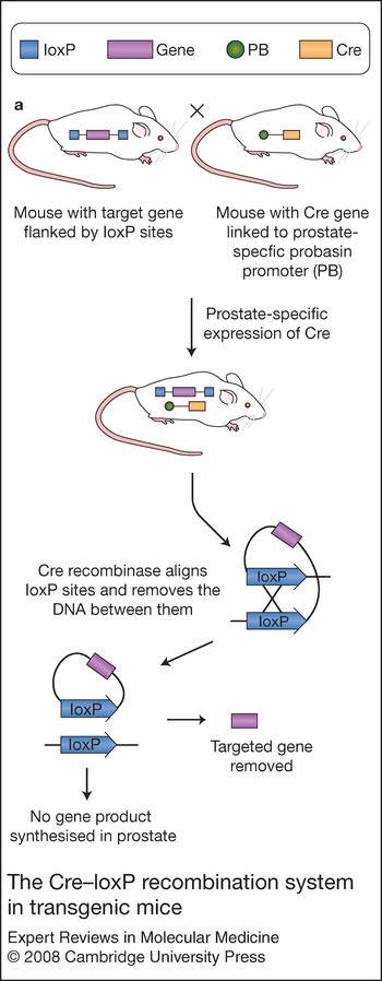
The loss of function of several genes has been implicated in human prostate cancer. There are currently two methods by which disruption of gene activity in the mouse models can be achieved. The traditional method is a genome-wide knockout of the gene in question (Ref. Reference Capecchi83). The major problem with this technique is that it does not allow evaluation of the gene function if it results in embryonic lethality or premature death. Also, because the gene is knocked out in all cell types, it is difficult to prove that the abnormal phenotype did not arise from a developmental defect (Ref. Reference Copp84). The second method is a conditional gene-knockout technique, which aims to overcome the above limitations. The Cre (causes recombination) –loxP (locus of X over P1) recombination system is one of these technologies and allows the study of conditional cell-type- and tissue-specific deletion of genes (Ref. Reference Nagy85) (Fig. 3). The bacterial Cre–loxP1 system is commonly used in mammalian cells (Ref. Reference Sauer and Henderson86). This system relies on a sequence-specific DNA Cre recombinase, which mediates intramolecular recombination and deletion of DNA between the 34 bp loxP sequences. Using gene targeting, loxP-flanked alleles are generated, allowing deletion of genes upon expression of the Cre recombinase. To permit gene deletion in the tissue of interest, tissue-specific Cre transgenes were generated and genes can now be deleted in almost every tissue of the mouse, including the prostate. One prostate-specific Cre system, the PB-Cre4 line has been widely reported (Ref. Reference Wu58). In this system, the nuclear-localised Cre recombinase is expressed from the ARR2 PB promoter. Using the conditional reporter gene R26R and its product, LacZ, it can be conclusively shown that Cre expression is postnatal and prostate-epithelium specific (Refs Reference Wu58, Reference Soriano87). Although Cre recombination has been detected in all lobes of the mouse prostate, there is a significant difference in expression levels between the prostate lobes: at 2 months, staining in the lateral lobe is almost complete (95%), in the ventral lobe is about 50%, but is much lower in the dorsal and anterior lobes (10% and 5%, respectively). However, because recombination is a unidirectional process, the frequency approaches 100% over time (approximately 8 months) (Ref. Reference Powell12).
The Cre–loxP recombination system in transgenic mice. The bacterial Cre–loxP (causes recombination–locus of X over P1) system relies on a sequence-specific DNA Cre recombinase, which mediates intramolecular recombination and deletion of DNA between the 34 bp loxP sequences. LoxP-flanked alleles are generated using gene targeting, allowing deletion of genes upon expression of Cre recombinase. To permit gene deletion in the prostate, tissue-specific probasin (Pb) and Cre transgenes have been generated.

Nkx3.1 displays prostate-specific expression and is deleted in 28% of primary tumours, 34% of hormone refractory tumours and 78% of metastatic tumours (and 20% of PIN lesions) by virtue of allelic deletion of the chromosome 8p21 (Refs Reference Bowen88, Reference Dong89). Nkx3.1 is androgen dependent and largely restricted to the luminal epithelial cells of the prostate (Ref. Reference Bieberich90). When the TRAMP model was assessed for Nkx3.1 activity, it appeared that lobe-specific cancer progression correlates with reduced Nkx3.1 protein and mRNA levels (Ref. Reference Bethel and Bieberich91). Both conventional and conditional knockout transgenic models have been produced (Refs Reference Abdulkadir36, Reference Bhatia-Gaur37, Reference Tanaka92). They suggest that Nkx3.1 regulates branching morphogenesis and the secretory phenotype of the prostatic epithelium. Mice lacking Nkx3.1 from the outset, as well as those with constitutional deletion of Nkx3.1, develop epithelial hyperplasia and PIN (Refs Reference Abdulkadir36, Reference Bhatia-Gaur37).
Pten (phosphatase and tensin homologue deleted on chromosome 10) is a tumour-suppressor gene that is frequently mutated in a variety of spontaneous cancers, including prostate cancer. It is a negative regulator of the PI3K–Akt pathway and mutations are reported in approximately 30% of primary prostate cancers (Ref. Reference Dahia93) and 63% of metastatic tissue samples (Ref. Reference Suzuki94). In an effort to define the role of PTEN, a hypomorphic mouse mutant series was generated by homologous recombination (Ref. Reference Trotman42). This allowed determination of the critical importance of subtle variations in the level of PTEN on prostatic tumourigenesis: the loss of PTEN is critical for prostate cancer initiation and the level of Pten expression is inversely associated with prostate tumourigenesis. Loss of one allele (Pten +/−) is associated with high-grade PIN, with incomplete penetrance after a long latency and no invasive adenocarcinoma (Refs Reference Suzuki94, Reference Di Cristofano95, Reference Podsypanina96, Reference Stambolic97). By contrast, in the hypomorphic mouse model (where the PTEN protein level is reduced to ~30%), invasive prostatic adenocarcinoma develops with incomplete penetrance. The loss of PTEN is embryonically lethal (Ref. Reference Di Cristofano95) and prostate-conditional loss of Pten expression results in invasive carcinoma with lymphovascular invasion within 12 weeks, which progresses to lung metastasis (Ref. Reference Wang43). This conditional PTEN-null mouse represented the first animal model in which deletion of a single endogenous gene leads to metastatic prostate cancer. Androgen ablation on these mice revealed that the cancer cells do respond to treatment, because there is a significant reduction in prostate volume. However, the cancer cells will continue to proliferate even after androgen withdrawal, contributing to hormone-resistant prostate cancer. A weakness of this model is that not all prostate tumours show a reduction in levels of PTEN protein, the reality is that multiple genetic ‘hits’ are required to progress to invasive tumour and metastasis in the human. Also, in humans, it is well known that the majority of metastases occur in bone, causing osteosclerotic lesions (Ref. Reference Mundy98). The counterpart lesions in the PTEN-null mouse have yet to be identified, despite evidence of significant bone-remodelling activity in the mature PTEN-null mice.
Alterations in the Wnt-signalling pathway have also been associated with prostate cancer. More specifically, changes in Apc expression have been identified in both primary and metastatic prostate cancers, including somatic alterations (Refs Reference Brewster, Browne and Brown99, Reference Phillips100) and promoter hypermethylation (Refs Reference Jeronimo101, Reference Yegnasubramanian102). An Apc-null transgenic line was created using the PB Cre-mediated system (Ref. Reference Bruxvoort44). The prostate tumours in these mice have higher levels of β-catenin protein and are highly proliferative, developing invasive adenocarcinoma within 7 months. No metastases were observed in this line. Surgical castration of 6-week-old mice inhibited tumour formation, and castration of mice with more advanced tumours resulted in partial regression. However, tumours returned within 2 months, suggesting that they are capable of growth under conditions of androgen depletion.
Compound mutational models
Carcinogenesis is classically thought to require multiple ‘hits’ of genetic mutations to develop invasive cancer that subsequently metastasises. Thus, transgenic models are being developed that reflect these multiple changes in an attempt to more accurately reflect the human pathophysiology. There are currently few mouse models that incorporate two or more genetic mutations for prostate tumourgenesis and even fewer are strictly prostate specific.
The INK4a/ARF locus encodes two distinct tumour suppressors, p16 INK4a and p19 ARF, which regulate the pRB and p53 pathways, respectively (Ref. Reference Sharpless and DePinho103). Inactivation is associated with a variety of human cancers (Refs Reference Sharpless and DePinho103, Reference Burri104). Transgenic mice with an altered dose of Ink4a/Arf in the Pten +/− background have been created (Ref. Reference You47). PIN develops earlier in the Ink4a/Arf +/−Pten +/− mice than in the Pten +/− controls, and the onset is further accelerated in Ink4a/Arf −/−Pten +/− mice. Progression to invasive carcinoma is not observed in this model.
Transgenic mice that are nullizygous for Pten and p53 in the prostate, develop a locally aggressive, lethal prostate cancer with complete penetrance (Ref. Reference Di Cristofano46). Complete loss of PTEN results in cellular senescence, in response to oncogenic stress, which occurs through a p53-dependent mechanism (Ref. Reference Chen105). This loss of PTEN results in an indolent prostate cancer; however, if p53 is lost as well, there is a dramatic acceleration of prostatic carcinogenesis.
The conventional knockout (nonspecific for prostate) mice for Nkx3-1 +/− and Pten +/− were crossed to examine their combined role in prostate carcinogenesis (Ref. Reference Kim49). The latency of PIN development in the Nkx3.1-insufficiency background was reduced by half in these mice. Loss of the Nkx3.1 protein, as well as loss of the second Pten allele, was seen in the prostate tumours of these mutants. Ageing beyond 1 year results in development of invasive prostate cancer and lymph node metastasis (Ref. Reference Abate-Shen106). This is consistent with other observations that there is a concurrent decrease in Nkx3.1 levels with the conditional inactivation of Pten alleles in the prostate (Ref. Reference Wang43). Recent work suggests that the Akt and Erk MAP-kinase-signalling pathways are activated in androgen-independent lesions of these mice (Ref. Reference Gao107). These prostate epithelial cells develop androgen-independent phenotypes long before they exhibit PIN or cancer phenotypes, suggesting that hormone refractory disease may arise in the early stages of prostate carcinogenesis (Ref. Reference Gao108).
Akt, the major downstream effector of PTEN, is hyperactivated when PTEN is inactivated. Pten +/− mice were crossed with Akt1 −/− mice to examine the ability of Akt deficiency to inhibit tumour development in Pten +/− mice (Ref. Reference Chen50). The deficiency was indeed sufficient to inhibit prostate neoplasia. A transgenic mouse containing a prostate-specific Fgf8b transgene and haploinsufficency in Pten has been developed (Ref. Reference Zhong51). Invasive prostatic adenocarcinoma was evident by 9 months, which ultimately led to lymph node metastasis in older animals. Mice with conditional inactivation of both p53 and Rb1 genes in the prostate epithelium rapidly developed prostatic adenocarcinoma with neuroendocrine differentiation and metastasis to liver, lung and regional lymph nodes (Ref. Reference Zhou52). Again, neither gene in isolation was sufficient for development of invasive carcinoma in the mouse model. The mice are resistant to androgen withdrawal from a relatively early stage and they also have alterations in loci that contained genes homologous to those implicated in human cancer, such as Nfib, L-myc and Nkx3-1.
Clinical implications and applications
The primary objectives of modelling human prostate cancer in mice are: (1) to improve our understanding of carcinogenesis in vivo; and (2) to develop relevant models for the evaluation of new, targeted therapies. The majority of the work carried out so far has utilised the TRAMP model.
Therapeutic applications of transgenic mouse models
Chemotherapy and chemoprevention
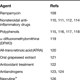
The various therapeutic agents that have been investigated within transgenic mouse models of prostate cancer are highlighted in Table 3. Several of these are explained below in further detail. The bacterial macrolide rapamycin has been shown to inhibit mTOR (mammalian target of rapamycin) kinase, which is a downstream effector of Akt related to tumourigenesis. A cohort of MPAKT mice treated with the mTOR inhibitor RAD001 (Ref. Reference Majumder109) provided evidence that expansion of tumour cells is mTOR dependent, and inhibition led to the complete reversal of the neoplastic phenotype in the prostate through apoptosis of epithelial cells. Nonsteroidal anti-inflammatory drugs have also been shown to reduce tumour progression in TRAMP mice. E-7869 (R-flurbiprofen) reduced the incidence of primary prostatic tumour and metastasis (Ref. Reference Wechter110) and celecoxib (Celebrex) was found to have similar effects, with a reduction in primary tumours and metastasis (Ref. Reference Gupta111). Further evidence shows a dose-dependent response for the preventative effects of celecoxib (Ref. Reference Narayanan112). The exact mechanism of action is uncertain, but is possibly due to changes in cyclooxygenase 2 (COX2) enzyme activity or mRNA levels (Ref. Reference Abdulkadir and Kim113). Recently, OSU03012, an orally bioavailable celecoxib-derived PDK1 inhibitor (which is COX2 inactive), revealed a reduction in the severity of PIN lesions but no significant decrease in the incidence of carcinoma or metastasis (Ref. Reference Sargeant114). PDK1 inhibits activation of Akt and thus appears to be an attractive therapeutic target.
Chemotherapeutic and preventative agents in prostate cancer
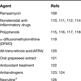
Genistein, the predominant phytoestrogen in soy food, has shown promise as a chemopreventative agent against androgen-dependent and -independent prostate cancers in the TRAMP model (Ref. Reference Wang, Eltoum and Lamartiniere115). The incidence of poorly differentiated prostate tumours decreased by 50% and 35%, in the androgen-dependent and -independent groups, respectively. Another polyphenol, resveratrol, is found in grape products, and in vitro studies have demonstrated its ability to halt carcinogenesis at the initiation, promotion and progression stages (Ref. Reference Chanvitayapongs, Draczynska-Lusiak and Sun116). An almost eightfold reduction in poorly differentiated prostate adenocarcinoma was found when adding resveratrol to the diet of TRAMP mice (Ref. Reference Harper117). These mice demonstrated a decrease in the expression of growth factor IGF1, downregulation of the downstream effector pERK1/2 and an increase in expression of the tumour suppressor ER-β. The role of green tea polyphenols in reducing the incidence of prostate cancer and eliminating metastasis in the TRAMP model has also been investigated (Ref. Reference Gupta118). An increase in the apoptosis of prostate cancer cells was found compared with levels in the water-fed controls. Earlier work reported that ornithine decarboxylase (ODC) activity, as well as protein expression, was markedly higher in prostates of TRAMP mice compared with controls (Ref. Reference Gupta119). As a result, the role of α-difluoromethylornithine (DFMO), an enzyme-activated irreversible inhibitor of ODC, was investigated in prostate cancer. None of the DFMO-fed TRAMP mice had any distant metastases to lymph node or lungs. E-cadherin and α- and β-catenin protein expression levels were restored in DFMO-fed animals. All-trans retinoic acid (ATRA) is able to slow prostate tumour cell proliferation, induce apoptosis, and block the emergence of the neuroendocrine phenotype (Ref. Reference Huss120).
Use of oral grape seed extract in TRAMP mice inhibited prostate cancer growth and progression via a strong suppression of cell-cycle progression, cell proliferation and an increase in apoptosis (Ref. Reference Raina121). This is thought to be due to a decrease in expression of cyclins and cyclin-dependent kinases (CDKs). Direct antioxidant treatment (vitamin E, selenium, lycopene) in the LADY model also reduces the incidence of invasive prostate cancer (~fourfold) and increases survival (Ref. Reference Venkateswaran122).
A beneficial response has also been noted with the antiandrogen flutamide and the tamoxifen derivative toremifene in the development of prostate cancer in the TRAMP model (Refs Reference Raghow123, Reference Raghow124). Tumours from high-dose flutamide-treated animals were more differentiated and retained much of the normal glandular architecture compared with those of the placebo group, whose tumours consisted of sheets of poorly differentiated cells. A later study found that toremifene was more effective than flutamide. Incidence of high-grade PIN was reduced and the animals had prolonged survival compared with placebo-treated animals.
Nexrutine® is a commercially available herbal extract from the bark of the Phellodendron amurense tree (Ref. Reference Mori125). This compound significantly suppresses palpable tumours (analysed using MRI) and progression of cancer in the TRAMP model (Ref. Reference Kumar126). There was also a reduction of the expression of Akt, cAMP-response element binding protein (CERB) and cyclin D1 in prostate tissue.
Immunotherapy
The majority of mouse models used in immunotherapy are actually mice injected with human prostate cancer cell lines – a method that does not display the natural kinetics of tumour induction within the anatomical site. True murine models have utilised the TRAMP system. An effective immune response against primary prostate tumours was observed in TRAMP mice using a combination of CTLA-4 blockade and an irradiated tumour cell vaccine (Refs Reference Hurwitz127, Reference Linsley128). Treatment resulted in a significant reduction in tumour incidence with lower-grade tumours.
Gene therapy
As with immunotherapy, the majority of gene therapy models are mice injected with human prostate cancer cell lines. TRAMP mice were treated with intraprostatic injections of OAdV220, an ovine adenovirus that expresses the purine nucleoside phosphorylase 1 gene (Pnp1) under the control of the Rous sarcoma virus promoter (Ref. Reference Martiniello-Wilks129). This was then followed by systemic prodrug fludarabine treatment. PNP is an E. coli enzyme capable of converting fludarabine phosphate into the toxic metabolite 2-fluoroadenine, which inhibits DNA and RNA synthesis. This PNP-directed enzyme prodrug therapy caused significant suppression of prostate cancer progression (significant reductions in prostate weight) and increased survival.
Antiangiogenic therapy is very promising as an adjuvant therapy and it overcomes some of the limitations of conventional therapies, such as antitumour effects. Sustained endostatin and angiostatin therapy has been attempted at different stages of tumour development in the TRAMP model (Ref. Reference Isayeva130). The treatment was delivered using the recombinant adeno-associated virus (rAAV) 6 vector to deliver systemically stable levels of the agent. Results showed prolonged survival only after the drug was given at early stages, before the onset of high-grade neoplasia. It arrested any further progression of moderately differentiated carcinoma.
Identification of novel markers
The generation of murine models has also aided in the identification of novel molecules and pathways that may have a role in the development of prostate cancer and thus offer potential therapeutic targets. The class III histone deacetylase SIRT1 may be an important mediator of lifespan extension by calorie restriction (Ref. Reference Kaeberlein, McVey and Guarente131). It is suggested that SIRT1 promotes cell survival via histone modifications that increase genome stability and silence gene expression, and via direct interactions with several apoptotic proteins (Refs Reference Brunet132, Reference Cohen133). SIRT1 is significantly elevated in calorie-restricted TRAMP mice with poorly differentiated adenocarcinoma compared with those with less-advanced disease. This led to SIRT1 being proposed as a novel tumour promoter for prostate cancer (Ref. Reference Huffman134).
Epigenetic silencing of tumour-suppressor genes by DNA methylation is also believed to play an important role in prostate cancer (Ref. Reference Bastian135). Indeed, the DNA methyltransferase enzyme DNMT1 may be upregulated in prostate cancer compared with levels in benign prostatic hyperplasia (Ref. Reference Patra136). Prostate lesions of TRAMP mice were found to have greatly elevated Dnmt1 mRNA and protein levels, which started in PIN and continued through to advanced prostate cancer and metastasis (Ref. Reference McCabe137). When these mice were treated with the DNA methyltransferase inhibitor 5-aza-2'-deoxycytidine, prostate enlargement and tumour progression slowed and development of lymph node metastasis was prevented, resulting in extended survival compared with the control group, offering another attractive therapeutic target.
Bioluminescent imaging
The in vivo imaging of tumours has revolutionised the way researchers manipulate murine models of cancer. The technology will allow disease progression, as well as the effects of therapeutic interventions, to be followed in a model system comparable with that of humans (Ref. Reference Weissleder138). The two most widely available imaging tools are bioluminescent enzyme firefly luciferase and green fluorescent protein (GFP). GFP provides the benefit that cells can be monitored directly, whereas luciferase only allows derived pseudolayered images to be studied. In addition, GFP does not require injection of a substrate. However, despite this, luciferase is much more sensitive and allows multiple mice to be monitored simultaneously (whereas GFP requires multiple excitation sources for multiple mice) (Refs Reference Massoud and Gambhir139, Reference Choy140).
Bioluminescent imaging (BLI) has been used to noninvasively monitor the growth of luciferase-expressing carcinoma cells in vivo. Transgenic models were developed to express firefly luciferase specifically in the prostate in an androgen-dependent fashion (Refs Reference Hsieh141, Reference Lyons142, Reference Ellwood-Yen, Wongvipat and Sawyers143). The luciferase technology has also been combined with the TRAMP model to allow the progression of tumour development to be followed in vivo (Ref. Reference Lyons142).
The combination of a PB-Cre4 Pten −/− mutant with conditional luciferase or enhanced GFP reporter line (Ref. Reference Liao144) provides a recombination mechanism that inactivates the Pten alleles whilst activating the reporter gene. Surgical castration of these animals results in a reduced bioluminescence signal, which was associated with tumour regression. In addition to the ability to monitor growth, regression or relapse, the emergence of androgen-independent cancer could also be detected using BLI (at 7–28 weeks post castration). Thus, the enhanced GFP technique provides an opportunity to locate or assess tumour bulk and isolate populations of cancer cells from tissues via fluorescence-based technologies. Furthermore, MRI assessment of TRAMP mice has recently been shown to allow the volume of the mouse prostate to be determined in a precise and reproducible manner, and thus is an excellent method to study the extent of regression and to monitor treatment over time (Ref. Reference Nastiuk145).
Research in progress and outstanding research questions
The currently available models have provided researchers with a wealth of information regarding the potential molecular mechanisms and pathways involved in prostate cancer. However, many limitations remain: no model has yet demonstrated metastasis to the skeletal system or hormone refractory disease. A transgenic mouse that recreated these events would be invaluable in preclinical therapeutic models. Why spontaneous osseous lesions have not been achieved remains to be elucidated, but could be due to a variety of reasons, including: the murine platform displaying fundamental pathophysiological differences (e.g. pelvic venous drainage); the inability to accurately detect the metastasis; the genetic defects in the current models being insufficient to initiate bone metastasis. There is also a need for further prostate-specific, but androgen-independent, promoters. This would allow careful study of the effects of castration on oncogene-related tumours, in isolation from the effect of oncogene inactivation secondary to transgene silencing in models generated with androgen-sensitive promoters. As further novel genes and markers are identified in human prostate carcinogenesis, it is essential that we base new murine models on these findings.
Cre–loxP systems, which are not only tissue specific but are also inducible in a time-specific fashion, have been available for some time now (Refs Reference Feil146, Reference Indra147, Reference Jonkers and Berns148). Transgenic mice have expressed a fusion of Cre and a mutated hormone-binding domain of human ER, which could be induced to undergo recombination in vivo at the site of topical application of 4-hydroxy-tamoxifen (Ref. Reference Bex149). In the prostate, this would mimic the multi-step tumourgenesis of human prostate cancer more closely. Currently there are no inducible systems in the field of prostate cancer, but models that carry out both temporal and spatial gene inactivation are likely to emerge soon and become the gold standard for transgenic models. These so-called ‘third-generation’ murine models will allow the study of the role of mutation reversal on cancer progression, as well as ultimate survival. Progress has been made with the delivery of adenovirus intraductally to the anterior lobe of the prostate, which generates prostate-specific Cre–loxP gene knockouts (Ref. Reference Leow, Wang and Gao150). Another exciting avenue is the prostatic delivery of short hairpin RNA (shRNA), which produces loss-of-function phenotypes by inhibiting gene function post transcriptionally. These have already been used with good effect in an inducible form in orthotopic models of prostate cancer (Ref. Reference Czauderna151).
As mentioned above, recent advances with in vivo imaging have given researchers new tools with which to monitor disease progression and the effect of treatment. Much work has been done with PET imaging of murine xenograft models of prostate cancer, revealing a variety of tracers that are useful in this setting (Refs Reference Oyama152, Reference Yang153, Reference Garrison154). However, this system does not reflect human prostate tumourigenesis as closely as the transgenic lines. In the future, this technology will hopefully become available, leading to multiple transgenic models of prostate cancer. There are few published results on therapeutic trials in any murine models other than the TRAMP model. As newer models mimic human prostate tumourgenesis more closely, they will offer attractive platforms on which to base such chemoprophylactic trials.
The role of the prostate microenvironment, and particularly the stroma, has been neglected in many of the murine models studies. However, the Fsp1 promoter has been used to disrupt gene expression in the stroma (Ref. Reference Bhowmick155), where the loss of Tgfbr2 expression promoted development of low-grade PIN. Another area for study in the future is the modelling of the effects of ageing, free radicals and infection (Ref. Reference Abdulkadir and Kim113). Recently, there has been interest in the use of bacteria to deliver specific molecular antitumour therapeutics in in vivo murine prostate tumours (Refs Reference Zhao156, Reference Zhang157). This has so far only been used in orthotopic models with human cancer cells, but the next stage would certainly be to trial this in transgenic mouse models. As highlighted by the consensus report on mouse models of prostate cancer (Ref. Reference Shappell7), there is a great need for the standardisation of nomenclature, as well as accurate and reproducible scoring systems.
In summary, murine models will continue to provide us with invaluable information about tumour initiation and progression, identification of molecular markers and further elucidation of molecular pathways. Mouse models are also essential for obtaining preclinical data regarding efficacy and tolerability of various anticancer agents, which would ultimately allow translation to the clinical setting. Human prostate tumourigenesis requires multiple hits, by which cells acquire multiple genetic mutations in a temporal sequence. The next generation of murine models will allow us to switch genes ‘on’ and ‘off’ in a prostate-specific manner at different times, and in different sequences, enabling us to more accurately model what is occurring in the human disease. This, in turn, will allow us to target treatment at a molecular level, initially within the murine model, but hopefully, eventually translating into human treatment.
Acknowledgements
We would like to thank the reviewers for their comments, which helped greatly in the preparation of this manuscript.