Introduction
Parasitic nematodes in humans and animals lead to infections that can cause significant health problems, sometimes with lethal outcomes. Intestinal nematode infections are particularly common in areas where unsatisfactory sanitary and economic conditions prevail. The neuromuscular system of the parasitic nematodes is the main target of action for many anthelmintic drugs. The most commonly used anti-nematodal drugs in human and veterinary medicine are cholinergic agonists: imidazothiazole (levamisole), tetrahydropyrimidines (pyrantel, morantel and oxantel), quaternary/tertiary amines (bephenium and tribendimidine), pyridines (methyridine) and the amino-acetonitrile derivatives (AADs; monepantel). This group of drugs produces spastic paralysis of the nematode by selectively gating acetylcholine receptor ion-channels at the synapses of motor neurons and interneurons, or at somatic muscle cells. The result is the opening of the sodium ion channel and depolarization, followed by a contraction (Holden-Dye et al., Reference Holden-Dye, Krogsgaard-Larsen, Nielsen and Walker1989). Another large group of anti-nematodal drugs is that of the avermectins. Avermectins interact with the γ-aminobutyric acid (GABA) receptor (which is most likely similar to the GABAA receptor in mammals), and the glutamate-gated chloride channel, which exists only in the pharynx of nematodes. This interaction leads to increased permeability of the postsynaptic membrane to chloride ions, causing hyperpolarization of the membrane and muscle relaxation, or atonic paralysis and death of parasites (Holden-Dye & Walker, Reference Holden-Dye and Walker2007; Wolstenholme, Reference Wolstenholme2011; Puttachary et al., Reference Puttachary, Trailovic, Robertson, Thompson, Woods and Martin2013).
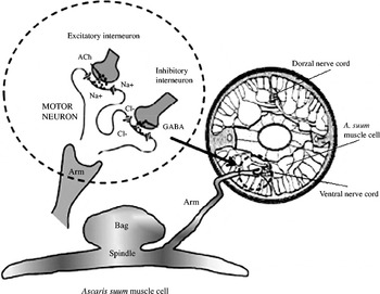
Ascaris suum (the large roundworm) is the most important gastrointestinal worm of pigs, and almost an ideal experimental model for investigations of the neurotransmitters and action of anthelmintic drugs in nematodes. Compared to the mammalian neuromuscular system, nematodes exhibit significant anatomical and physiological differences. Ascaris somatic muscle cells differ in structure from those of their vertebrate counterparts. Muscle cells of A. suum are composed of three distinct regions: the spindle or fibre, the muscle bag or belly, and the arm (fig. 1). The spindle contains actin and myosin and is the contractile part of the cell. Actin and myosin are arranged in overlapping rows that produce striations, as in vertebrate skeletal muscle. However, in nematode body-wall muscles, the striations are not aligned perpendicularly to the long axis of the muscle. Instead, the muscle is obliquely striated, which enables extensibility with high-speed contractions. The bag region of the cell contains the nucleus and is enriched with glycogen and mitochondria. It therefore acts as the energy store for the muscle cell (Martin et al., Reference Martin, Purcell, Day, Robertson, Marr and Nilsen2003). There is also an argument that the muscle bag provides support. In the intact parasite, the bag bulges inwards towards the gut. Harris & Crofton (Reference Harris and Crofton1957) proposed that the turgor of the muscle bags supports the nematode, acting as an endoskeleton. The muscle arm is the connection from the muscle cell to the nerve cord. Thus, in contrast to mammals where the nerve axons come from the spinal cord to the muscle, in the nematodes muscle cells send extensions to the nerve cord (fig. 1). This phenomenon is not completely understood, especially because each single muscle cell has one or more arms extending from the muscle bag towards the nerve cord in the hypodermis. At their termination, each muscle arm branches into processes that join with those from other arms to form a syncytium, or a functional network around the nerve cord. The individual processes are separated from each other by tight junctions, which ensure the electrical coupling between muscle cells. Synapses are seen at the junction between the terminal division of the muscle arm and the nerve (del Castillo et al., Reference del Castillo, Rivera, Solórzano and Serrato1989). The nervous system of A. suum, as in other nematodes, is consistent and relatively simple. It contains about 300 neurons (sensory and motor) which are mainly concentrated in the head and tail regions. The circumpharyngeal nerve ring is located in the head region, around the pharynx. It is some kind of a primitive brain, from which the dorsal, lateral and ventral nerve cords begin, and run along the animal to the tail. Muscle cells receive innervation from the dorsal and ventral nerves, while the lateral nerve is connected with the excretory canal. The largest of these three nerves is the ventral (Martin et al., Reference Martin, Purcell, Day, Robertson, Marr and Nilsen2003). Between motor neurons and the ventral nerve cord, relay cells or interneurons are arranged. Acetylcholine (ACh) is a neurotransmitter in the excitatory synapses, while GABA is the neurotransmitter in the inhibitory synapses between interneurons and motor neurons (fig. 1). These interneurons could be related evolutionarily to mammalian Renshaw cells, and are potentially a very attractive drug target (Trailović & Varagić, Reference Trailović and Varagić2007). In addition to receptors on nerve–muscle synapses, it was proved that functional GABA and nicotinic receptors exist extrasynaptically, in the bag region of Ascaris muscle cells (Martin, Reference Martin1982, Martin et al., Reference Martin, Purcell, Day, Robertson, Marr and Nilsen2003).
The innervation of the Ascaris suum muscle cell.

The aim of this study was to determine the role of postsynaptic nicotinic acetylcholine (nACh) and GABA receptors in contractions in the bag region of A. suum muscle cells, as well as the role of synaptic receptors between interneurons and motor neurons in the dorsal and ventral nerve cords. We are interested to find out whether a potential drug target is located here. In our investigations we used innervated and denervated somatic muscle flaps of A. suum for contraction assays.
Materials and methods
Preparation of muscle flaps from adult Ascaris suum
Adult female A. suum were collected weekly from the slaughterhouse at Vrčin, Belgrade, Serbia. Worms were maintained in Locke's solution (composition (mm): NaCl 155, KCl 5, CaCl2 2, NaHCO3 1.5 and glucose 5) at a temperature of 37°C. The Locke's solution was changed twice daily and each batch of worms was used within 4 days of collection.
Ascaris body muscle flaps for contractions were prepared by dissecting the anterior part of the worm, 2–3 cm caudal to the head. The resulting tube was then cut along one of the lateral lines and pinned on to a Sylgard chamber (filled with Ascaris perienteric fluid (APF) at 37°C) cuticle side down. Under 10 × magnification, the gut was removed easily, using fine forceps. Thereafter the ventral and dorsal nerve cords appeared white at the mid-points between the lateral lines. Innervated flaps were excised from this section by cutting along both lateral lines. For denervated muscle flaps, the dorsal or ventral cord was removed from the muscle flap. The muscle flap was cut on the inner side of the nerve cords so that only body-wall muscle (consisting of half of the ventral muscle field and half of the dorsal muscle field) between the nerve cords remained. At the same time, two flaps from a single piece of worm were prepared: innervated and denervated, or dorsal and ventral. Then, the two flaps from the same worm were placed in two experimental baths and tested at the same time. Each flap (always the same length of 1 cm) was monitored isometrically by attaching a force transducer in an experimental bath maintained at 37°C, containing 20 ml APF-Ringer (composition (mm): NaCl, 23; Na-acetate, 110; KCl, 24; CaCl2, 6; MgCl2, 5; glucose, 11; HEPES, 5; pH 7.6) and bubbled with room air. After dissection, the preparations were allowed to equilibrate for 15 min under an initial tension of 0.5 g. Different concentrations of ACh were then added to the preparation (1, 3, 10, 30 and 100 μm), and the maximum contraction was observed, before washing and the subsequent application of the next concentration of ACh. The responses for each concentration were expressed in grams (g), produced by each individual flap preparation.
The effects of GABA (3 μm) on control ACh dose–response plots were determined for dorsal and ventral flaps.
Contractions were monitored on a PC computer, using a two-channel BioSmart interface and eLAB software (El Unit, Belgrade). The system allows real-time recording, displaying and analysis of experimental data. Sigmoid dose–response curves for each individual flap preparation were described by the Hill equation.
Intact or completely denervated muscle flaps of A. suum were stimulated by electrical field stimulation (EFS). With a pair of platinum electrodes, placed parallel to the muscle flap, EFS was performed by the application continuous electrical pulses (widths 1 or 2 ms, frequency 100 Hz, voltage 25 V and duration 2 s). The EFS was generated by the digital stimulator BioSmart with software control (El Unit). We examined the effects of the nicotine antagonist mecamylamine and GABA on the contractions caused by EFS in innervated and denervated muscle flaps.
ACh, GABA and mecamylamine were obtained from Sigma-Aldrich Co. (St Louis, Missouri, USA), and were dissolved in the APF-Ringer.
Data analysis
In the studies presented here, sigmoid concentration dose–responses were described by the equation: % response = 1/[1+ (EC50/Xa)nH], where EC50 is the concentration of agonist (Xa) producing 50% of the maximum response and nH is the Hill coefficient (slope). Prism 5.0 (GraphPad Software, San Diego, California, USA) was used to estimate the constants EC50 and nH, by non-linear regression for each preparation. We determined mean contraction responses to each concentration of ACh (control dose–response: CR[Ach]) and mean responses to each of the concentrations of ACh in the presence of GABA. Paired t-tests were used to test differences between control contractions that were followed by test contractions on the same muscle flap. One-way analysis of variance (ANOVA), was applied for the analysis of the differences between the different preparations. Differences were considered significant when the P value was < 0.05.
The statistical analysis was done using Graphpad Prism software, while all values are expressed as mean ± standard error (SE).
Results
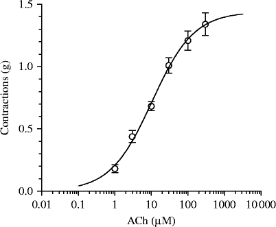
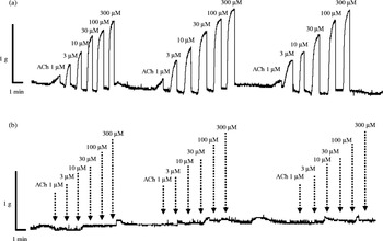
At the very beginning of the study, we chose to examine the role of nerve cords in the isometric muscle contractions of the A. suum muscle flap induced by ACh (1, 3, 10, 30, 100 and 300 μm) or EFS. The mean effective concentration of ACh (EC50) for contractions of intact neuromuscular preparations of A. suum was 10.88 μm (95% confidence interval (CI) = 5.75–19.17 μm). The maximal contractile effect observed in this study was 1.44 g, with 95% CI from 1.17 to 1.71 g (n= 15) (figs 2 and 3a). In contrast to the innervated muscle flaps, which responded with dose-dependent contractions to increased doses of ACh, in denervated flaps contractions were completely absent (fig. 3b).
The control concentration–response plot for acetylcholine (ACh) fitted with non-linear regression, with mean contractions in g ( ± SE) from 12 readings; EC50 for ACh = 10.88 μm.

(a) Isometric contractions of an innervated A. suum muscle flap, produced by increasing concentrations of acetylcholine (ACh) from 1 to 100 μm. (b) Lack of contractions in a denervated muscle flap of A. suum, following the application of similar concentrations of ACh.

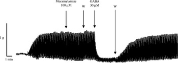
Continuous EFS (1 ms, 100 Hz, 25 V, 2 s) produced stable contractions of innervated Ascaris muscle flaps, with a mean amplitude of 0.76 ± 0.09 g. These contractions were not sensitive to mecamylamine. In the presence of mecamylamine (100 μm), the mean amplitude of the contractions was 0.77 ± 0.06 g. However, EFS-induced contractions were sensitive to GABA. The amplitude of the contractions was reduced significantly in the presence of 30 μm GABA (P < 0.001), to 0.15 ± 0.04 g. This inhibitory effect of GABA was reversible and, after washing, the mean value of the amplitude of the contractions reached 0.75 ± 0.05 g (n= 5) (fig. 4).
Representative recording of contractions of A. suum muscle flap induced by continuous EFS (1 ms, 100 Hz, 25 V, 2 s) and the effect of mecamylamine (100 μm) and GABA (30 μm) on these contractions. W, washing of the preparation.

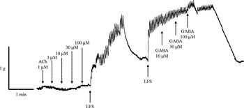
EFS with the same parameters of stimulation (1 ms, 100 Hz, 25 V, 2 s) did not cause muscle contractions in the denervated muscle flaps. However, EFS with a wider pulse (2 ms, 100 Hz, 25 V, 4 s) in the denervated muscle flaps caused an increase of tone and irregular contractions, the mean amplitude of which was 0.27 ± 0.05 g, obviously less than that in the innervated muscle flaps. EFS-induced contractions and increase of tone were completely insensitive to the effect of GABA (10, 30 and 100 μm) (fig. 5).
Denervated muscle flap of A. suum: lack of contractions after application of increasing concentrations of acetylcholine (1, 3, 10, 30 and 100 μm) and contractions caused by EFS (2 ms, 100 Hz, 25 V, 4 s), which were completely insensitive to GABA (10, 30 and 100 μm).

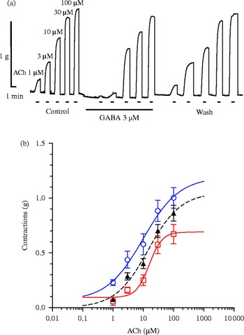
In the second part of the study, we tested the effects of ACh and GABA on the dorsal and ventral muscle flaps of A. suum. For this investigation we used the dorsal and ventral parts of the same worm segment. The value of the control EC50 of ACh for the dorsal muscle flaps was 9.83 μm (95% CI: 1.33–72.74 μm). Addition of 3 μm GABA had a significant inhibitory effect on contractions induced by 1, 3, 10, 30 and 100 μm ACh (P= 0.034, P= 0.022, P= 0.027, P= 0.009 and P= 0.045) (table 1). In the presence of GABA, the EC50 of ACh increased to 15.92 μm (95% CI: 7.70–32.90 μm), while after washing the EC50 was 13.14 μm (95% CI: 2.89–59.62 μm). The control value of the maximal effect (R max) was 1.19 g (95% CI: 0.11–2.27 g), in the presence of GABA it decreased to 0.69 g (95% CI: 0.52–0.86 g), while after washing the R max was 1.05 g (95% CI: 0.54–1.56 g) (fig. 6a, b).
Mean contractions in g (± SE) of five of each of the dorsal and ventral muscle flaps in Ascaris suum, relative to increasing concentrations of acetylcholine (ACh) from 1 to 100 μm and the inhibitory effect of GABA (3 μm).

Level of significance compared to the control: *P < 0.05, **P < 0.01.
(a) Isometric contractions of dorsal A. suum muscle flap produced by increasing concentrations of acetylcholine (ACh, 1–100 μm) and inhibition of contractions induced by GABA (3 μm). (b) A concentration–response plot for ACh with mean contractions ± SE (bars) from five readings: control (blue circles, blue line); GABA 3 μm (red squares, red line) and after washing the preparation (black triangles, black dotted line).

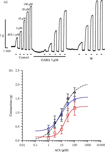
In the experiments on Ascaris ventral muscle flaps, the control EC50 value of ACh was 9.45 μm (95% CI: 1.38–64.84 μm) and the R max was 1.56 g (95% CI: 0.51–2.50 g). As in experiments on dorsal muscle flaps, GABA significantly decreased the value of ACh-induced contractions (P= 0.030; P= 0.0088; P= 0.0013; P= 0.0100; P= 0.0400) (table 1). In the presence of GABA, the EC50 was increased to 18.07 μm (95% CI: 5.11–63.86 μm), while the maximum effect decreased to 1.22 g (95% CI: 0.53–1.90 g). An interesting finding was observed after the washing of GABA-treated flaps. The EC50 was returned to near the control level (10.37 μm; 95% CI: 1.24–86.65 μm); however, the maximal effect was increased above the initial value, reaching a 2.07 g (95% CI: 0.23–3.09 g) (fig. 7a, b).
(a) Isometric contractions of ventral A. suum muscle flap produced by increasing concentrations of acetylcholine (ACh, 1–100 μm) and inhibition of contractions induced by GABA (3 μm). (b) A concentration–response plot for ACh with mean contractions ± SE (bars) from five readings: control (blue circles, blue line); GABA 3 μm (red squares, red line) and after washing the preparation (black triangles, black dotted line).

Discussion
According to Stretton et al. (Reference Stretton, Fishpool, Southgate, Donmoyer, Walrond, Moses and Kass1978), somatic muscle cells in Ascaris are arranged longitudinally and can be divided into two groups. The dorsal mass of somatic muscles is innervated by the motor axons in the dorsal nerve cord, while the ventral mass receives innervation from the motor component of the ventral nerve cord. Due to this arrangement, waves of contractions are propagated along the nematode's body in the dorsoventral plane. This type of innervation allows the worm to move forwards and backwards.
The use of denervated muscle flaps of Ascaris allows us to determine whether or not the observed physiological response is nerve-cord dependent (Halton et al., Reference Halton, Behnke and Marshall2001). To prepare denervated preparations, the muscle segments were not trimmed along the lateral lines (as outlined above), but were cut on either side of the ventral and dorsal nerve cords (these appear white at the mid points between the lateral lines). According to this method, two very narrow flaps that contain the ventral and dorsal nerve cords were produced and were discarded. These dissections are more difficult to perform than those in standard innervated muscle flap preparations.
Based on our results, obtained in both Ascaris segments (dorsal and ventral), the presence of the nerve cord is essential for the contractile effects of acetylcholine. The EC50 value of ACh was 10.88 μm and somewhat higher than the value that we published previously (about 8 μm, Trailović et al., Reference Trailović, Marjanović, Nedeljković Trailović, Robertson and Martin2015); however, in the present series of experiments we applied six concentrations of ACh, where the highest was 300 μm (usually the maximum concentration applied is 100 μm ACh). Unlike intact (innervated) preparations, there was no contraction of the muscle flaps when the nerve cord (either the dorsal or ventral) was mechanically removed. Muscle flaps without the nerve cord did not respond, even to 300 μm ACh. Continuous EFS produced stable contractions of innervated Ascaris muscle flaps, but they were not sensitive to mecamylamine. The absence of the antagonistic effect of mecamylamine could be explained by the possibility that EFS induced direct contractions. We have already recorded such an absence of the mecamylamine effect in mammalian skeletal muscle contractions (Trailović et al., Reference Trailović, Ivanovic and Varagić2011). Indirect contractions caused by EFS are the consequence of the release of the excitatory neurotransmitter (ACh) and may be blocked by nicotinic antagonists (blockade of the postsynaptic nicotinic receptors). Direct muscle contractions occur as a result of the direct effect of the electrical field on the contractile machine. Therefore, these contractions cannot be blocked by nicotine receptor antagonists. Whether EFS causes direct or indirect contractions, depends mainly on the width of the electrical pulse. Here, we used the parameters of the electrical field that causes indirect contractions of the isolated muscle of mammals (Trailović et al., Reference Trailović, Robertson and Nedeljković-Trailović2009). However, a completely unexpected result was that GABA significantly inhibited the EFS-induced contractions. Although it is probably a non-competitive, physiological antagonism, this could indicate that EFS induces indirect contractions of the Ascaris muscle flaps. This assumption was confirmed in the next part of the study, of denervated muscle flaps. The EFS, with the same characteristics, did not cause muscle contractions, but EFS with a wider pulse (2 ms, 100 Hz, 25 V, 4 s) induced the increasing of tone and irregular contractions with less amplitude. These contractions were completely insensitive to the effect of GABA, which confirmed the assumption that they are induced by a direct stimulation of contractile machinery. However, the question remains why EFS-induced contractions are not sensitive to mecamylamine and are sensitive to GABA. We hypothesize that EFS releases ACh presynaptically, while mecamylamine only affects extrasynaptic nACh receptors, located on the bag region of muscle cells. On the other hand, GABA acts on synaptic and extrasynaptic GABA receptors, and therefore is able to inhibit the contractions caused by EFS.
It was particularly interesting to investigate whether a difference in sensitivity to ACh and GABA exists between the dorsal and ventral Ascaris segments. The value of control EC50 of acetylcholine for the dorsal segments was 9.83 μm, and for the ventral segments it was 9.45 μm, which is in accordance with our previously published data (Puttachary et al., Reference Puttachary, Trailovic, Robertson, Thompson, Woods and Martin2013; Trailović et al., Reference Trailović, Marjanović, Nedeljković Trailović, Robertson and Martin2015). The results obtained indicate that the EC50 for ACh did not differ between the dorsal and ventral segments. The presence of GABA (3 μm) led to an increase in the EC50 in dorsal segments by about 60% (to 15.92 μm), while the EC50 of ACh in the ventral segments was doubled (to 18.07 μm). Different results were recorded for the maximal effects. The R max of ACh in the dorsal preparations decreased by 40% in the presence of GABA, from the control value of 1.19 g to 0.70 g. A smaller inhibition of the maximal effect was observed in the ventral preparations, from 1.56 g to 1.22 g, or a decrease by about 22%. GABA inhibits the ACh-induced contractions by binding to specific GABA receptors. It would be expected that GABA consequently exhibits non-competitive antagonism, which does not significantly change the EC50 value, but only reduces the maximal effect (Katzung et al., Reference Katzung, Masters and Trevor2012). In our investigations, GABA exhibited features of competitive and non-competitive antagonists, regardless of whether it acted on the dorsal or ventral segments of Ascaris. Therefore, the presence of GABA increased the value of the EC50 of ACh and decreased the R max. The non-competitive component of the inhibitory effect of GABA on the contractions induced by ACh is completely clear, because they act on different receptors. However, the potential explanation for the competitive component of the effect suggests the existence of physiological antagonism. The presynaptic action of GABA, through inhibitory interneurons, decreases the activity of excitatory interneurons and the postsynaptic membrane of motor neurons, thereby inhibiting excitatory effects when ACh is applied. But the question about the role of the extrasynaptic receptors in the bag region of Ascaris muscle cells remains for further investigations. Maybe, they present just a part of an exclusively non-competitive inhibitory mechanism of contraction, exhibited by GABA. GABA exerted a more intense competitive antagonism on the ventral nerve cord. The ventral nerve cord is larger than the dorsal, and therefore has a denser syncytium around the nerve cord (Martin et al., Reference Martin, Purcell, Day, Robertson, Marr and Nilsen2003). This could be the reason for the existence of a large number of synapses between motor neurons and interneurons and, therefore, a more intense manifestation of physiological antagonism.
It is interesting that after the washing of GABA-treated flaps, the ACh EC50 was normalized more intensively in the ventral (pronounced competitive antagonism) than in the dorsal segments (pronounced non-competitive antagonism). On the other hand, after the washing of GABA-treated flaps, the maximal effect of ACh recorded on the ventral segments was higher by 33%, compared to the control value (2.07 g compared to 1.56 g). This phenomenon, of the increasing of the contractile effect of ACh, was also observed and described after washing following treatment with the nematode neuropeptide AF2, and certainly deserves additional research (Trailović et al., Reference Trailović, Clark, Robertson and Martin2005).
Our investigations have shown the neuromuscular system of parasitic nematodes, while relatively anatomically simple, exhibits complex physiological activity. The mechanism of action of the two largest groups of anti-nematodal drugs (agonists of nACh and GABA receptors) is based on their interaction with excitatory and inhibitory components of this system. It is obvious that the drugs will be more effective if they act on both the synaptic and extrasynaptic nACh and GABA receptors. In the development of new anti-parasitic drugs it is also necessary to obtain substances that are capable of performing a ‘chemical’ denervation of somatic muscles in nematodes, which would increase drug efficiency and reduce the possibility of development of resistance in parasites.
Acknowledgements
The authors would like to thank to Dr Nebojša Lazić for the supply of live adult worms.
Financial support
Funding for this study was from the Ministry of Education and Science, Republic of Serbia, Grant No TR31087.
Conflict of interest
None.