Implications
Scientists are working to develop individual animal-based measurements that can be used to detect harmful situations that may affect animal welfare and meat quality. In this context, the mechanisms underlying the adverse effects of certain animal management procedures on postmortem muscle metabolism need to be elucidated to understand how animal stress may influence the conversion of muscle to meat. In this study, we demonstrate that mixing unfamiliar animals at the farm and/or before slaughter produces increased oxidative stress in the muscle tissue and triggers autophagy. Therefore, biomarkers of these processes could be used as tools for detecting inappropriate strategies that may induce animal stress and affect meat quality.
Introduction
Pre-slaughter handling conditions such as transportation, lairage and mixing unfamiliar animals have been shown to affect animal stress and, therefore, pork quality (Warriss et al., Reference Warriss, Brown, Nute, Knowles, Edwards, Perry and Johnson1995; Hambrecht et al., Reference Hambrecht, Eissen, Newman, Smits, Verstegen and den Hartog2005). Mixing unfamiliar animals is a common practice in pig production, but it may lead to social stress within the group and is usually followed by fighting to create a new hierarchy (Fàbrega et al., Reference Fàbrega, Puigvert, Soler, Tibau and Dalmau2013). Therefore, mixing animals may be an important ante- and peri-slaughter stressor that could affect pig welfare, and thus some pork quality traits such as pH, color and eating quality (Warriss et al., Reference Warriss, Brown, Nute, Knowles, Edwards, Perry and Johnson1995 and Reference Warriss, Brown, Knowles, Edwards, Kettlewell and Guise1998; Terlouw et al., Reference Terlouw, Porcher and Fernandez2005).
The mechanisms underlying the adverse effects of animal stress on the muscle tissue have not been completely elucidated so far. Stress is a general term that describes the inadequate physiological response of an organism to any mental, emotional or physical demand, whether real or imagined (Schiavone et al., Reference Schiavone, Jaquet, Trabace and Krause2013). The cellular mechanisms that trigger stress are unknown, but a relationship between stressors and oxidative stress has been described, because oxidative changes occur in situations such as road transport, weaning or heat stress (Burke et al., Reference Burke, Fiehn and Moran2010). The increase in the activity of individuals subjected to stress requires greater ATP production, resulting in increased oxygen consumption by the mitochondria. This situation leads to increased production of free radicals, which provokes an imbalance between reactive oxygen species (ROS) and the system’s ability to readily detoxify reactive intermediates and/or repair the resulting damage (Lardone et al., Reference Lardone, Álvarez-García, Carrillo-Vico, Vega-Naredo, Caballero, Guerrero and Coto-Montes2006). The oxidation process has been reported to be a major cause of deterioration in the quality of muscle foods (Trefan et al., Reference Trefan, Bunger, Bloom-Hansen, Rooke, Salmi, Larzul, Terlouw and Doeschl-Wilson2011). Furthermore, previous studies by our group have shown that oxidative stress is directly related to meat tenderization at early postmortem stages (Coto-Montes et al., Reference Coto-Montes, Caballero, Sierra, Vega-Naredo, Tomás-Zapico, Hardeland, Tolivia, Ureña and Rodríguez-Colunga2004; Caballero et al., Reference Caballero, Sierra, Vega-Naredo, Tomás-Zapico, Rodríguez-Colunga, Tolivia, Hardeland, Oliván and Coto-Montes2006). However, the link between physiological stress and cellular oxidative stress in postmortem muscle still needs to be elucidated.
At slaughter, the process of exsanguination deprives cells and tissues of nutrients and oxygen, leading to massive accumulation of ROS, halting ATP production and causing cytoplasmic acidification and calcium dysregulation (Caballero et al., Reference Caballero, Sierra, Oliván, Vega-Naredo, Tomás-Zapico, Álvarez-García, Tolivia, Hardeland, Rodríguez-Colunga and Coto-Montes2007). These stimuli may trigger different cellular responses including changes in autophagy (García-Macia et al., Reference García-Macia, Sierra, Palanca, Vega-Naredo, de Gonzalo-Calvo, Rodríguez-González, Oliván and Coto-Montes2014). Autophagy is considered a specialized form of lysosomal proteolysis, where a portion of the cytoplasm or entire organelles are sequestered into double-membrane vesicles known as autophagosomes, which then fuse with lysosomes, resulting in the degradation of their contents by lysosomal cathepsins (Caballero and Coto-Montes, Reference Caballero and Coto-Montes2012; Coto-Montes et al., Reference Coto-Montes, Boga, Rosales-Corral, Fuentes-Broto, Tan and Reiter2012).
In the light of these findings, the aim of this study was to determine the effect of animal stress caused by mixing unfamiliar animals at different times in the production process (at farm and/or during transport and lairage before slaughter) on the presence and evolution of the main biomarkers of autophagy and oxidative stress in pork meat during early postmortem aging. In addition, their relationship with muscle pH will be evaluated. This study is part of a broader investigation on the effect of animal mixing strategies on animal welfare and performance (Fàbrega et al., Reference Fàbrega, Puigvert, Soler, Tibau and Dalmau2013).
Material and methods
Animal management and sampling procedure
Entire male ((Large White×Landrace)×Duroc) pigs were reared from birth (April) to slaughter (October) on an experimental farm at IRTA-Monells (Spain), and were subjected to different management treatments that may promote stress, such as mixing unfamiliar animals on the farm and/or during transport and lairage before slaughter. These pigs were part of a bigger experiment in which 96 animals were managed under two rearing treatments during the fattening period: 48 of them were in a wean-to-finish regime – that is, no mixing after the initial experimental group formation (at weaning) – and the other 48 followed the normal routine of creating new groups at different stages of the rearing period (21 and 73 days of age), without taking into account their litter.
Each fattening pen measured 4×2.7 m (0.9 m2/pig regardless of the size of the pig), had a partly slatted floor comprising 60% solid concrete and 40% slatted and had one drinking bowl. A wooden board was attached to some pen divisions to avoid visual contact between animals from different pens. Each pen was equipped with an IVOG®-station (Insentec, Marknesse, The Netherlands) for monitoring the individual feed intake. All pigs were fed the same commercial diet (3365.75 Kcal DE/kg, 17.9% CP, 7% crude fat, 1.95% lysine and 6.55% ash on dry matter basis).
When the pigs reached a weight of 120 kg, they were slaughtered at an experimental abattoir located 1 km away from the farm. Pre-transport fasting time was 8 h. The trip duration from the farm to the abattoir was about 5 min; the transport vehicle was a double-deck lorry with natural ventilation located along the full length of its body, and a hydraulic elevator was used for loading and unloading, so that the animals were gently loaded and unloaded in order to avoid additional stress. Pigs were slaughtered after a lairage time ranging from 30 min to 2 h.
In this step of the experiment, two different pre-slaughter treatments were used: some animals were not mixed with animals from other pens during transport and lairage before slaughter (unmixed group during transport and lairage, UTL), whereas others were mixed with unfamiliar animals on the lorry and during lairage (mixed group during transport and lairage, MTL). For this study, a total of 15 animals, slaughtered in the same batch, were randomly selected from the whole experiment. These animals belonged to the following three treatments (n=5/treatment):
-
∙ UF-UTL (unmixed at the farm-unmixed during transport and lairage), which represents the control group. They were not mixed either at the farm or during transport and lairage.
-
∙ MF-UTL (mixed at the farm-unmixed during transport and lairage), which represents the group with stress caused by mixing unfamiliar animals at the farm (twice: at 21 and 73 days of age), but not during transport and lairage.
-
∙ MF-MTL (mixed at the farm-mixed during transport and lairage), which represents the group with stress caused by mixing unfamiliar animals at the farm (twice: at 21 and 73 days of age) and during transport and lairage.
The pH of the Longissimus dorsi (LD) muscle was recorded at 45 min (pH45) and 24 h (pH24) after slaughter, on the left-half carcass, at the last rib level, using a Crison portable meter (Crison–Hach Lange, Barcelona, Spain), equipped with a xerolyt electrode and coupled with a temperature probe. In addition, muscle samples were taken from the LD muscle of each animal at 4, 8 and 24 h postmortem. These samples were immediately snap-frozen in liquid nitrogen and stored at −80ºC until analysis. Muscle samples were homogenized at a ratio 1 : 10 ml (muscle : buffer) in a homogenization buffer (pH 7.4) containing 10 mM potassium phosphate buffer, 50 mM sodium chloride and 0.1% Triton using an Ultra-Turrax T25 Mixer (IKA, Staufen, Germany) (Sierra, Reference Sierra2003). The homogenates were centrifuged at 1500×g for 6 min at 4ºC. Supernatants containing proteins were collected, aliquoted and frozen at −80ºC until further analysis.
Antioxidant activities
Total antioxidant activity (TAA) was determined using the ABTS/H2O2/HRP method (de Gonzalo-Calvo et al., Reference de Gonzalo-Calvo, Neitzert, Fernández, Vega-Naredo, Caballero, García-Macia, Suárez, Rodríguez-Colunga, Solano and Coto-Montes2010). The results are expressed in equivalents of mg Trolox/mg protein. Superoxide dismutase (SOD; EC 1.15.1.1) activity was measured according to Martin et al. (Reference Martin, Dailey and Sugarman1987). This enzyme inhibits hematoxylin auto-oxidation to the colored compound hematein. The results are expressed as µmol hematein/mg protein×min. Catalase (CAT; EC 1.11.1.6) activity was assayed according to Lubinsky and Bewley using H2O2 as a substrate (Lubinsky and Bewley, Reference Lubinsky and Bewley1979). The results are expressed as µmol H2O2/mg protein×min. Lipid peroxidation was measured by determining the contents of the reactive aldehydes malondialdehyde (MDA) and 4-hydroxy-2-(E)-nonenal (4-HNE), which are the end-products of the lipid peroxidation cascade. The amounts of MDA and 4-HNE were determined in the muscle using an LPO Assay Kit (lipid hydroperoxide (LPO), Assay Kit, No. 437634, Calbiochem; Merck Millipore, Billerica, MA, USA) based on the condensation reaction of the chromogene 1-methyl-2-phenylindole with either MDA or 4-HNE. The results are expressed as µmol (MDA+4-HNE)/g protein.
Western blot immunoassay
Aliquots of muscle homogenate samples containing 100 µg of protein per sample were mixed with Laemmli sample buffer (BioRad Laboratories, Inc., Hercules, CA, USA) and denatured. The samples were then fractionated through SDS-PAGE and subsequently transferred to polyvinylidene fluoride membranes (Immobilon TM-P; Millipore Corp., Bedford, MA, USA). The membranes were blocked overnight and incubated with anti-Beclin 1 (sc-10086; Santa Cruz Biotechnology, Santa Cruz, CA, USA) and anti-LC3 (PD014; Medical & Biological Laboratories Co., Ltd, Naka-ku Nagoya, Japan), which were previously diluted. After washing, the membranes were incubated with the corresponding horseradish peroxidase-conjugated secondary antibody (Sigma Aldrich, Saint Louis, MO, USA), diluted and developed using a chemiluminescent horseradish peroxidase substrate (WBKLS0500; Merck Millipore). The levels of proteins were quantitatively analyzed using Quantity One 5.5.1 software. The results were normalized to Ponceau as a loading control.
Statistical analysis
Data were analyzed via two-way ANOVA, with a model including the effects of the mixing treatment (UF-UTL, MF-UTL, MF-MTL), postmortem time (4, 8 and 24 h) and their interaction as main effects. Once the interaction between the mixing treatment and time was demonstrated, the effect of postmortem time (with animal as the random factor) was tested, and differences between individual means were analyzed with the Bonferroni post hoc test. Multivariate analysis was applied to explore the complex relationships between meat quality (pH45) and biochemical variables (biomarkers of autophagy and oxidative stress) and between the variables and treatments. Subsequently, principal component analysis (PCA) was performed, with extraction of four factors and varimax rotation. Statistical analyses were performed with GraphPad Prism 5 software (GraphPad Software, Inc., La Jolla, CA, USA) and XLStat (2013).
Results
The pH45 of the LD muscle of pigs subjected to MF-UTL and MF-MTL treatments showed lower values (6.51 and 6.54, respectively) compared with meat from animals of the unmixed (control) group (UF-UTL=6.66), although the difference was not significant, probably due to the small sample size and high individual variability in the rate of pH decline within every group. However, LD muscles showed same pH values at 24 h postmortem (pH24=5.4 to 5.5) in all the groups.
Antioxidant activities
As shown in Table 1, TAA was affected by the interaction between mixing treatment and sampling postmortem time (P<0.001). Muscle TAA increased from 4 to 8 h postmortem in the muscles of animals subjected to the double-mixing treatment (MF-MTL; P<0.05) followed by a decrease at 24 h postmortem (P<0.05). However, in the group of animals mixed only at the farm (MF-UTL), there was a slower increase of TAA in the muscles, with maximum values at 24 h postmortem (P<0.05). In the group of animals without mixing (UF-UTL), TAA showed moderate values, with a maximum at 8 h postmortem (P<0.05).
Effect of the mixing group (G), postmortem time (T) and their interaction (G×T) on biomarkers of oxidative stress measured in the LD muscles of pigs

UF-UTL=unmixed at the farm-unmixed during transport and lairage; MF-UTL=mixed at the farm-unmixed during transport and lairage; MF-MTL=mixed at the farm-mixed during transport and lairage; TAA=total antioxidant activity (mg Trolox/mg protein); SOD=superoxide dismutase activity (µmol hematoxylin/mg protein×min); CAT=catalase activity (µmol H2O2/mg protein×min); LPO=lipid peroxidation (µmol MDA+4-HNE/g protein).
For a given mixing group, means in the same row followed by different letters are significantly different at P<0.05.
**P<0.01, ***P<0.001.
The activity of SOD was influenced by the interaction (P<0.01). In both mixing treatments (MF-UTL and MF-MTL), there was maximal SOD activity at 8 h postmortem (P<0.05), with further decrease at 24 h postmortem, when the SOD activity in the muscles of control animals (UF-UTL) was greater (P<0.05). The CAT enzyme also showed increased activity at 8 h postmortem in the UF-UTL and MF-UTL groups (P<0.05) and at 4 h postmortem in the MF-MTL group.
The interaction between mixing treatment and sampling postmortem time also affected LPO variation (P<0.001), with LPO showing the greatest (P<0.05) value at 4, 8 and 24 h postmortem in the LD muscles of MF-MTL, UF-UTL and MF-UTL pigs, respectively.
Autophagy
Our results clearly show that autophagy activity takes place during pig meat maturation, as two main autophagy biomarkers (Beclin 1 and LC3-II) were present during the postmortem period under all the studied conditions.
Beclin 1 showed higher (P<0.05) expression at the early postmortem time point (4 h) in the LD muscles of MF-UTL and MF-MTL pigs, and a decrease at 24 h postmortem. However, in the UF-UTL group, the maximum values were detected at 8 h postmortem (P<0.05) (Table 2).
Effect of the mixing group (G), postmortem time (T) and their interaction (G×T) on biomarkers of autophagy (optical density, in arbitrary units) measured in the Longissimus dorsi muscles of pigs

UF-UTL=unmixed at the farm-unmixed during transport and lairage; MF-UTL=mixed at the farm-unmixed during transport and lairage; MF-MTL=mixed at the farm-mixed during transport and lairage.
For a given mixing group, means in the same row followed by different letters are significantly different at P<0.05.
**P<0.01, ***P<0.001.
In all the treatments, LC3-I showed a decreasing pattern, with maintained values between 4 and 8 h postmortem and a clear decline at 24 h postmortem (Table 2). LC3-II always showed lower values than LC3-I, mainly due to the important role that LC3-I (known as LC3) plays in the cytoplasm of muscle cells as one of the three light chains (LC1, LC2 and LC3) associated with purified MAP1A and MAP1B (Tanida et al., Reference Tanida, Ueno and Kominami2008). The pattern of LC3-II expression was analogous to that of LC3-I (Table 2). The autophagy flux, monitored through the relationship between LC3-II and LC3-I, was maintained in the control group within the studied postmortem time range, whereas the flux was reduced over time in the two mixing treatments – MF-UTL and MF-MTL (Table 2).
Multivariate analysis
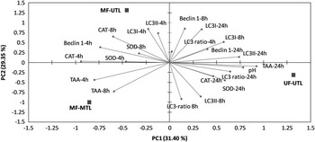
Multivariate analysis was applied to obtain a synthetic assessment of the complex relationships between the biomarkers of meat quality (pH45), autophagy (Beclin 1, LC3-I, LC3-II, autophagy flux) and antioxidant defense (TAA, SOD and CAT), together with the different mixing treatments. The bi-plot obtained via PCA (Figure 1) showed that PC1 and PC2 explained 61% of the variability in the data. On the positive side, PC1 distinguished meat samples displaying a normal postmortem pH decline (higher pH at 45 min in the LD muscle) together with a higher antioxidant ability (SOD, CAT) at 24 h postmortem, in addition to higher LC3-II levels and even a higher LC3-II/LC3-I ratio at 24 h. These samples corresponded to animals from the UF-UTL treatment – that is, with no animal mixing; thus, showing no effect of stress on the postmortem muscle metabolism. However, on the negative side of the bi-plot, there were meat samples from the MF-UTL and MF-MTL treatments (Figure 1), which showed higher antioxidant activity (TAA, CAT and SOD) and higher Beclin 1 levels at short (4 h) postmortem times. These results show clearly that greater pre-slaughter stress induced by mixing unfamiliar groups of pigs produced an earlier activation of autophagy in the muscle cells as an adaptative strategy for counteracting oxidative stress.
Principal component analysis bi-plot of variables (pH, TAA, SOD, CAT, Beclin 1, LC3-I, LC3-II and LC3 ratio) and mixing treatments (UF-UTL, MF-UTL and MF-MTL). TAA=total antioxidant activity; SOD=superoxide dismutase; CAT=catalase; and LC3 ratio=LC3-II/LC3-I; UF-UTL=unmixed at the farm-unmixed during transport and lairage; MF-UTL=mixed at the farm-unmixed during transport and lairage; MF-MTL=mixed at the farm-mixed during transport and lairage; PC=principal component.

Discussion
Variations in animal emotions and stress perception due to different management strategies on the farm and at the slaughterhouse can have an effect on meat quality (Gispert et al., Reference Gispert, Faucitano, Oliver, Guardia, Coll, Siggens, Harvey and Diestre2000). Different stress factors and the individual responses of animals experiencing the same conditions need to be understood, as they could partly explain the high variations in meat quality found in the market.
It is known that psychological stress results in oxidative damage in the muscle (Li et al., Reference Li, Zhang, Chen, Wang, Huang and Liu2011). Our data support this conclusion, as we observed an increase in the antioxidant defense, which was proportional to the stress suffered. Thus, during the postmortem maturation period, an increase in TAA as well as antioxidant enzymes was induced in the muscle tissue to counteract the ROS produced by stress. Previous data from our group showed that in muscle cells after slaughter, the activity of various enzymes can be induced, mainly those of antioxidant enzymes (Coto-Montes et al., Reference Coto-Montes, Caballero, Sierra, Vega-Naredo, Tomás-Zapico, Hardeland, Tolivia, Ureña and Rodríguez-Colunga2004; Caballero et al., Reference Caballero, Sierra, Vega-Naredo, Tomás-Zapico, Rodríguez-Colunga, Tolivia, Hardeland, Oliván and Coto-Montes2006) and cathepsins (Caballero et al., Reference Caballero, Sierra, Oliván, Vega-Naredo, Tomás-Zapico, Álvarez-García, Tolivia, Hardeland, Rodríguez-Colunga and Coto-Montes2007), and thus the increases in antioxidant enzymes observed at 4 and 8 h could be expected. These increased activities ran in parallel over time, which implies a correct oxidative balance, because although CAT and SOD activities were expressed in different units they followed an analogous pattern, maintaining a delicate SOD/CAT balance for detoxification (Pinho et al., Reference Pinho, Andrades, Oliveira, Pirola, Zago, Silveira, Dal-Pizzol and Moreira2006). However, at 24 h postmortem, these antioxidant enzymes showed cellular degradation effects, losing their cellular synthesis ability, which was a repeated finding for all the studied proteins.
Even with this antioxidant activity, LPO was unavoidable in all cases. Thus, at 4 h, muscle tissue from all the animals studied showed similar levels of lipid damage, which implies that in the early postmortem period, slaughtering was the main stress-inducing factor. However, during meat maturation, the increasing antioxidant defense observed in the muscles of animals subjected to ante- and peri-mortem mixing stressors had clear effects on the damage induced by free radicals, such as LPO, with a significant decrease in LPO being observed in the MF-UTL and MF-MTL animals at 8 h postmortem, whereas LPO remained low in the group subjected to double-mixing (MF-MTL) at 24 h postmortem. Again, the loss of the cellular synthesis ability at 24 h was manifested under all the studied conditions.
Autophagy triggered by oxidative stress has been amply documented in transformed and cancer cells (Chen et al., Reference Chen, McMillan-Ward, Kong, Israels and Gibson2008), although the relationship between psychological and/or physical stress and autophagy is still unknown. Recently, our group showed that autophagy may be directly related to the oxidative stress status of the muscle cells in beef (García-Macia et al., Reference García-Macia, Sierra, Palanca, Vega-Naredo, de Gonzalo-Calvo, Rodríguez-González, Oliván and Coto-Montes2014). Therefore, the possibility of autophagy modulation by ante- or peri-mortem stress could be expected when this stress produces alterations in the oxidative balance, as shown in the present study.
As a metabolic pathway, autophagy is highly sensitive to changes in the environment (Scherz-Shouval and Elazar, Reference Scherz-Shouval and Elazar2011), and the relationship between ROS and autophagy has been fully documented (Azad et al., Reference Azad, Chen and Gibson2009). Our results are in agreement with these data, because the main autophagy markers – Beclin 1 and LC3 – showed an increase in activity at early postmortem times (from 4 to 8 h) in the muscle samples showing an alteration of oxidative balance. However, these increases must be carefully interpreted, as they could be the results of two very different stimuli that have to be considered independently: slaughtering stress and stress induced by ante- or peri-mortem animal mixing.
It has been shown that slaughtering stress together with anoxic conditions caused by the sudden cut-off of blood flow is able to induce autophagy as a survival mechanism in the muscle tissue (García-Macia et al., Reference García-Macia, Sierra, Palanca, Vega-Naredo, de Gonzalo-Calvo, Rodríguez-González, Oliván and Coto-Montes2014), and this situation was observed in all the animals in our study, even in those from the control group without animal mixing (UF-UTL), in addition to those subjected to different animal mixing stressors (MF-UTL and MF-MTL). However, based on the data obtained from this study, a certain psychological stress component involved in autophagy induction should be considered, because mixing stress affected the time-scale evolution of autophagy biomarkers in the muscle. In fact, both experimental conditions showing stress caused by mixing unfamiliar animals (at the farm or at the farm and during transport and lairage) resulted in a significant increase in Beclin 1 in the muscle tissue at a very short (4 to 8 h) postmortem period. This mammalian ortholog of yeast Atg6 plays a central role in autophagy as a main contributor to autophagy initiation (Kang et al., Reference Kang, Zeh, Lotze and Tang2011). Its value was even higher in the MF-UTL group (animal mixing at farm) in comparison with the MF-MTL group (animal mixing at farm and during transport and lairage). This greater increase in the group subjected to animal mixing at the farm, where an intermediate level of induced stress due to animal mixing could be expected compared with the double-mixing treatment group (at the farm and during transport and lairage), appears to reflect high inter-individual variability in the animals’ susceptibility to stress, thus showing that autophagy triggering depends strongly on the level of psychological stress in every individual animal. This is in agreement with previous reports describing that the stress levels of animals depend indirectly on the situation and directly on the animals’ evaluation of the situation (Terlouw, Reference Terlouw2005). It is for this reason that scientists are working on the development of individual animal-based biomarkers of animals’ emotional state.
Given the role of ROS in inducing autophagy, antioxidants can serve as natural downregulators of this process (Scherz-Shouval and Elazar, Reference Scherz-Shouval and Elazar2011). Several recent reports have demonstrated the modulation of autophagy by antioxidants. TP53-induced glycolysis and apoptosis regulator (TIGAR), which modulates the glycolytic pathway and decreases intracellular ROS levels, is able to inhibit autophagy through constitutive activity in the cellular antioxidant defense system (Bensaad et al., Reference Bensaad, Cheung and Vousden2009). Likewise, under conditions of prolonged starvation/glucose deprivation, over-expression of both SOD and CAT inhibits autophagy (Chen et al., Reference Chen, Azad and Gibson2009). Glucose deprivation and abrupt hypoxia show several common effects including increases in ROS. In our results, we observed that antioxidant enzymes were increased under both types of stress (slaughter stress and animal mixing-induced stress); however, although at 4 h postmortem these increases were proportional to the induced stress, at 8 h this tendency changed. This significant antioxidant activity appears to inhibit autophagy if we take into account the essential role in autophagy played by the autophagy flux, measured as the LC3-II/LC3-I ratio.
LC3 is the most frequently used marker of autophagy. LC3 is initially synthesized in an unprocessed form, proLC3, which is converted into the proteolytically processed form LC3-I, lacking amino acids from the C-terminus, and is finally modified into a phosphatidylethanolamine-conjugated form – LC3-II (Skop et al., Reference Skop, Cahova, Papackova, Palenickova, Dankova, Baranowski, Zabielski, Zdychova, Zidkova and Kazdova2012). LC3-II is the only protein marker that is reliably associated with phagophores, sealed autophagosomes and mature autophagosomes/lysosomes (Rubinsztein et al., Reference Rubinsztein, Cuervo, Ravikumar, Sarkar, Korolchuk, Kaushik and Klionsky2009). Nevertheless, LC3-II localizes to both the luminal and cytosolic sites of the autophagosome and undergoes rapid degradation within the lysosome (Skop et al., Reference Skop, Cahova, Papackova, Palenickova, Dankova, Baranowski, Zabielski, Zdychova, Zidkova and Kazdova2012). To determine the real autophagy flux, it is necessary to determine the LC3-II/LC3-I ratio and also increased LC3-I and LC3-II levels. In the present study, we estimated the autophagy flux taking into account all possible LC3 transformations, and we observed that LC3-I is consequently increased together with Beclin 1. However, LC3-II showed a decrease at 8 h in the group of animals mixed at the farm (MF-UTL), when SOD and CAT were overproduced, but showed a decrease later (24 h) in the other treatments. This decrease may be a consequence of either low formation of LC3-II due to the blockage of autophagy by antioxidant activity as well as increased degradation due to increased lysosomal activity. Both actions are possible and could even be accumulative. However, after 8 h of meat maturation, the highest antioxidant activity was observed in terms of SOD and CAT activity, which can induce a dramatic reduction in LC3-II formation. Further studies will be necessary to elucidate which factor has a greater influence regarding autophagy inhibition.
In the present study, mixing unfamiliar animals led to ante- and peri-slaughter stress in the pigs and had an effect on the postmortem muscle metabolism. Furthermore, multivariate analyses highlight a correlation between animal mixing treatments and, therefore, the resulting mixing-induced stress, with an earlier postmortem activation of antioxidant defense and the autophagic–lysosomal pathway in the muscle tissue. Thus, autophagy appears again to play an important role in the regulation of postmortem muscle metabolism. From these results, we propose that monitoring the evolution of the main biomarkers of autophagy (Beclin 1, LC3-II/LC3-I ratio) and muscle antioxidant defense (TAA, SOD and CAT) in the muscle tissue within the first 24 h postmortem may help in detecting animal stress and its potential effect on the process of conversion of muscle into meat.
Acknowledgments
The authors are members of the INPROTEOLYS and INEUROPA network. This work was supported by projects AGL2011-30598-C03-03, FISS-13-RD12/0043/0030, MICINN10-BFU2010-20919, RTA2007-00049-00-00 and FEDER funds. Financial support from the University of Oviedo is also acknowledged.