I. INTRODUCTION
The effect of high magnetic field processing on microstructure evolution and performance enhancement in steels is a growing research topic. High magnetic field can modify the Gibbs energy of ferromagnetic phase and boundaries (A1 and A3 line) in Fe–C phase diagram. Reference Joo, Choi, Kim, Shin and Koo1–Reference Guo and Enomoto4 Influence of high magnetic field on the solid transformation of steels has been shown in many aspects such as martensite transformation, Reference Fields and Graham5 bainite transformation, Reference Ohtsuka6 proeutectoid ferrite transformation, Reference Ludtka, Jaramillo, Kisner, Nicholson, Wilgen, Mackiewicz-Ludtka and Kalu7–Reference Enomoto, Guo, Tazuke, Abe and Shimotomai9 pearlite transformation, Reference Zhou, Zhang and Wu10–Reference Zhang and Enomoto12 recrystallization, Reference He, Zhang, Zhao, Zuo, He, Watanabe, Zhang, Nishijima and Esling13–Reference Masahashi, Matsuo and Watanabe15 and carbide precipitation in temper. Reference Zhang, Zhao, Bozzolo, He, Zuo and Esling16–Reference Xia, Zhang, Lan, Liu and Yang18 However, many researchers Reference Ohtsuka6,Reference Ludtka, Jaramillo, Kisner, Nicholson, Wilgen, Mackiewicz-Ludtka and Kalu7,Reference Zhang, Esling, Gong, Vincent, Zhao and Zuo11 investigated the influence of magnetic field on the austenitic decomposition by applying high magnetic field during both austenitization and following cooling process. However, this experimental procedure confuses the understanding of effects of magnetic field because it is difficult to separate the coupling effect of magnetic field on the austenitization and following continuous cooling, and even the effect of magnetic field on austenitization is neglected. Therefore, it is helpful to analyze accurately the mechanism of magnetic field effects by investigating the effect of magnetic field on austenitization.
Fully pearlitic steel wires exhibit ultrahigh strength and have important applications where high strength and wear resistance are required. The hypereutectoid compositions present a promising potential for increasing the mechanical properties, especially the strength. Reference Bae, Nam and Lee19,Reference Choi and Park20 The austenitization condition strongly influences the proeutectoid cementite and pearlite transformation. Reference Li, Godfrey and Liu21 Therefore, the magnetic field application to change the austenite state may provide a new way for developing customized microstructure and enhanced performance.
Investigation for effects of magnetic field on the austenitization is of importance for scientific research and industrial processing. Austenitization can be divided into two stages: austenite formation and austenite grain coarsening. The former stage includes pearlite to austenite (P → γ) transformation and cementite dissolution. The magnetic field was applied during the different stages to contrastively investigate the effects of magnetic field on the austenite formation and grain size.
II. EXPERIMENTAL PROCEDURES
The material used in this study is a hypereutectoid plain carbon steel with chemical composition (wt.%) C 1.11%, Cr 0.18%, Si 0.17%, Mn 0.28%, P 0.009%, S 0.007%, and bal. Fe. The as-received steel wire rod was machined into cylindrical samples with dimensions of 10 mm in height and 4 mm in diameter.
The samples treated without and with 10 T magnetic field were austenitized at 950 °C for different times and then quenched into water quickly out of magnetic field. Standard grinding and electropolishing (10% perchloric acid in ethanol) procedures were used to prepare samples. The samples were immersed into 4% nital etchant to reveal the microstructure. Then the transformed microstructures were observed using optical microscopy (OM) and scanning electron microscopy (SEM). The fraction of austenite (martensite after quenching) was determined through point-counting method. Reference Li, Miyamoto, Yang and Furuhara22 The finishing time of cementite dissolution can be determined from the SEM observation and this time for samples treated without magnetic field is designated as X min.
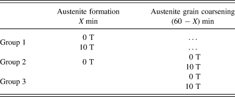
Three groups were designed to investigate the effects of magnetic field on the austenite grain size by applying magnetic field during austenite formation, austenite grain coarsening, and the whole austenitization process, respectively. Group 1 is to investigate the influence of magnetic field on the austenite grain size during the austenite formation stage by holding the samples treated without and with 10 T magnetic field at 950 °C for X min; Group 2 is to investigate the effects of magnetic field on the austenite grain size during the austenite grain coarsening stage by holding the samples treated without magnetic field at 950 °C for X and then transferring the samples into 0 T/10 T magnetic field at 950 °C for (60 − X) min; Group 3 is to investigate the effects of magnetic field on the austenite grain size during the whole austenitization process by holding the samples treated without and with 10 T magnetic field at 950 °C for 60 min. The details of the three groups are summarized in Table I. All of the samples after heat treatment at 950 °C were quenched into water and then etched by picric acid to reveal the prior austenite grain boundary. Reference Elwazri, Wanjara and Yue23 The austenite grains were observed using OM. The linear intercept method was used to measure the grain size.
Summary of experimental procedures for investigation of austenite grain size.

III. RESULTS AND DISCUSSION
A. Effects of magnetic field on the austenite formation
The initial microstructure of the experimental material is shown in Fig. 1 with different magnifications and consists of typical lamellar pearlite (the mixture of lamellar ferrite and cementite) with proeutectoid cementite distributed along the prior austenite grain boundary.
Initial microstructure of the experimental material. (a) low magnification; (b) high magnification.

The austenite was found to nucleate in the samples heated for 30 s at 950 °C regardless of treatment without and with magnetic field. Several samples have been examined carefully after holding for less than 30 s at 950 °C, but the austenite has never been found. Figure 2 shows the OM images of samples held at 950 °C for 30 s. The bright points are the austenite (martensite after quenching) particles which present the austenite nucleation sites. It is seen obviously that the bright points in the samples treated with magnetic field are much less than those of the samples treated without magnetic field.
OM images of the microstructure in samples held at 950 °C for 30 s without (a) and with (b) 10 T magnetic field treatment.

Figure 3 shows the SEM images of the microstructure shown in Fig. 2. The austenite fraction in the samples treated with magnetic field is much lower than that of the samples treated without magnetic field. The sizes of austenite particles in the two samples are similar, but the number of austenite particles in the sample treated with magnetic field is much less, which coincides with the comparison of OM images in Fig. 2. From the experimental results shown in Figs. 2 and 3, it can be concluded that magnetic field decreases the nucleation rate of austenite largely.
SEM images of the microstructure in samples held at 950 °C for 30 s without (a) and with (b) 10 T magnetic field treatment.

SEM images in Figs. 4(a)–4(d) show microstructure evolution during P → γ transformation after holding the samples treated without and with magnetic field at 950 °C for 34 s and 38 s. It can be seen clearly that the austenite fraction in the samples treated with magnetic field is much lower.
SEM images of microstructure evolution in the samples held at 950 °C for 34 s (a, b) and 38 s (c, d) without (a, c) and with (b, d) 10 T magnetic field treatment.

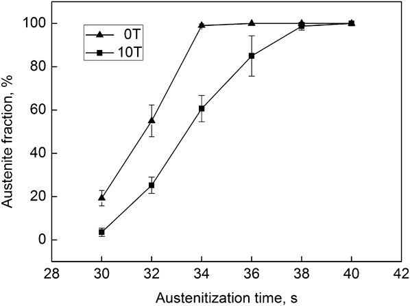
The variation of austenite fraction against holding time is shown in Fig. 5 and the effects of magnetic field on the P → γ transformation kinetics can be clearly seen. The P → γ transformation lasts 6 s (from 30 to 36 s) in the samples treated without magnetic field, but the same process lasts 10 s (from 30 to 40 s) if magnetic field is applied. The rate of transformation (slope of the curve) is smaller for the samples treated with magnetic field. Therefore, the P → γ transformation of samples treated with magnetic field spent more time and finished later. This difference of P → γ transformation rate shown above should be resulted from the nucleation rate change induced by magnetic field.
Variation in fraction of austenite in the samples treated without and with magnetic field.

The mechanism about the effects of magnetic field on the nucleation rate is analyzed in the following way. An applied magnetic field influences the thermodynamic state of the phases in steels directly. This is clearly shown in Eq. (1) (Refs. Reference Guo and Enomoto4 and Reference Enomoto, Guo, Tazuke, Abe and Shimotomai9) for calculating the Gibbs free energy change induced by magnetic field:
 $$\Delta G = - \int_0^H {M{\mathop{\rm d}\nolimits} H} \quad {\rm{,}}$$
where ΔG M is the Gibbs free energy change by magnetic field, M is magnetization, and H is the applied magnetic field strength.
The austenite and cementite are paramagnetic phases at 950 °C and cannot be influenced by the magnetic field with respect to Gibbs free energy. Ferrite transforms into ferromagnetic state above 770 °C (Curie temperature), but the high magnetic field contributes to the ferrite stabilization even above the Curie temperature Reference Enomoto, Guo, Tazuke, Abe and Shimotomai9 and the Gibbs free energy of ferrite is decreased at 950 °C by the high magnetic field. Reference Liu, Fang, Wang, Ma and Peng24 For the austenitization process: ferrite (α) + cementite (θ) → austenite (γ), the transformation of Gibbs energy change is decreased by the applied magnetic field.
According to the relationship between nucleation rate of diffusion transformation and Gibbs free energy change as shown in Eq. (2), Reference Lange, Enomoto and Aaronson25 the smaller Gibbs free energy change leads to lower nucleation rate.
 $$I = A \cdot D\exp \left( { - {{\Delta G} \over {kT}}} \right) = A \cdot D\exp \left[ { - {{K{\sigma ^2}} \over {\left( {\Delta {g_1} + \Delta {g_2}} \right)kT}}} \right]\quad {\rm{,}}$$
where I is the nucleation rate, A is complex constant independent of temperature, D is the diffusivity of carbon in austenite, kT has its usual meaning, ΔG is the driving force for nucleation, K is the shape factor of nucleus, σ is the interfacial energy between parent and new phases, Δg 1 is the volume Gibbs free energy change associated with nucleation, namely, ΔG M in Eq. (1), Δg 2 is the volume strain energy of nucleation. Lower nucleation rate and invariant growth rate of austenite grains lead to a slower transformation rate and longer transformation time for P → γ transformation as shown in Fig. 5.
Cementite does not dissolve during the P → γ transformation and is left to dissolve in the austenite matrix. Figures 6(a)–6(f) present the cementite dissolution process in the samples after P → γ transformation. It is seen clearly that the cementite particles in the samples treated with magnetic field dissolved more slowly than those of the samples treated without magnetic field. For the samples treated without magnetic field, the cementite dissolution finished after heating for 120 s, but the same process needed 125 s when magnetic field was applied. The delayed time of cementite dissolution induced by magnetic field is about 5 s which is similar to the time difference (4 s) of the P → γ transformation time between the samples treated without and with magnetic field. Therefore, the delay of cementite dissolution should be just caused by the longer P → γ transformation time, and high magnetic field has no effect on the cementite dissolution. Cr diffusion in high magnetic field can be used for explaining this experimental result.
SEM images of cementite particles dissolution in the samples held at 950 °C for 50 s (a, b), 120 s (c, d), and 125 s (e, f) without (a, c, e) and with (b, d, f) 10 T magnetic field.

The high content of Cr in cementite which is calculated to be 11 wt% by means of Thermal-Calc software according to the chemical composition of the material and the lower diffusion mobility of Cr determine that the cementite particles dissolution should be controlled by Cr diffusion. Some researchers Reference Shtansky, Nakai and Ohmori26 also suggest that the kinetics of austenitization is controlled by Cr diffusion in the later stage. Nakamichi Reference Nakamichi, Tsurekawa, Morizono, Watanabe, Nishida and Chiba27 found that there is no noticeable influence of a high magnetic field on the Ti diffusion with the vacancy mechanism in γ-iron and pointed out that the lattice stiffening due to field-induced magnetic ordering cannot influence the Ti diffusion through vacancy mechanism; this is because the vacancy in the neighborhood of Ti atom causes a relaxation. Additionally, the contribution of the applied field to the vacancy formation energy can be negligible. Reference Nakamichi, Tsurekawa, Morizono, Watanabe, Nishida and Chiba27 Therefore, the Ti diffusion in γ-iron is not affected by the external applied magnetic field. This result suggests that the magnetic field most likely would not influence the Cr diffusion either, since the diffusion of both Ti and Cr obeys vacancy mechanism. Reference Porter and Easterling28 It can be seen that Cr diffusion controls the cementite dissolution and is not affected by the applied magnetic field. Therefore, the magnetic field has no effect on the cementite dissolution.
Based upon the above discussion, the high magnetic field slows down the austenite formation process, which is just caused by the longer time consumption for P → γ transformation in the field treated samples; the high magnetic field has no effect on the cementite dissolution.
B. Effects of magnetic field on the austenite grain size
According to the above experimental results, the finishing time of cementite dissolution without magnetic field treatment was determined to be 120 s (2 min), namely, the value X described in the experimental procedures is 2 min. The following three groups were designed to investigate the effects of magnetic field on the austenite grain size during austenite formation, grain coarsening, and the whole austenitization process.
Figure 7 shows the austenite grain microstructure treated without and with magnetic field only during austenite formation. The austenite grain sizes were determined to be 84.2 ± 3.9 μm and 96.6 ± 5.5 μm, respectively, for samples treated without and with magnetic field. High magnetic field applied during the austenite formation increases the austenite grain size. As discussed above, austenite nucleation rate is reduced obviously by high magnetic field, so the austenite grains have longer time for growing into larger size before impingement. The grains in Figs. 7(a) and 7(b) show equiaxed morphology which means that the magnetic field has no influence on the austenite grain shape. The proeutectoid ferrite grains formed under magnetic field treatment are obviously elongated along the field direction, which can be qualitatively explained by dipolar interaction between atomic magnetic moments carried by iron atoms in ferrite grains. Reference Zhang, Esling, Muller, He, Zhao and Zuo29 In our case, austenite grain shape cannot be changed by magnetic field because austenite is paramagnetic.
OM images of austenite grains after heating at 950 °C for 2 min without (a) and with (b) 10 T magnetic field treatment.

Figure 8 shows the austenite grain microstructure treated without and with magnetic field only during austenite grain coarsening. The grain sizes were tested to be 94.3 ± 5.1 μm and 93.1 ± 4.9 μm, respectively, for samples treated without and with magnetic field, so the grain sizes almost keep invariant by applying magnetic field only during austenite grain coarsening. Interface energy strongly influences the grain size which determines the amount of interface. Higher interface energy promotes the grain coarsening to decrease the amount of grain boundary; conversely, lower interface energy suppresses the grain coarsening. If no magnetic field is applied, the interface energy is determined by crystal structure and composition between the two neighboring grains. If there is an applied magnetic field, the interface energy difference resulting from the magnetization between the two grains should be considered. The ferrite/cementite interface energy is increased by high magnetic field, which is caused by the Gibbs energy reduction for ferrite and cementite on both sides of the interface due to magnetization. Reference Zhang, Gey, He, Zhao, Zuo and Esling30 Weiss molecular field theory calculation also proves this effect of magnetic field on the ferrite/cementite interface energy. Reference Xia, Zhang, Lan, Liu and Yang18 Magnetic field also increases the ferrite/ferrite energy in a Fe–Sn alloy. Reference Tsurekawa, Okamoto, Kawahara and Watanabe31 If there is no ferromagnetic phase on any side of the interface, magnetic field cannot influence the interface energy through Gibbs energy change due to magnetization. The Gibbs energy of austenite which is in a paramagnetic phase does not be influenced by magnetic field. Therefore, high magnetic field has no effect on the austenite interface energy and thus cannot change the austenite grain size through interface energy.
OM images of austenite grains in the samples after heating at 950 °C for 2 min out of magnetic field and then heating for 58 min without (a) and with (b) 10 T magnetic field treatment.

Figure 9 shows the austenite grain microstructure treated without and with magnetic field during the whole austenitization process (including austenite formation and grain coarsening) at 950 °C. The grain sizes were measured to be 93.5 ± 4.5 μm and 106.3 ± 5.9 μm, respectively, for samples treated without and with magnetic field. High magnetic field increases the austenite grain size by applying magnetic field during the whole austenitization process. It is shown from the above experimental results that magnetic field increases the austenite grain size by applying magnetic field only during austenite formation and does not change the grain size by applying magnetic field only during grain coarsening. Therefore, this change shown in Fig. 9 is caused by the influence of magnetic field during austenite formation stage and has nothing to do with that of austenite grain coarsening. The larger austenite grains cause the lower transformation temperature and thus refine the pearlitic microstructure Reference Li, Godfrey and Liu21 during the following continuous cooling. The finer pearlite is helpful for the processing Reference Choi and Park20 and mechanical Reference Elwazri, Wanjara and Yue32 properties enhancement of steels. Zhang Reference Zhang, Zhang, Gong, Esling, Zhao and Zuo33 investigated the magnetic-field-induced microstructural features in a high carbon steel and found that high magnetic field increases the amount of abnormal structure. The proposed origin is that the magnetic field may increase the austenite grain size and thus promote the transformation of carbon-depleted austenite to ferrite. Zhang's speculation is consistent with our experimental results. As can be seen here, the investigation of magnetic field effects on the austenitization is important for understanding the magnetic-field-induced behavior in steels.
OM images of austenite grains in the samples after heating at 950 °C for 60 min without (a) and with (b) 10 T magnetic field treatment.

IV. CONCLUSIONS
The effects of high magnetic field on the kinetics of austenite formation and grain size have been investigated. The chief results obtained are as follows.
-
(1) High magnetic field retards the P → γ transformation by decreasing the nucleation rate of austenite and extends the transformation time, while there is no effect of magnetic field on the cementite dissolution.
-
(2) High magnetic field slows down the austenite formation process, which is just caused by the longer P → γ transformation time due to magnetic field.
-
(3) The austenite grain size is increased by magnetic field applied during austenite formation process, while the austenite grain size almost keeps invariant by applying high magnetic field during austenite grain coarsening. If high magnetic field is applied during the whole austenitization process, the austenite grain size is also increased.