INTRODUCTION
Fasciola hepatica is a digenean parasite that causes disease of economic importance in sheep and cattle, and as a zoonosis it is classed by the World Health Organisation as a neglected tropical disease (Hopkins, Reference Hopkins1992; Rim et al. Reference Rim, Farag, Sornmani and Cross1994; Hillyer and Apt, Reference Hillyer and Apt1997; WHO, 2015).
Reports of the ploidy of F. hepatica vary, yet determining ploidy is important to enhance our understanding of parasite biology and is essential to increase our knowledge of parasite genetics, reproduction and the level of gene flow from one population to another. Such studies are necessary to better understand the genetic evolution and spread of drug resistance within parasite populations. Knowledge of ploidy is also vital for facilitating our understanding of genetic diversity of F. hepatica populations, genome assembly (Cwiklinski et al. Reference Cwiklinski, Dalton, Dufresne, La Course, Williams, Hodgkinson and Paterson2015) and for design of gene knockdown experiments in downstream functional analyses such as RNA interference (McGonigle et al. Reference McGonigle, Mousley, Marks, Brennan, Dalton, Spithill, Day and Maule2008).
In the UK, F. hepatica was first reported to be diploid with 10 bivalent chromosomes (2n = 2x = 20) by Sanderson (Reference Sanderson1953). Since then there have been several reports describing fluke with 10 bivalent pairs of chromosomes including the triclabendazole resistant laboratory isolate of F. hepatica Sligo (n = 5) originally isolated from sheep in the Republic of Ireland (Fletcher et al. Reference Fletcher, Hoey, Orr, Trudgett, Fairweather and Robinson2004), wild-type F. hepatica (n = 15) from cattle livers in Northern Ireland (Fletcher et al. Reference Fletcher, Hoey, Orr, Trudgett, Fairweather and Robinson2004) and F. hepatica (n = 10) obtained from a cattle liver in Slovakia (Reblánová et al. Reference Reblánová, Špakulová, Orosová, Králová-Hromadová, Bazsalovicsová and Rajský2011). In contrast, the triclabendazole susceptible laboratory isolate Cullompton (n = 10), originally isolated from sheep in the UK, was shown to be triploid with 30 univalent chromosomes (2n = 3x = 30; Fletcher et al. Reference Fletcher, Hoey, Orr, Trudgett, Fairweather and Robinson2004).
To analyse ploidy in F. hepatica, the rapidly dividing sperm cells can be used to visualize chromosomes. Spermatogenesis in F. hepatica follows a series of three mitotic divisions (from one primary spermatogonia to two secondary spermatogonia to four tertiary spermatogonia to an 8-cell rosette of primary spermatocytes) followed by two meiotic divisions to produce 16 secondary spermatocytes and then 32 haploid spermatozoa (John, Reference John1953; Gresson, Reference Gresson1965; Stitt and Fairweather, Reference Stitt and Fairweather1990). The triploid Cullompton parasites were aspermic due to a failure of the first meiotic division (Fletcher et al. Reference Fletcher, Hoey, Orr, Trudgett, Fairweather and Robinson2004), and were therefore assumed to undergo parthenogenesis to reproduce (Hanna et al. Reference Hanna, Edgar, Moffett, McConnell, Fairweather, Brennan, Trudgett, Hoey, Cromie, Taylor and Daniel2008).
Most of the studies on Asian Fasciola spp. do not differentiate between F. hepatica and its sister species Fasciola gigantica because intermediate forms of the parasite exist (Itagaki et al. Reference Itagaki, Tsutsumi, Ito and Tsutsumi1998, Reference Itagaki, Sakaguchi, Terasaki, Sasaki, Yoshihara and Van Dung2009; Terasaki et al. Reference Terasaki, Noda, Shibahara and Itagaki2000; Peng et al. Reference Peng, Ichinomiya, Ohtori, Ichikawa, Shibahara and Itagaki2009). Diploid and triploid Fasciola spp. have been isolated from both cattle and deer livers in Japan (1 diploid and 18 triploid; Terasaki et al. Reference Terasaki, Moriyama-Gonda and Noda1998, Reference Terasaki, Noda, Shibahara and Itagaki2000; 3 triploid; Itagaki et al. Reference Itagaki, Tsutsumi, Ito and Tsutsumi1998; 1 diploid and 11 triploid; Itagaki et al. Reference Itagaki, Kikawa, Sakaguchi, Shmo, Terasaki, Shibahara and Fukuda2005a ); from cattle livers in Vietnam (19 diploid and 22 triploid; Itagaki et al. Reference Itagaki, Sakaguchi, Terasaki, Sasaki, Yoshihara and Van Dung2009); and from cattle livers in Korea (65 diploid and 19 triploid; Terasaki et al. Reference Terasaki, Noda, Shibahara and Itagaki2000; 1 diploid and 1 triploid; Itagaki et al. Reference Itagaki, Kikawa, Terasaki, Shibahara and Fukuda2005b ). In Korea an additional level of complexity has also been reported as in addition to diploid (n = 143) and triploid (n = 23) Fasciola spp., mixoploid (n = 46), also called mosaic or chimera, Fasciola spp. were found. The mixoploid parasites have a mixture of cells, some with a diploid number of chromosomes and some with a triploid number (2x/3x; Rhee et al. Reference Rhee, Eun and Lee1987). Usually the Asian triploids showed abnormal spermatogenesis (Itagaki et al. Reference Itagaki, Tsutsumi, Ito and Tsutsumi1998, Reference Itagaki, Sakaguchi, Terasaki, Sasaki, Yoshihara and Van Dung2009). However diploid Fasciola spp. with no sperm and triploid Fasciola spp. with small numbers of sperm have also been found (Terasaki et al. Reference Terasaki, Moriyama-Gonda and Noda1998, Reference Terasaki, Noda, Shibahara and Itagaki2000).
To date, studies on the ploidy of F. hepatica in the UK have been limited to small numbers (5–15 parasites; Fletcher et al. Reference Fletcher, Hoey, Orr, Trudgett, Fairweather and Robinson2004). Due to the differing reports of ploidy in this parasite, here we determined the ploidy of a larger number of F. hepatica isolated from naturally infected sheep and cattle in Great Britain. The results of this study may also be relevant to other areas where F. hepatica exists in isolation from F. gigantica.
MATERIALS AND METHODS
Fasciola hepatica collection
F. hepatica was recovered from the livers of 66 naturally infected lambs between November 2012 and February 2013, from three different abattoirs located in Wales, North West England and Central England. Based on the catchment areas of these abattoirs, these samples represent field populations of F. hepatica from England, Wales and Scotland. F. hepatica was recovered from 35 naturally infected cattle livers between October 2013 and January 2014, from an abattoir located in Wales. Based on ear tag information, these samples represent field populations of F. hepatica from England and Wales, and the cattle were a mixture of beef and dairy breeds with a mean age of 7·4 years (range 1·6–15·1).
Sheep livers were transported to the University of Liverpool, where adult parasites were isolated from the bile ducts. Parasites from cattle livers were isolated in situ at the abattoir. To purge intestinal contents and eggs, the parasites were incubated, for a minimum of 2 h at 37 °C in 1–2 mL of Dulbecco's Modified Eagle's Media (Sigma-Aldrich) with 120 μg mL−1 of gentamicin (Sigma-Aldrich) and 120 μg mL−1 of amphotericin B (Sigma-Aldrich). After incubation, each individual parasite was washed in Dulbecco's Phosphate Buffered Saline (PBS). A total of 565 F. hepatica were recovered from sheep with 1–12 parasites obtained from each liver, and 150 F. hepatica were recovered from cattle with 1–7 parasites from each liver.
Determining ploidy of Fasciola hepatica
An additional 129 F. hepatica collected from sheep were used to optimize methods of chromosome visualization adapted from Fletcher et al. (Reference Fletcher, Hoey, Orr, Trudgett, Fairweather and Robinson2004) and Reblánová et al. (Reference Reblánová, Špakulová, Orosová, Králová-Hromadová, Bazsalovicsová and Rajský2011). While it was preferable to use fresh material and perform an aceto-orcein squash immediately after a 2 h purge of adult F. hepatica, the practicalities of using large numbers of parasites necessitated purging for 2 h followed by incubation in 0·025% (w/v) colchicine (Sigma-Aldrich) in PBS, pH 7·4 for 1 h at room temperature (RT), followed by incubation in 75 mm potassium chloride (VWR) for 1 h at RT, and fixing (3:1, ethanol: acetic acid). Aceto-orcein squash could then be performed at a later date. Briefly, for aceto-orcein squash a section of the distal two-thirds of the parasite (approximately 1 mm3) was macerated in a drop of 3% (w/v) orcein (Sigma–Aldrich) in 45% (w/v) acetic acid on a glass slide, and then squashed under a cover slip using filter paper to soak up the excess stain. All the samples of F. hepatica from sheep were examined in this manner but all parasites from cattle were examined immediately after purging i.e. using fresh material.
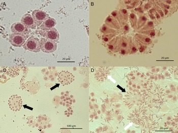
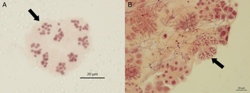
Preparations were examined using a Zeiss Axio Imager M2 microscope and Zen 2011 software. The stages of spermatogenesis were identified: rosettes (8-cell stage); 16-cell stage; 32-cell stage and spermatids and sperm (Fig. 1). The number of chromosomes was counted in well-spread cells (Fig. 2); they were most clearly identified in the first meiotic division from the rosette to the 16-cell stage. Triploid F. hepatica is aspermic or has very few abnormally developed sperm, and do not undergo the same stages of spermatogenesis as diploid parasites (Terasaki et al. Reference Terasaki, Noda, Shibahara and Itagaki2000; Fletcher et al. Reference Fletcher, Hoey, Orr, Trudgett, Fairweather and Robinson2004; Hanna et al. Reference Hanna, Edgar, Moffett, McConnell, Fairweather, Brennan, Trudgett, Hoey, Cromie, Taylor and Daniel2008; Itagaki et al. Reference Itagaki, Sakaguchi, Terasaki, Sasaki, Yoshihara and Van Dung2009). In previous studies the ploidy of F. hepatica was determined at the same time as determining the presence or absence of sperm (Fletcher et al. Reference Fletcher, Hoey, Orr, Trudgett, Fairweather and Robinson2004; Hanna et al. Reference Hanna, Edgar, Moffett, McConnell, Fairweather, Brennan, Trudgett, Hoey, Cromie, Taylor and Daniel2008; Itagaki et al. Reference Itagaki, Sakaguchi, Terasaki, Sasaki, Yoshihara and Van Dung2009). Therefore, here the presence of sperm was used as a proxy to determine diploidy and a parasite was deemed to be diploid if either 10 bivalent chromosomes or sperm were observed.
The different stages of spermatogenesis in Fasciola hepatica: (A) the 8-cell or rosette stage; (B) the 16-cell stage; (C) the 32-cell stage (black arrows); (D) the nuclei from the 32-cell stage elongate to spermatids (black arrow) and then sperm (white arrows).

Examples of chromosomes from Fasciola hepatica: in well-spread cells (arrows) chromosomes can be counted: (A) F. hepatica from a sheep liver; (B) F. hepatica from a cow liver.

Statistical programs
The StatCalc function of Epi Info 7 (http://wwwn.cdc.gov/epiinfo/7/) was used to calculate sample size. Calculations were initially based on an expected proportion of 50% triploid individuals (to give the highest possible sample size required). A sample size of 384 individuals gave 95% confidence level with 5% confidence limits (precision).
RESULTS
Ploidy was determined for 565 and 150 of the F. hepatica parasites isolated from naturally infected sheep and cattle, respectively. All 715 parasites were diploid. The proportion of triploids identified was 0% (95% CI 0–0·49%; Hanley and Lippman-Hand, Reference Hanley and Lippman-Hand1983). No difference between the ploidy of parasites isolated from sheep and cattle was found.
Ten bivalent chromosomes (Fig. 2) and sperm were observed in 335 (46·9%) of all F. hepatica samples; sperm alone were observed in 380 (53·1%) of the samples. Different stages of spermatogenesis: rosettes (8-cell stage); 16-cell stage; 32-cell stage and spermatids and/or sperm were observed in 93·3, 34·7, 72·9 and 100% of samples, respectively (Fig. 1; Table 1). All stages of spermatogenesis were observed in 144 F. hepatica from sheep and 53 F. hepatica from cattle. Sperm cells were commonly associated with the rosette and 32-cell stages; fewer 16-cell stages were observed. If the 16-cell stages were excluded, all other stages of spermatogenesis (rosette, 32-cell and sperm) were seen in 360 F. hepatica from sheep and 146 from cattle (Table 1).
Ploidy of wild British Fasciola hepatica from sheep and cattle, a breakdown of the spermatic stages observed and details of parasite burden

a Total enumeration of parasites was not determined for 19 of the sheep livers, therefore these figures are based on 47 of the 66 sheep.
DISCUSSION
The F. hepatica samples analysed in this study were isolated from naturally infected sheep and cattle from England, Wales and Scotland. All 715 parasites were diploid as shown by detection of 10 bivalent chromosomes, or sperm. These results are consistent with other studies of ploidy in wild populations of F. hepatica. In Northern Ireland a smaller scale study identified diploid parasites (n = 15) isolated from naturally infected cattle (Fletcher et al. Reference Fletcher, Hoey, Orr, Trudgett, Fairweather and Robinson2004) and similarly a study in Slovakia which analysed 10 parasites from a cattle liver found exclusively diploid organisms (Reblánová et al. Reference Reblánová, Špakulová, Orosová, Králová-Hromadová, Bazsalovicsová and Rajský2011). Overall the results from this study suggest that triploidy in F. hepatica populations, at least in Great Britain, is a rare occurrence. The presence of sperm in all of the mature parasites studied here is in contrast to Fasciola spp. in Asian populations which have frequently been described as aspermic or, triploid and aspermic, and are assumed to reproduce by parthenogenesis (Itagaki et al. Reference Itagaki, Tsutsumi, Ito and Tsutsumi1998, Reference Itagaki, Sakaguchi, Terasaki, Sasaki, Yoshihara and Van Dung2009; Terasaki et al. Reference Terasaki, Moriyama-Gonda and Noda1998, Reference Terasaki, Noda, Shibahara and Itagaki2000). The results here suggest that sexual reproduction, rather than parthenogenesis, is the most frequent means of reproduction in F. hepatica populations from Great Britain.
Different isoenzyme electrophoretic patterns provide evidence that triploidy in Japanese Fasciola spp. has arisen independently on more than one occasion (Agatsuma et al. Reference Agatsuma, Terasaki, Yang and Blair1994). Sequence comparison of the mitochondrial NADH dehydrogenase subunit I (NDI) and cytochrome c oxidase subunit I (COI) from triploid Fasciola spp. from Japan showed identity to F. gigantica from Zambia but were different to F. hepatica from Uruguay (Itagaki et al. Reference Itagaki, Tsutsumi, Ito and Tsutsumi1998). However, when comparing the ribosomal DNA internal transcribed spacer (ITS) 2 sequence, 6 of 7 Japanese triploids were almost identical to F. hepatica from Uruguay, while one parasite showed greater identity to F. gigantica from Indonesia (Itagaki and Tsutsumi, Reference Itagaki and Tsutsumi1998). Interestingly chimeric sequences (with nucleotide regions common to F. hepatica and F. gigantica) have been observed in the ITS-2 of Korean Fasciola spp. (Agatsuma et al. Reference Agatsuma, Arakawa, Iwagami, Honzako, Cahyaningsih, Kang and Hong2000) as well as the ITS-1 of triploid Fasciola spp. from Vietnam and aspermic Fasciola spp. from China and Bangladesh (Itagaki et al. Reference Itagaki, Sakaguchi, Terasaki, Sasaki, Yoshihara and Van Dung2009; Peng et al. Reference Peng, Ichinomiya, Ohtori, Ichikawa, Shibahara and Itagaki2009; Mohanta et al. Reference Mohanta, Ichikawa-Seki, Shoriki, Katakura and Itagaki2014). The majority of sequencing evidence therefore supports the hypothesis that triploids are the result of hybridization between F. hepatica and F. gigantica in areas where their distribution overlap. Since F. hepatica and F. gigantica have been found in the same definitive host (Amer et al. Reference Amer, Dar, Ichikawa, Fukuda, Tada, Itagaki and Nakai2011), this presents the opportunity for hybridization between the two species and confirmation of the ability of F. hepatica and F. gigantica to cross-fertilise using experimental systems supports this hypothesis (Itagaki et al. Reference Itagaki, Ichinomiya, Fukuda, Fusyuku and Carmona2011).
There is one report of triploidy in F. hepatica in the UK, in the Cullompton laboratory isolate. This isolate was derived in 1998 from multiple eggs obtained from the bile ducts of several sheep, which were used to infect snails, and then passaged twice through sheep and once through rats. The analysis of ploidy on this isolate was not performed until between 2001 and 2003 (Fletcher et al. Reference Fletcher, Hoey, Orr, Trudgett, Fairweather and Robinson2004). Since UK populations of Fasciola spp. are restricted to F. hepatica it is unlikely that triploids formed by hybridization, and our results suggest that triploidy in the Cullompton isolate is an artefact of isolation and laboratory passage of this isolate, rather than being a true reflection of what is occurring in the field. It is reported the Cullompton isolate was passaged several times following isolation; potentially creating a bottleneck. Passage was also through rats, a non-natural host of F. hepatica with far smaller livers able to support far fewer individual fluke, compared with sheep and cattle, which may have increased stress on the parasites. While we do not know definitively the effect long term laboratory passage has had on the parasite, these stresses may have led to the induction of triploidy in the Cullompton isolate. It is possible to induce polyploidy in helminths: heat shock induced polyploidy in Caenorhabditis elegans producing tetraploids from diploids, probably via a triploid (Madl and Herman, Reference Madl and Herman1979). Other stressors have also been shown to alter ploidy in vertebrates. For example, in bovine embryos the chemical cytochalasin B induced changes in ploidy (Bai et al. Reference Bai, Liu, Liu, Wu, Cheng, Bou and Li2011), and in leopard frogs, Rana pipiens, hydrostatic pressure has a similar affect (Dasgupta, Reference Dasgupta1962). In plant species, wounding and water or nutrient stress have also been shown to alter ploidy (Ramsey and Schemske, Reference Ramsey and Schemske1998). If one of these stressors produced a diploid female gamete, a triploid would be produced following fertilization with a haploid sperm. In order to maintain triploidy over successive generations, the initial creation of an accidental triploid would have to be derived from a parthenogenic diploid (Terasaki et al. Reference Terasaki, Noda, Shibahara and Itagaki2000). Fletcher et al. (Reference Fletcher, Hoey, Orr, Trudgett, Fairweather and Robinson2004) also suggested that triploidy could be produced by the fertilization of a haploid egg with two haploid sperm. However, in another study of F. hepatica, while more than one sperm became enclosed within the capsule of the egg, more than one sperm was never observed in the ooplasm (Sanderson, Reference Sanderson1959). A further possibility is the introduction of triploids, from an endemic area into the UK. Although there is evidence that triploid Fasciola spp. can stabilize within a population once introduced (Itagaki et al. Reference Itagaki, Kikawa, Sakaguchi, Shmo, Terasaki, Shibahara and Fukuda2005a ), the UK does not routinely import from such endemic areas.
Despite being triploid and aspermic, the infectivity of the Cullompton isolate has not been affected, since recent experiments have demonstrated that the isolate will successfully infect sheep and cattle (Hanna et al. Reference Hanna, Edgar, Moffett, McConnell, Fairweather, Brennan, Trudgett, Hoey, Cromie, Taylor and Daniel2008). However laboratory isolates are often not representative of what is happening in the field and knowledge of the provenance of isolates, their maintenance within the laboratory in terms of passage and continued assessment of their infectivity, pathogenicity and resistance status is an essential part of studies on F. hepatica but is frequently overlooked (Hodgkinson et al. Reference Hodgkinson, Cwiklinski, Beesley, Paterson and Williams2013).
Since the study reported here has shown that all the F. hepatica, from naturally infected sheep and cattle in Great Britain, were diploid, triploidy, if it exists, is rare. These results suggest that triploidy is most likely to develop from hybridization between F. hepatica and F. gigantica. If this is the case, it means that the results of this study may be extrapolated to other countries where, like the UK, the only species present is F. hepatica. This, taken with the observation that sexual reproduction was more frequent than parthenogenesis (since all the parasites studied here contained sperm) will enable further genetic and molecular studies of F. hepatica populations. Knowledge that F. hepatica populations in the UK are diploid facilitates the successful silencing of target genes using RNA interference (McGonigle et al. Reference McGonigle, Mousley, Marks, Brennan, Dalton, Spithill, Day and Maule2008) and is essential to support assumptions about inheritance patterns and for the calculation of observed and expected allele frequencies that are fundamental to population genetic studies (Dufresne et al. Reference Dufresne, Stift, Vergilino and Mable2014). Most recently the diploid nature of F. hepatica was an important consideration in the assembly of our 1·3 Gb genome (Cwiklinski et al. Reference Cwiklinski, Dalton, Dufresne, La Course, Williams, Hodgkinson and Paterson2015).
ACKNOWLEDGEMENTS
The authors would like to thank Mrs Catherine Hartley and Miss Katherine Allen for their support in the laboratory; the AHVLA for their assistance with cattle ear tag information; Dr Eleni Michalopoulou for her assistance identifying abattoirs; and the abattoirs, their workers and members of the food standards agency that assisted with the collection of samples for this project.
FINANCIAL SUPPORT
Ms Nicola Beesley is a PhD student funded by the Institute of Infection and Global Health, University of Liverpool. We are grateful for funding from the European Union (D. J. L. W., grant number KBBE-2010-4-265862: PARAVAC) and the Biotechnology and Biological Sciences Research Council (J. H., D. J. L. W., grant number BB/I002480/1).
ETHICAL APPROVAL
Ethical approval was received from the University of Liverpool's Veterinary Research Ethics Committee (VREC106 and VREC145).