Introduction
The incidence of tuberculous otitis media had fallen dramatically since the beginning of the twenty-first century, when 3–5 per cent of cases of otitis media were due to tubercle bacilli.Reference Mills, Adams and Cinnamond 1 More recent reports indicate that tuberculous otitis media has become less common.Reference Mills, Adams and Cinnamond 1 However, there has been a recent resurgence in tuberculosis, sometimes co-occurring with acquired immune deficiency syndrome (AIDS). The incidence of tuberculous otitis media in India has remained high. Similar to pulmonary tuberculosis, tuberculous otitis media may be acquired by contact with infected sputum; in the case of the middle ear, it can be contracted via the Eustachian tube by coughing up sputum laden with tubercle bacilli.Reference Slattery, Glasscock and Shambaugh 2 Tuberculous otitis media is typically characterised by an insidious, painless, afebrile onset and an enlarging perforation or multiple perforations with pale granulations, accompanied by hearing damage out of proportion to other symptoms and findings. Complications are more severe, including facial palsy, mastoiditis, sensorineural hearing loss and labyrinthitis. Recently, a large number of cases have had atypical presentation. Otolaryngologists should suspect tuberculous otitis media in any atypical case of chronic suppurative otitis media that does not respond to conventional treatment, particularly in at-risk groups. Investigations such as smear tests, sputum culture, polymerase chain reaction and histopathological examination have both advantages and drawbacks. This study emphasises the importance of clinical criteria, together with diagnostic tests, in the prompt diagnosis and appropriate management of tuberculous otitis media.
Materials and methods
A total of 502 chronic otitis media patients underwent tympanoplasty with mastoidectomy over a two-year period (January 2011 to December 2012) at Madras ENT Research Foundation, a tertiary care otolaryngology facility in South India. Each patient provided a detailed history and underwent a clinical examination including tuning fork tests and otomicroscopic, haematological, audiological and radiological examinations, including a chest X-ray and temporal bone computed tomography (CT). Granulations from the mastoid antrum were analysed by acid-fast bacilli smear test, bacterial culture, histopathological examination and polymerase chain reaction for the presence of Mycobacterium tuberculosis. For the smear test, purulent discharge from the external auditory meatus was analysed by hematoxylin and eosin staining and acid-fast bacilli staining were used. Pus from the external auditory canal was used for culturing M tuberculosis. For each patient, specimens were collected in three sterile containers: (1) containing formalin for histopathological analysis; (2) containing normal saline for acid-fast bacilli smear test and culture; and (3) for DNA purification using the QIAamp DNA mini kit (Qiagen, New Delhi, India) followed by polymerase chain reaction. The DNA mini kit can purify nucleic acids via enzymatic lysis (without mechanical homogenisation) from tissue samples, swabs, cerebrospinal fluid, blood or cells washed from urine samples.
In all, 25 patients (5 per cent) were positive for M tuberculosis in one or more diagnostic modality. All patients diagnosed with tuberculous otitis media were given post-operative antituberculous therapy, comprising four drugs (5 mg/kg isoniazid, 10 mg/kg rifampicin, 15 mg/kg ethambutol and 15–30 mg/kg pyrazinamide) for the first two months and then isoniazid and rifampicin only for the next four months.
Results
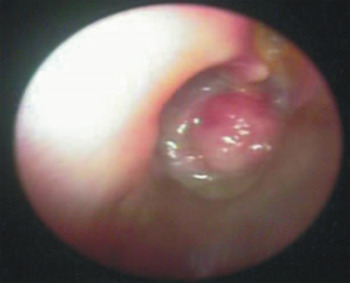
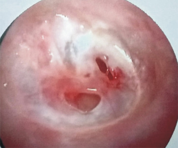
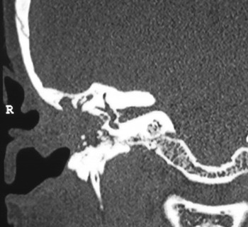
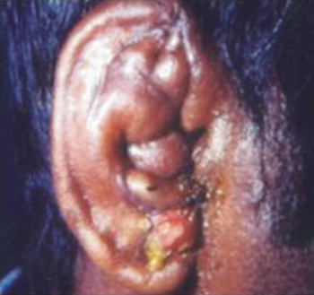
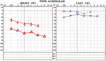
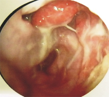
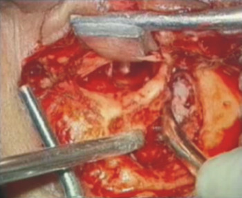
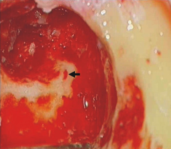
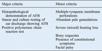
Of the 502 chronic otitis media patients, 60 per cent underwent canal wall down and 40 per cent underwent canal wall up mastoidectomy. A total of 25 patients (5 per cent) had a positive diagnosis of tuberculosis: 56 per cent were male and 44 per cent were female. Presenting symptoms included ear discharge (96 per cent), hearing impairment (68 per cent), otalgia (20 per cent), tinnitus (20 per cent), vertigo (16 per cent), headache (12 per cent) and facial palsy (8 per cent). Otomicroscopic examination findings of central perforation (68 per cent), aural polyp (12 per cent; Figure 1), cholesteatoma (8 per cent), masked mastoiditis (4 per cent), atelectatic tympanic membrane (4 per cent) and more than one perforation (4 per cent; Figure 2) were obtained. In two patients (8 per cent) with pre-operative facial paralysis, CT revealed extensive disease (Figure 3): one patient had a post-aural fistula (Figure 4) and the other had perichondritis of the pinna (Figure 5). In all, 64 per cent of patients had conductive hearing loss and 36 per cent had mixed hearing loss (Figure 6). Five patients (20 per cent) presented with bilateral disease and all others had unilateral disease. All patients underwent bilateral disease clearance even though antituberculous treatment was started after first-side surgery. Nine patients (36 per cent) had previously undergone surgery elsewhere and presented with disease recurrence (Figure 7). These patients underwent a revision procedure. No patient showed any evidence of pulmonary tuberculosis upon chest X-ray or had human immunodeficiency virus (HIV) infection. All patients had middle-ear or mastoid granulations (Figure 8), with osteomyelitis in 32 per cent, cholesteatoma in 12 per cent, an aural polyp in 12 per cent, ossicular chain erosion in 64 per cent, facial canal involvement in 24 per cent and lateral semicircular dehiscence in 4 per cent (Figure 9). Only 1 the 25 tuberculosis-positive patients had an extradural abscess (Figure 10). Tegmen defects in two patients were successfully repaired using tragal cartilage (Figures 11 and 12). Patients were diagnosed with tuberculous otitis media based on clinical criteria and positive findings in at least one test. Diagnostic criteria were classified as major or minor (Table I). Major criteria were histopathological demonstration of acid-fast bacilli (Figure 13), presence of acid-fast bacilli in ear discharge smear and culture tests (Figures 14 and 15), and positive polymerase chain reaction findings. Minor criteria were multiple tympanic membrane perforations, abundant pale granulations, severe hearing loss (with cochlear involvement), bony sequestra, constitutional symptoms and facial palsy. One major and two minor criteria were considered necessary for diagnosis. In the absence of a positive major criterion, the presence of three or more minor criteria indicated a high level of suspicion for tuberculous otitis media. Of the 25 tuberculosis-positive patients, 8 (32 per cent) had positive polymerase chain reaction findings, 5 (20 per cent) had positive smear findings, 5 (20 per cent) were culture positive and 15 (60 per cent) had positive histopathological findings (some had positive findings in more than one test).
Otoscopy image showing middle-ear granulations in tuberculous otitis media.

Otoscopy image showing multiple tympanic membrane perforations in tuberculous otitis media.

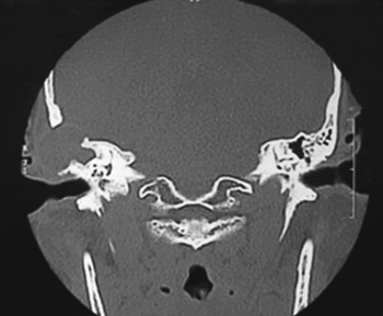
Coronal temporal bone computed tomography image of a patient with right tuberculous otitis media and facial palsy.

Photograph showing a left post-aural fistula.

Photograph showing perichondritis of the right pinna.

Pure tone audiograms for a tuberculous otitis media patient, showing mixed hearing loss.

Otoendoscopy image of a mastoidectomy cavity, showing post-mastoidectomy recurrence of granulation tissue.

Photograph showing pale granulations seen in tuberculous otitis media intra-operatively.

Photograph showing lateral semicircular canal fistula (indicated by arrow).

Coronal temporal bone computed tomography image of a patient with right-sided tuberculous otitis media and an extradural abscess.

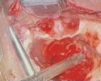
Photograph showing a tegmen defect during tympanomastoidectomy.

Intra-operative photograph showing repair of a tegmen defect with tragal cartilage.

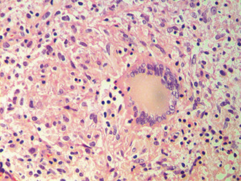
Photomicrograph showing tuberculous otitis media histopathology, including tubercle bacilli (H&E; 400×).

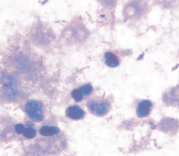
Photomicrograph showing a smear with tubercle bacilli (modified Ziehl–Neelsen stain, oil immersion microscopy; 100×).

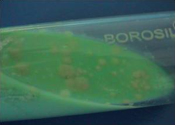
Photograph showing bacterial culture in Lowenstein Jensen medium.

Diagnostic criteria for tuberculous otitis media

AFB = acid-fast bacilli
All patients diagnosed with tuberculous otitis media were given post-operative antituberculous therapy. Facial paralysis recovered within three months of surgery and commencement of antituberculous treatment. This case series was followed up for 12 months to 3 years. All patients currently have completely healed ears with no evidence of active disease.
Discussion
Tuberculosis is a major public health problem in India, with a reported incidence of 1.5 per 1000 population; 4 per 1000 population are bacteriologically positive. The prevalence increases with poorly controlled diabetes mellitus or a compromised immune system, for example resulting from HIV infection. Tuberculous otitis media may be contracted via infected sputum or, for middle-ear infection via the Eustachian tube, by coughing up sputum laden with tubercle bacilli.Reference Slattery, Glasscock and Shambaugh 2 The disease also occurs in children without pulmonary tuberculosis via drinking unpasteurised milk from tubercular cows.
Jean Louis Petit first described tuberculosis of middle-ear cleft in the early eighteenth century.Reference Birrel 3 In 1853, Wilde clinically recognised tuberculosis of the mastoid bone. Ménière was the first to describe the effect of tuberculosis on the tympanic membrane in 1882, before the discovery of Koch's bacillus in the same year. In 1883, Eschle described the bacteria in otitis media. In 1915, Turner and Fraser implicated tuberculosis in 2–8 per cent of otitis media cases in children aged under 15 years and in 50 per cent of otitis media cases in children aged under 1 year.Reference Burns, Meyerhoff, Paparella and Shumrick 4 In the early twenty-first century, 1.3–18.6 per cent of all chronic otitis media cases were reportedly caused by tuberculosis, but more recent studies report tuberculous otitis media rates of 0.05–0.9 per cent.Reference Nadol, Merchant, Cummings, Harker and Krause 5 , Reference Grewal, Baser, Shahani and Khanna 6 Tuberculous otitis media is more common in children, and the frequency is now much higher than anticipated.Reference Palva, Palva and Karja 7
The pathogenesis of tuberculosis otitis media is variable. The tubercle bacilli gain access to otic structures via three routes: haematogenous dissemination, lymphatic channels or direct extension. Mycobacterial infection of the temporal bone begins in the marrow space of the temporal bone, within the tympanic membrane or within middle-ear mucosa and mastoid air cells.Reference Burns, Meyerhoff, Paparella and Shumrick 4 As delayed hypersensitivity to the bacterium develops, multiple foci of cheese-like (or caseous) necrosis develop. Erosion of the ossicular chain and fallopian canal may occur, with resultant conductive hearing loss and facial nerve injury. The granulomatous inflammatory process may extend ultimately into the temporal bone labyrinth via the round and oval windows. In untreated patients, extension of the granulomatous inflammatory process into the overlying skin and regional lymph nodes may produce persistent fistulae, often in the post-auricular region.
The classic clinical features of tuberculous otitis media were first described in 1953 by Wallmer as painless otorrhoea, multiple tympanic membrane perforations, pale granulation tissue, ipsilateral facial nerve paralysis, early severe hearing loss and bone necrosis, including sequestrum formation.Reference Wallmer 8 However, this combination of features is rarely observed today. Severe hearing loss occurs in 90 per cent of patients, often at an early stage, and may be conductive, sensorineural or mixed type. Middle-ear mucosa is hyperaemic and may contain pale granulations. However, single perforations are now predominant, usually with bulging granulation or aural polyposis. Moreover, facial nerve paralysis is reported in only 30–45 per cent of patients.Reference Burns, Meyerhoff, Paparella and Shumrick 4 Facial paralysis is much more common in children than in adults, and appears to be due to pressure from granulation tissue and bone sequestration around the facial canal.Reference Ormerod 9 Mastoid air cells are replaced by tuberculous granulations, which may perforate the mastoid cortex leading to post-aural abscess and subsequent (often multiple) fistula formation. Tuberculous mastoiditis can also present with sagging of the posterior meatal wall. Tuberculous otitis media may cause osteomyelitis, labyrinthitis, petrositis, lateral sinus thrombophlebitis and thrombosis, meningitis, brain abscess and tuberculoma of the brain.
However, the classic features are rarely observed today. A serious problem associated with tuberculosis of the middle ear is delayed diagnosis, stemming from non-specific signs and symptoms. In addition, most patients do not have coincident pulmonary tuberculosis. In 1980, Plester et al. reported that a dull, deep-seated, aching pain attributable to tension or mild pressure exerted by granulation tissue was diagnostically important.Reference Plester, Pusalkar and Steinbach 10 The presence of bulging granulation tissue in the mastoid, sequestrum of the mastoid bone or fallopian canal erosion should alert the surgeon to the possibility of tuberculous mastoiditis.Reference Grewal, Hathiram, Kameswaran and Kameswaran 11
A clinical examination including otomicroscopy, radiology (including chest X-ray and high-resolution temporal bone CT), and audiological and haematological investigations is recommended. Mantoux tuberculin tests may be unreliable for diagnosis. Tuberculous otitis media occurs in only 2 per cent of active pulmonary tuberculosis cases.Reference Arya, Dixit, Paramez, Sharma and Rathore 12 Smear findings are positive in 20 per cent of patients and cultures are positive in 5–35 per cent. Skin tests, however, are not very reliable. Histopathological examination of granulation tissue demonstrating acid-fast bacilli remains the cornerstone of definitive diagnosis. False negative rates of up to 10 per cent in the first histopathological tests are reported. DNA amplification by the polymerase chain reaction was originally developed by Mullis and Saiki.Reference Mullis and Saiki 13 This technique requires knowledge of the nucleic acid sequence of the region to be analysed, but is extremely rapid (single day), easy to automate, relatively cheap and extremely specific.Reference Mullis and Saiki 13 It can differentiate M tuberculosis from atypical Mycobacteria. Ideally, automated real-time nucleic acid amplification technology for the rapid, simultaneous detection of tuberculosis and rifampicin resistance (Xpert MTB/RIF assay) should be performed in all adults and children with suspected tuberculous otitis media.Reference Mahajan, Agarwal, Singh and Gadre 14 According to Cousins et al., the polymerase chain reaction has a sensitivity of 98.2 per cent and specificity of 97.4 per cent.Reference Cousins, Wilton, Francis and Gow 15 In 2003, Do et al. reported the findings of a comparative study of polymerase chain reaction and histopathological analyses.Reference Do, Kim, Choi, Lee, Lee and Sol 16 These authors found that 53 out of 81 patients had chronic granulomatous inflammation; of these, 32 per cent had positive findings for tuberculosis via acid-fast bacilli and 68 per cent had positive findings via polymerase chain reaction. They concluded that the polymerase chain reaction is a rapid, sensitive method for diagnosing tuberculosis in patients with suspected disease.Reference Do, Kim, Choi, Lee, Lee and Sol 16
Surgery may involve biopsy or a canal wall up or down procedure, depending on the pathology. Surgery is also recommended for functional reconstruction, removing bony sequestra and treating complications.Reference Vital, Printza and Zaraboukas 17 Facial nerve decompression may sometimes be necessary. First-line antituberculous treatment comprising a combination of isoniazid, rifampicin, ethambutol and pyrazinamide provides the highest efficacy with acceptable toxicity. A six-month course of these drugs (isoniazid, rifampicin, pyrazinamide and ethambutol are given for two months, followed by isoniazid and rifampicin for four months) is successful for most patients. If there is extensive middle-ear involvement, mastoid or bony involvement with sequestrate formation or osteomyelitis with sinus formation, treatment can be extended to nine months. Occasionally, because of microbial resistance or patient-related factors such as HIV infection or AIDS, second-line drug therapy may be necessary, usually after determining the sensitivity pattern. In the present study, no patient required second-line drug therapy.
-
• Tuberculous otitis media is a chronic granulomatous disease of the temporal bone
-
• Diagnosis is difficult due to a lack of reliable diagnostic tests
-
• Demonstration of bacteriological positivity is the exception rather than the rule
-
• The polymerase chain reaction test is recommended for suspected tuberculous otitis media
-
• New diagnostic criteria for tuberculous otitis media incorporating both diagnostic tests and clinical symptoms are proposed
In patients in the present series, hearing loss out of proportion to clinical findings, the presence of complications, post-mastoidectomy recurrence of granulation tissue, slow wound healing, persistent otorrhoea and formation of bony sequestra were all highly suggestive of tuberculous otitis media. Diagnosis of this disease can be difficult because of atypical presentation, and multiple investigative modalities may be required. A high index of suspicion is essential for diagnosis. Granulations from all patients who undergo mastoidectomy should be sent for acid-fast bacilli smear and culture tests, histopathological examination and M tuberculosis polymerase chain reaction, especially in endemic zones of the world. The Quantiferon-TB Gold Test is reported to be a useful diagnostic tool for both latent and active tuberculosis.Reference Simpson, Fox, Crouse and Field 18 Multi-drug resistant tuberculosis can be a challenge: aminoglycosides used to treat multi-drug resistant tuberculosis are potentially ototoxic and may cause loss of hair cells, supporting cells and spiral ganglion cells in the cochlea.Reference Duggal and Sarkar 19 The high incidence of tuberculous otitis media in the present series suggests that this disease is underdiagnosed in India.
Conclusion
Tuberculosis is a common infectious disease in developing countries, including India. A resurgence of tuberculous otitis media has been suspected in recent years. Recurrent otitis media could be due to a failure to detect tuberculous otitis media at the time of previous surgery. The main problems for diagnosis are non-specific signs and symptoms because the classic features are very rarely observed today. The lack of sensitivity and specificity of available diagnostic modalities adds to the problem. Hence, a combination of clinical criteria and diagnostic tests is now essential for diagnosing tuberculous otitis media. In areas of the world where tuberculous otitis media is making a comeback, otolaryngologists should consider this disease when treating chronic otitis media patients. This study suggests criteria for diagnosing tuberculous otitis media.