Endoscopes are essential in various medical specialties because they allow minimally invasive diagnostic and therapeutic procedures. To minimize the risk of transmitting pathogens, reusable endoscopes must be reprocessed and subjected to high-level disinfection at least. 1 Endoscope reprocessing is a worldwide challenge due to their highly complex design. Many outbreaks related to endoscopes have been reported, Reference Balan, Sfarti, Chiriac, Stanciu and Trifan2,Reference Humphries, Yang and Kim3 even when the recommendations for reprocessing were followed. Reference Ross, Baliga, Verma, Duchin and Gluck4,Reference Ofstead, Wetzler, Heymann, Johnson, Eiland and Shaw5
Build-up biofilm (BUB) develops gradually with repeated rounds of hydrated phases (ie, endoscopy procedures, point-of-use before cleaning, cleaning, liquid chemical disinfection/sterilization, and rinsing) and with dehydrated phases (ie, drying and storage). Reference Alfa and Singh6 BUB a key factor in persistent contamination of endoscope channels; it is more compact, more adherent, and more resistant to disinfectants Reference Alfa and Howie7 than traditional biofilm. These factors are particularly relevant for endoscope channels of smaller diameter that cannot be cleaned by brushing. Reference Pajkos, Vickery and Cossart8,Reference Johani, Hu and Santos9
The kinetics of BUB formation on endoscope channels suggests that the longer the endoscope is used, the greater the likelihood of BUB formation. Reference Alfa and Howie7 An endoscope can remain in service for up to 10 years, Reference Lee, Kim and Kim10 and its frequency of use varies according to the type of device and the healthcare service. Reference Vickery, Ngo, Zou and Cossart11 However, “older” devices are exposed to more opportunities for contamination and damage; thus, the longer these devices are used, the greater the risk of contamination of their channels.
Considering the negative impact of BUB for endoscope reprocessing and the absence of studies investigating the contamination of new endoscope channels in clinical use over a controlled length of time, we prospectively assessed the accumulation of protein and biofilm on the inner surfaces of new flexible gastroscope channels after multiple rounds of patient use and full reprocessing after 30 and 60 days of clinical use.
Material and methods
Setting
This study was performed at the endoscopy service of a public hospital (235 beds) in the midwestern region of Brazil, where procedures on adults and children are performed under any type of sedation or anesthesia. 12 The average number equipment reprocessing cycles during the study was ˜60 per month.
Reprocessing of flexible gastroscopes
For reprocessing the flexible gastroscopes (FGs), a protocol was assessed by 7 Brazilian experts in the field, selected through the Lattes Curriculum website (http://lattes.cnpq.br/). Reference Azevedo13 The protocol describes the following stages of reprocessing flexible endoscopes: before cleaning; leak testing; manual cleaning, rinsing, and drying; automated cleaning, disinfection (2% glutaraldehyde; Laboratoires Anios, Argentina), rinsing and drying (Endoclear, Brazil); manual drying and rinsing with 70% alcohol (this in case of storage); and storage. 1 The 2% glutaraldehyde was a ready-to-use, neutral pH, with a 10-minute contact time at room temperature, and the solution was reused for up to 30 days, associated with the dipstick test result (according to the manufacturer’s instructions). The reprocessing area has water, detergent, and air guns, with pedal operation. The storage cabinets have ultraviolet lamps and air filters (bottom and top). These cabinets did not provide air purging of the channels.
The reprocessing protocol was implemented as part of the routine service after the nursing personnel completed training over 3 weeks, based on the problematizing methodology of the Charles Maguerez Arc method. Reference Berbel14,Reference Primo, Guadagnin, Azevedo, Costa Luciano, Costa and Tipple15
Sampling
Three flexible video-gastroscopes (GIF-Q150, Olympus, Japan) were selected for the study because they were used most frequently. The gastroscopes were divided into groups according to the time they were included in the study (phases 1–3).
Phase 1: Baseline (before the implementation of the reprocessing protocol). One FG in clinical use for ˜5 years was included in the study before the implementation of the revised reprocessing protocol, when reprocessing was performed according to the Brazilian Society of Nursing in Endoscopy 16 and Brazilian legislation. Reference Azevedo13 The 3 internal channels of this gastroscope were replaced by new ones by a specialized company. The 3 channels that were replaced were sent to the microbiological laboratory for analysis.
Phase II: After 30 days of use in clinical practice. One month after implementing the reprocessing protocol, 3 FGs were selected for the study. The 9 channels (air, water, and biopsy channels) were removed and replaced by new ones. The gastroscopes were then returned to the endoscopy service for routine use in clinical practice and reprocessing for 30 days. Then, the channels of these 3 devices were replaced by new ones. The removed channels were transported to the microbiology laboratory for analysis.
Phase 3: After 60 days of use in clinical practice. The same 3 FG selected for phase 2 underwent channel replacement as previously described and were returned to clinical use reprocessing for 60 days. After the 60 days of use, the channels of these 3 devices were replaced by new ones. The removed channels were transported to the microbiology laboratory for analysis.
Procedures for removal and replacement of the FG channels
The channels were collected using aseptic procedures. Guidance was provided to the technicians of the accredited company regarding hand hygiene (before and after wearing gloves) and proper use of personal protective equipment (sterile gloves, hat, and mask), preparation of the workbench (cleaning, disinfection with 70% alcohol, and use of sterile surgical drape), and decontamination of the tools (cleaning, and wiping with 70% alcohol). Upon disengaging the ends of the channels, they were sealed with the aid of hemostats. The channels were transported to the microbiological laboratory for analysis in a disinfected plastic container with a lid.
Cutting flexible gastroscope channels
The external surface of the channels was cleaned with detergent and disinfected with 70% alcohol, and the ends remained sealed with hemostats. The channels were then placed in a Class II Biological Safety Cabinet (Pachane, Brazil), on a sterile drape. Channel segments were cut using a sterile blade, and sterile gloves were used to handle the channels.
Scanning electron microscopy
Fragments of the channels (1 cm long) were cut lengthwise, totaling 28 fragments, including those from the air–water junction. Each fragment was cut into 2 parts. Fragments of new (ie, unused) air, water, and biopsy channels, of the same model and submitted to 1 reprocessing cycle, were subjected to scanning electron microscopy (SEM).
The fragments were fixed in 2.5% glutaraldehyde (Sigma-Aldrich, St Louis, MO) and dehydrated in an increasing series of ethanol (30%, 50%, 70%, 80%, 90%, and 100%). Then they were dried in hexa-methyl-disilazane (Sigma-Aldrich) and assembled with a concave side facing upward prior to coating with gold (20 nm; Denton Vacuum, Moorestown, NJ). Finally, they were analyzed using a JSM-6610LA (JEOL, Japan). SEM for all fragments was performed in straight vertical lines from top to bottom, back and forth.
Protein test
Each channel fragment was subjected to protein extraction using the alkaline hydrolysis procedure, as previously described Reference Li, Webb, Kjelleberg and Rosche17,Reference Chowdhury, Rahman, Hu, Jensen, Deva and Vickery18 with modification. Briefly, the channel fragments were filled with a solution of ice-cold 2M-morpholino-ethane sulfonic acid (MES) (20 mM) in 0.9% saline containing NaOH (30%). The solution was subjected to sonication, spinning in a vortex mixer, incubation at 30°C for 30 minutes, and immersion in boiling water for 15 minutes. After cooling down, the extracted solution from each channel was transferred to individual plastic tubes and 32% HCl was added. The solution was then subjected to centrifugation (13,000 rpm for 5 minutes). A 150 μL aliquot of the of extracted sample from each channel was used to quantitate protein (Pierce micro-BCA protein Assay, ThermoFisher Scientific, Waltham, MA) at 560 nm. The protein concentration was calculated in µg/mL, using a standard curve according to the manufacturer’s instructions, optimized for use with dilute protein samples from 0.5 µg/mL. The protein per cm2 was calculated as follows: (µg/mL × volume of extracted sample)/cm2 inner surface area of segment extracted = µg/cm2.
Bacterial culture
Each channel segment was positioned in a “U” shape, and tryptic soy broth (TSB) (KASVI, Italy) was injected into the channel. The ends of the channels were then sealed with the aid of hemostats and were sonicated for 10 minutes (40 kHz; UNIQUE, Brazil). Subsequently, the ends of the channels were sealed with sterilized metal clips and were then incubated at 35°C per 48 hours. The TSB was then removed from the channels and transferred to 1.5-mL microtubes. The channels were immediately filled with the same volume of TSB and incubated under the same conditions, totaling 96 hours of incubation. The TSB removed from the channels after 48 and 96 hours of incubation was seeded (20 μL) onto blood agar (Laborclin, Brazil) was then incubated at 35°C for up to 72 hours or more in case of slow growth.
Colonies were evaluated using traditional microbiology techniques, including Gram staining and Ziehl-Neelsen staining. Bacterial identification and antimicrobial susceptibility testing were performed using the Vitek2 Compact system (BioMérieux, Durham, NC) and were defined according to standard break points. 19 Micrococcus spp, gram-positive bacilli and Mycobacterium spp were not subjected to antimicrobial susceptibility testing. The acid-fast bacteria, suggestive of mycobacteria, underwent genotypic identification by partial sequencing using Sanger methodology of the 16S rRNA gene (3130xL Genetic Analyzer Applied Biosystems, Foster City, CA) by a specialized company. The similarity analysis was restricted to the fragment limited by positions 4221781 and 4222788 of the Escherichia coli genome (GenBank CP001368.1). The same similarity index (99.38%) was obtained when the sequence was compared to those of isolated bacteria, including Mycolicibacterium phocaicum, M. mucogenicum, and M. llatzerense.
Results
Scanning electron microscopy
The air–water junction channels (n = 7) were included, totaling 28 fragments. The presence of organic residuals was detected in all channel fragments (n = 28 of 28) (Table 1). At the time of the fragment cuts, 3 channels (biopsy, air, and water) of FG #1 (phase 2) contained residual moisture.
Structural Damage, Biofilm and Debris Detected on the Luminal Surface of Flexible Gastroscope Channels in Use in Clinical Practice, Before and After (30 and 60 days) Implementation of a Reprocessing Protocol

Note. FG, flexible gastroscope; FG #1, flexible gastroscope no. 1; FG #2, flexible gastroscope no. 2; FG #3, flexible gastroscope no. 3. NV, channel surface not visible due to the thick layer of biofilm and/or other residuals; +, positive; ~, suggestive of biofilm.
a Channels positive for bacterial growth.
Structural damage (ie, holes, cracks, and grooves) was detected on the luminal surfaces of 20 of 28 fragments of channels submitted to SEM: 2 fragments from phase 1; 11 from phase 2; and 7 from phase 3 (Table 1). All 7 fragments of biopsy channels analyzed by SEM showed structural damage (Table 1). Structural damage was not verified on the 8 fragments of the remaining water and air channels (n = 8 of 28) due to the thick layer of biofilm and/or residual matter coating the inside of the channels (Table 1). Structural damage was also verified on the new air, water, and biopsy channels that had been reprocessed only once.
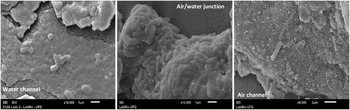
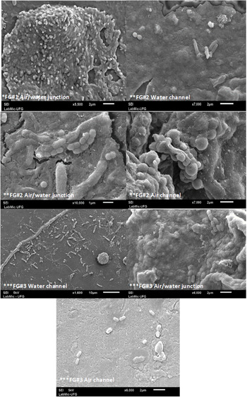
Extensive biofilms containing bacilli or rods and/or cocci were detected in the inner surfaces of 18 of 28 channels fragments, including those from the air–water junction. In phase 1, biofilms were detected on 1 water channel, 1 air channel, and at the air–water junction (Table 1 and Fig. 1). In phase 2, biofilms were detected on 2 water channels, 2 air channels, and 3 fragments from the air–water junction (Table 1 and Fig. 2). In phase 3, biofilms were detected on 3 water channels, 2 air channels, and 3 air–water junctions (Table 1 and Fig. 3).
Extensive biofilm, with bacilli or rods and/or cocci cell shape, on the luminal surface of flexible gastroscope channels before the implementation of a reprocessing protocol (phase 1). Images obtained by scanning electron microscopy.

Extensive biofilm, with bacilli or rods and/or cocci cell shape, on the luminal surface of flexible gastroscope channels after 30 days of use in clinical practice (phase 2) and implementation of a reprocessing protocol. Images obtained by scanning electron microscopy. *FG#1, flexible gastroscope no.1; **FG#2, flexible gastroscope no.2; ***FG#3, flexible gastroscope no. 3.

Extensive biofilm, with bacilli or rods and/or cocci cell shape, on the luminal surface of flexible gastroscope channels after 60 days in use in clinical practice (phase 3) and implementation of a reprocessing protocol. Images obtained by scanning electron microscopy. *FG#1, flexible gastroscope no. 1; **FG#2, flexible gastroscope no. 2; *** FG#3, flexible gastroscope no. 3.

As for the FG channels that showed positive cultures (n = 8), the presence of biofilm was detected in more than half (62.5%) of the fragments corresponding (paired) to those submitted to SEM (n = 5 of 8) (Table 1).
Amount of protein
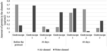
Residual protein was detected in all of the air and water channels of all of the FGs, with the exception of 1 channel in use for 30 days (FG #1, phase 2) (Fig. 4). Protein was not detected (<0.5 µg/mL) in any of the biopsy channels.
Amount of protein (µg/cm2) on the luminal surface of flexible gastroscope channels in use in clinical practice before and after (30 and 60 days) implementation of a reprocessing protocol. *FG#1, flexible gastroscope no. 1; **FG#2, flexible gastroscope no. 2; ***FG#3, flexible gastroscope no. 3.

Bacteriological culture
Bacterial growth was detected in 8 of 21 channels evaluated. Most growth was found in the FGs in use for 30 days after the implementation of the protocol (n = 6 of 8), of which 3 channels were from the same FG (#1, phase 2). The air channel showed the highest frequency of bacterial growth (n = 5 of 8); 1 was the FG channel in use before the protocol implementation (phase 1), 3 were FG channels in use for 30 days; and 1 was the FG channel in use for 60 days.
In total, 9 bacterial isolates were recovered from the channels, most were genus Mycolicibacterium spp (n = 5) and were detected in the air and/or water channels of 1 or more FGs in each phase evaluated (phases 1–3). Gram-negative bacteria (Pseudomonas aeruginosa, P. putida, and Aeromonas hydrophila/caviae) were isolated from a single FG (#1) in use for 30 days (phase 2). No bacteria was resistant to the tested antimicrobials.
Discussion
The results of this study reveal that the luminal surface of all air–water junction channels, and most of water and air channels of FGs were contaminated by BUB following 30 and 60 days of clinical use and manual and automated reprocessing according to a revised protocol. Similar results were reported for endoscopes in clinical use and reprocessed using an automated method. Reference Johani, Hu and Santos9,Reference Lee, Kim and Kim10,Reference Ren-Pei, Hui-Jun and Ke20 The rapid establishment of the BUB (30 and 60 days) on the channels evaluated in this study can be attributed to the fact that the channels on the connector tube (umbilical), were not replaced, thus allowing the recolonization of the new channel segments. Furthermore, the water and air channels and their junction are not accessible for brushing or friction during the cleaning step and, in the automated process, the force of the irrigation jets in the channel (shear stress) is not sufficient to remove strongly adhered microorganisms Reference Vickery, Ngo, Zou and Cossart11 and other organic and inorganic matter. These factors can also explain the detection of protein in most air and water channels in this study, demonstrating that the essential principle of cleaning, removing organic and inorganic matter, 1,21 is not possible to achieve. Brushing and friction comprise a critical parameter for biofilm removal during endoscope cleaning. Reference Ribeiro, Graziano, Olson, França and Alfa22
Structural damage was identified in the channels evaluated in this study, even in the new channels (n = 20 of 28). Damaged channels provide shelter to microorganisms and can protect them from disinfection. Structural damage on new endoscopes Reference Pajkos, Vickery and Cossart8 and after <4 weeks in use Reference Thaker, Kim, Sedarat, Watson and Muthusamy23 has been reported in previous studies and can be attributed to the use of accessories, such as biopsy forceps. Reference Hervé and Keevil24 In 8 of 28 sample fragments, a thick layer of debris or biofilm prevented the visualization of the surface of the other channels. This occurrence was also reported for channels used in endoscopy services in Australia. Reference Pajkos, Vickery and Cossart8 Protein, carbohydrates, and even dead microorganisms inside these channels can act as a conditioning film for the adhesion of microorganisms and formation of BUB and can also interfere in the action of cleaning agents and disinfectants. Reference Vickery, Ngo, Zou and Cossart11,Reference Hervé and Keevil24,Reference Costa, Lopes, Hu, Tipple and Vickery25 This condition can result in a sequence of accumulation of matter and microorganisms (BUB), inadequate disinfection, and death or partial inhibition of microorganisms, which can result in outbreaks of infections related to endoscopic procedures. Reference Kovaleva, Peters, Van der Mei and Degenera26 It also reinforces the need to reflect on the finitude and the definition of criteria for the safe use of endoscopes and/or changing their channels beyond cases of loss of functionality/integrity. Reference Vickery, Ngo, Zou and Cossart11,Reference Thaker, Kim, Sedarat, Watson and Muthusamy23,Reference Costa, Lopes and Tipple27
Within biofilm, bacteria have a low metabolic rate and can enter in a “viable, but not culturable” stage. Reference Li and Zhao28 Johani et al Reference Johani, Hu and Santos9 verified viable bacteria by confocal laser scanning microscopy on 4 endoscopes channels, in which 3 were culture negative and most of the isolated bacteria were environmental contaminants. In this study, most isolates, mainly from water and air channels, belonged to the genus Mycolicibacterium. These are environmental contaminants, and they have been suggested as primary colonizers for biofilm formation, particularly in humid environments, possibly due to the hydrophobicity of their cell wall Reference Hall-Stoodley and Lappin-Scott29 and the fact that they are oligotrophic. Reference Falkinham30 For clinical application, the genus Mycobacterium has not been subdivided into separate genera, as Mycolicibacterium. Reference Tortoli, Brown-Elliott and Chalmers31 Thus, environmental bacteria can be the primary BUB-forming and incorporate pathogenic agents. M. fortuitum were isolated from air and water channels from gastroscopes in clinical use in southeastern Brazil. Reference Ribeiro and Oliveira32
Gram-negative bacteria were isolated only from the channels in use for 30 days in which moisture was detected on the luminal surface (FG #1). Residual moisture in endoscope channels favors the proliferation of microorganisms, Reference Pajkos, Vickery and Cossart8,Reference Kovaleva33,Reference Perumpail, Marya, McGinty and Mathusamy34 mainly due to failures in drying before storage. Reference Alfa and Singh6 Moisture may also have favored the biofilm formation on the channels. Several challenges are inherent in the reprocessing cycle: the complex design of endoscopes, the absence of a method for evaluating the drying of endoscope channels in health services, and the lack of parameters to determine the appropriate “level” of drying for these channels. Reference Alfa and Singh6 Storage cabinets with adapters for flushing air through the channels have been used as a strategy to prevent moisture on endoscopes. 21,Reference Perumpail, Marya, McGinty and Mathusamy34 This type of cabinet promoted the drying of endoscope channels that had not previously been flushed with alcohol within an hour. In contrast, in a cabinet without ventilation accessories, there was incomplete drying of the channels after 24 hours of storage. Reference Perumpail, Marya, McGinty and Mathusamy34 These results indicate that the use of alcohol in the drying stage is not necessary when the circulation of air inside the channels is adequate. Furthermore, the European guidelines 21,Reference Beilenhoff, Biering, Blum and Brljak35 do not recommend the use of alcohol for drying endoscope channels, due to the organic-matter–fixing property of this disinfectant. The difficulty in removing organic matter (eg, blood) from stainless steel after application of alcohol in different concentrations has been demonstrated. Reference Costa, Lopes and Tipple27
This study has several limitations. The analytical tests were performed on different fragments of the channels, although paired. The bacterial culture did not include friction to remove adhered organic matter, addition of tensioactive or neutralizing agents, nor sample concentration. Sonication is a recommended but may not have been sufficient to lift the biofilm. Samples from the patient on whom the endoscopic procedure was performed were not collected; thus, it was not possible to confirm the transmission from the investigated gastroscopes. Finally, despite the previous training of workers and automated reprocessing, it was impossible to guarantee that the reprocessing protocol was strictly followed by all workers at all times.
In conclusion, the short time before damage and biofilm accumulation in the channels suggests that improvements in the endoscope design are needed. Although some recommendations have been in place to improve endoscope reprocessing practices, better reprocessing methods, such as allowing physical cleaning of air-water channels and channel maintenance, need to be investigated. Improving design, maintenance, and reprocessing of endoscopes worldwide will ensure safe use of these devices.
Acknowledgments
The authors acknowledge the Laboratório Multiusuário de Microscopia de Alta Resolução (LabMic) and the Laboratório de Análises Microbiológicas em Saúde (Lamsa) of the Federal University of Goiás for the Scanning Electron Microscopy imagens and the bacteriological analysis, respectively.
Financial support
This study was funded by the Fundação de Amparo à Pesquisa do Estado de Goiás, in partnership with the Ministério da Saúde, Conselho Nacional de Desenvolvimento Científico e Tecnológico, and Secretaria de Estado da Saúde de Goiás (protocol no. 201810267000105), and Coordenação de Aperfeiçoamento de Pessoal de Nível Superior (protocol no. 88882.306482/2018-01) (D.M.C. was in receipt of a scholarship from the National Program of Postdoctorage - Programa Nacional de Pós-doutorado).
Conflicts of interest
All authors declare no conflicts of interest related to this article.