Introduction
The Heterostraci are a widespread group of armored jawless vertebrates that are particularly associated with shallow-marine and brackish environments of the Old Red Sandstone Continent from the early Silurian to the Late Devonian (Janvier, Reference Janvier1996). Branson and Mehl (Reference Branson and Mehl1930, Reference Branson and Mehl1931) and Bryant (Reference Bryant1932, Reference Bryant1934, Reference Bryant1935) first reported their presence in the western United States in their studies on the Early Devonian rocks of Utah and Wyoming. Subsequently Denison (Reference Denison1952, Reference Denison1953, Reference Denison1958, Reference Denison1967, Reference Denison1970) provided detailed descriptions of the heterostracan fauna. Early Devonian faunas in eastern Nevada, western Utah, and in Death Valley, California, have been used to develop an extended correlation scheme for the western United States (Elliott and Johnson, Reference Elliott and Johnson1997), although endemism has restricted the extension of this scheme outside the area and its correlation with the western European vertebrate zonal scheme. However, recent publications have demonstrated a connection between the western United States fauna and that of the Canadian Arctic (Elliott and Blieck, Reference Elliott and Blieck2010; Elliott and Petriello, Reference Elliott and Petriello2011) with the cyathaspid Poraspis being the first genus to be documented in both areas. The cyathaspids described here provide additional support for this connection.
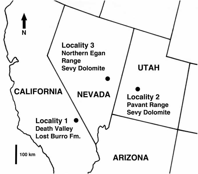
Almost all of the heterostracans described from this region to date have been pteraspids (Elliott and Ilyes, Reference Elliott and Ilyes1996 and references therein), Other taxa are the enigmatic heterostracan Cardipeltis (Denison Reference Denison1953, Reference Denison1966), the heterostracans Allocryptaspis (Elliott et al., Reference Elliott, Reed and Loeffler2004 and references therein) and Ctenaspis (Elliott and Blieck, Reference Elliott and Blieck2010), and the cyathaspids Poraspis and Lechriaspis (Elliott and Petriello, Reference Elliott and Petriello2011). Despite this, many non-pteraspid heterostracan taxa are known to be present in sediments of Early Devonian age in the western United States. This paper continues the documentation of these organisms by describing three species of cyathaspids from Early Devonian sediments in Death Valley, California; west-central Utah; and east-central Nevada (Fig. 1).
Index map showing the geographical setting for the new cyathaspid localities.

The Death Valley locality was first reported by Snow (Reference Snow1990) and subsequently by Elliott and Ilyes (Reference Elliott and Ilyes1992). The vertebrates occur in a terrigenous clastic unit, identified as the Lippincott Member of the Lost Burro Formation, on the south side of Trail Canyon, located in the east-central part of the Panamint Mountains (Fig. 1). The pteraspids from this locality were described by Elliott and Ilyes (Reference Elliott and Ilyes1996), and the cyathaspid Poraspis thomasi by Elliott and Petriello (Reference Elliott and Petriello2011). An arthrodire and acanthodians await description, together with the cyathaspid described here.
The localities in west-central Utah and east-central Nevada are in the Sandy Member of the Sevy Dolomite. The Utah locality is in the Pavant Range near the town of Kanosh (Davis, Reference Davis1983). None of the vertebrates has been described previously, but the fauna of heterostracans, acanthodians, and arthrodires is similar to that found in the Sevy Formation of the Northern Egan Range, east-central Nevada, which has now mostly been published: the pteraspids by Ilyes and Elliott (Reference Ilyes and Elliott1994); a ctenaspid by Elliott and Blieck (Reference Elliott and Blieck2010); the acanthodians by Burrow (Reference Burrow2007); and the arthrodire by Johnson et al. (Reference Johnson, Elliott and Wittke2000).
Geologic setting
The material described here was collected from the lower part of the Lippincott Member of the Lost Burro Formation near Trail Canyon, Death Valley, California, the Sandy Member of the Sevy Dolomite in the Northern Egan Range, east-central Nevada, and the Pavant Range west-central Utah (Fig. 1).
In the Death Valley region, sediments of Early Devonian age consist of the upper part of the Hidden Valley Dolomite (Silurian and Early Devonian) and the overlying Lost Burro Formation (Early to Late Devonian) (McAllister, Reference McAllister1974). The vertebrate-bearing unit is located in a complexly faulted sequence of Paleozoic rocks and is represented by small patches of almost vertical faulted outcrop that are aligned in a north-south direction. The upper part of the unit is covered by Tertiary volcanics, while the lower boundary with the Hidden Valley Dolomite is an erosional unconformity that cuts down into the Ordovician Ely Springs Dolomite in the southern part of the outcrop of the vertebrate-bearing unit. The type Lippincott Member is characterized by sandy and silty dolomite, sandstone, quartzite interbedded with some dolomite, and nodular chert (McAllister, Reference McAllister1974), while the vertebrate-bearing unit contains interbedded dolomitic siltstones and sandstones, quartzite, and pebble conglomerates with clasts of dolomite. The vertebrate-bearing unit has a very low content of pure dolomite beds and nodular chert compared to the type Lippincott Member, and the abundant pebble conglomerates with rounded clasts of dolomite characteristic of the lower part of the vertebrate-bearing unit, are totally absent in the Lippincott Member. The vertebrate-bearing unit is here considered to be a variant of the lower part of the Lippincott Member (cherty argillaceous unit of Johnson et al., Reference Johnson, Sandberg and Poole1988) and is interpreted as channel-fill deposits laid down in an estuarine environment with low or fluctuating salinity, and therefore differing from the marine Lippincott Member. The deposits seem to have a limited lateral extent and to be between 50 and 100 m thick based on lithostratigraphy. A late Emsian age (inversus-serotinus Zones) is suggested for this unit based on conodont data and lithostratigraphy (Johnson et al., Reference Johnson, Sandberg and Poole1988).
The Sevy Dolomite is a widespread carbonate unit of Silurian to Early Devonian age exposed in east-central Nevada and west-central Utah. It is part of a thick carbonate sequence deposited within the Cordilleran region during the early Paleozoic (Osmond, Reference Osmond1954, Reference Osmond1962). The Sevy Dolomite is divided into a lower dolomitic unit devoid of sand and an upper sandy unit containing floating quartz grains, some chert, sand stringers, and thin to thick dolomitic sandy beds (the Sandy Member). The occurrence of chert in the lowermost part of the Sandy Member and the abundant sandy and silty layers throughout suggest it is a lithological correlative of Johnson et al.’s (Reference Johnson, Sandberg and Poole1988) cherty argillaceous unit in the Lippincott Member of the Lost Burro Formation—a correlation supported by the vertebrate fauna (Elliott and Johnson, Reference Elliott and Johnson1997). The locality in the Pavant Range consists of a completely overturned sequence in a complexly faulted area. The vertebrates occur in a thin dolomitic sandstone complex in the lowermost part of the sandy unit. This part of the sequence has been interpreted as ephemeral fluvial deposits on a carbonate flat (Smith, Reference Smith1989). However, lingulids are found in both localities with the vertebrates, indicating a marine environment, although the lack of other invertebrates indicates that the environment was one in which there were fluctuations in salinity (Emig, Reference Emig1997). The fauna is dated as late Emsian (inversus-serotinus Zones) based on correlation of the vertebrates with localities containing spore data (Beartooth Butte Formation; Tanner, Reference Tanner1983) or conodont data (cherty argillaceous unit; Johnson et al., Reference Johnson, Sandberg and Poole1988).
Materials and methods
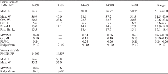
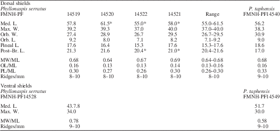
The specimens from Death Valley are mostly preserved in dolomitic sandstones altered by low-grade metamorphism caused by Tertiary intrusions. The specimens from Utah and Nevada were preserved in sandy dolomites. All were prepared by mechanically removing matrix using a vibrotool with a tungsten-carbide bit or by the use of 10% acetic acid and a needle. Some specimens were reversed on plastic using the technique of Toombs and Rixon (Reference Toombs and Rixon1950). The measurements and ratios employed (Tables 1, 2) are those standardized by Denison (Reference Denison1964).
Measurements (mm) and ratios for Phyllonaspis laevis n. gen. n. sp. (* denotes an estimated measurement.)

Measurements (mm) and ratios for Phyllonaspis serratus n. gen. n. sp. and P. taphensis n. gen. n. sp. (* denotes an estimated measurement.)

Repositories and institutional abbreviations
FMNH-PF, Field Museum of Natural History, Chicago.
Systematic paleontology
Class Pteraspidomorphi Goodrich, Reference Goodrich1909
Subclass Heterostraci Lankester, Reference Lankester1868
Order Cyathaspidiformes Berg, Reference Berg1937
Family Cyathaspididae Kiaer, Reference Kiaer1932
Subfamily Boothiaspidinae new subfamily
Type genus
Boothiaspis Broad, Reference Broad1973. Lower Member of the Peel Sound Formation, Prince of Wales Island, Arctic Canada. late Silurian.
Other genera
Alainaspis and Phyllonaspis n. gen.
Diagnosis
Large- to medium-sized taxa with broad, oval to quadrangular, flattened, dorsal shields without epitega. A variably developed lateral brim is present and is serrated in some species. Dorsal ornament of fine, flat-topped ridges; oriented longitudinally, but forming whorls laterally.
Remarks
The three new species described here are clearly members of the Cyathaspididae because they show all the characteristics of the family (Denison, Reference Denison1964), including the presence of a branchial plate that forms the lower margin of the branchial opening. The Cyathaspididae has been divided into subfamilies based particularly on characters of the ornamentation and the presence or absence of growth areas on the dorsal shield, termed epitega (Denison, Reference Denison1964; Greeniaus and Wilson, Reference Greeniaus and Wilson2003). Because these new species do not show epitega and exhibit an ornament of fine, continuous, longitudinal dentine ridges on the dorsal shield, they appear to fit within the Poraspidinae—a cyathaspid subfamily in which the epitega are indicated faintly or not at all, and the dentine ridges are long and mainly longitudinal in orientation, although they may radiate on the anterior and be oriented diagonally on the lateral parts of the dorsal shield (Denison, Reference Denison1964). Phyllonaspis is congruent with these characters, but it differs from typical poraspidinids in having a lateral brim that forms a variably developed flange along the lateral margin of the dorsal shield. This feature is otherwise only seen within the Poraspidinae (and the Cyathaspididae as a whole) in Boothiaspis and Alainaspis, two genera of arctic cyathaspids (Broad, Reference Broad1973; Elliott and Dineley, Reference Elliott and Dineley1985, Reference Elliott and Dineley1991). Boothiaspis includes three species from the Early Devonian Douro and Peel Sound formations of Somerset and Prince of Wales islands and the Devon Island Formation of Ellesmere Island, and was originally described as being a member of the Amphiaspidiformes, an order of distinctive heterostracans otherwise only known from the Lower Devonian of the north-west Siberian Platform and south-western Taimyr (Broad, Reference Broad1973). Elliott and Dineley (Reference Elliott and Dineley1985) described a related genus, Alainaspis, from the Somerset Island Formation and the lower Member of the Peel Sound Formation on Somerset and Prince of Wales islands. They demonstrated that neither Alainaspis nor Boothiaspis were amphiaspids, although the morphology of their shields shows a superficial similarity to some of the later amphiaspids (Elliott and Dineley, Reference Elliott and Dineley1985, Reference Elliott and Dineley1991). In both these genera however, a somewhat flattened dorsal shield with a lateral brim is combined with a very fine ornament of continuous dentine ridges that break up into laterally directed whorls along the margins of the shield, indicating a close relationship between them (Elliott and Dineley, Reference Elliott and Dineley1985, Reference Elliott and Dineley1991). Although branchial plates are not known for either of the arctic species, the reconstruction of Alainaspis platyrhina (Elliott and Dineley, Reference Elliott and Dineley1991, fig. 3.1) shows a lateral opening, labeled by them as the branchial opening, but more likely (from its size) to represent the position of the branchial plate. Phyllonaspis also shows similarities to the other two genera in the presence of a flattened dorsal shield with a lateral brim and in the development of whorls in the ornament along the lateral margins of the dorsal and ventral shields. Alainaspis and Boothiaspis can, however, be distinguished from Phyllonaspis by the presence of a distinct preorbital suture, which was considered by Elliott and Dineley (Reference Elliott and Dineley1985) to possibly represent the last vestige of the demarcation between the lateral and rostral epitega.
Genus Phyllonaspis new genus
Type species
Phyllonaspis laevis n. sp., described herein.
Other species
Phyllonaspis serratus n. sp. and P. taphensis n. sp.
Diagnosis
Medium-sized cyathaspid with no epitega; flattened, oval to quadrangular dorsal shield and convex ventral shield. Narrow, downwardly directed lateral laminae are separated from the rest of the dorsal shield by narrow lateral brims that are serrated posteriorly in some species. No median rostral process. Ornament of very fine (8–10 per mm), flat-topped ridges; generally longitudinal on dorsal shield, but forming whorls laterally against a lateral band of coarser ridges (6–7 per mm). Single median oral plate is keystone shaped.
Etymology
From Greek phyllon, leaf referring to the shape of the dorsal shield, and Greek aspis, shield.
Occurrence
The Sandy Member of the Sevy Dolomite, Northern Egan Range, east-central Nevada and Pavant Range, west-central Utah; Lippincott Member of the Lost Burro Formation, Panamint Mts., Death Valley, California. These localities are all dated to the late Emsian.
Remarks
Phyllonaspis n. gen. is raised to accommodate the three new species from the late Emsian of the western United States described here. The presence of a lateral brim distinguishes this genus from all other cyathaspids except Boothiaspis and Alainaspis from the late Silurian and Early Devonian of the Canadian Arctic. However, Boothiaspis is considerably larger and has a distinct pre-orbital suture that is not present in Phyllonaspis. Alainaspis is also larger and has a pre-orbital suture, but additionally has an enclosed orbit, which distinguishes it from all other cyathaspids. Thus Phyllonaspis is distinct from the Arctic taxa but does show characters that indicate a close relationship to them.
Phyllonaspis laevis new species
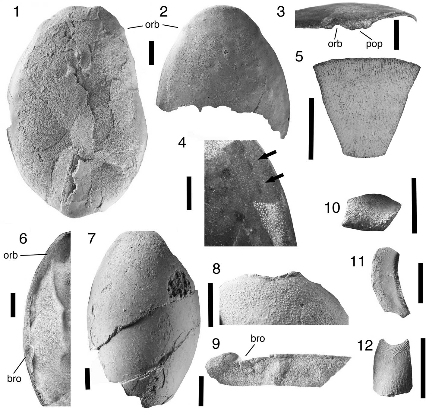
Figures 2.1–2.12, 3, 6.2, Table 1
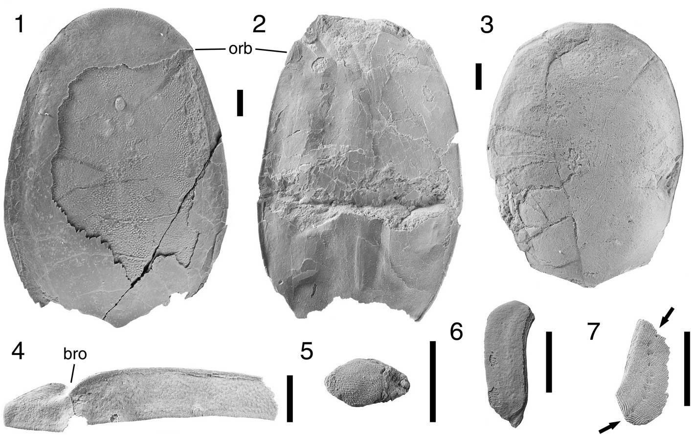
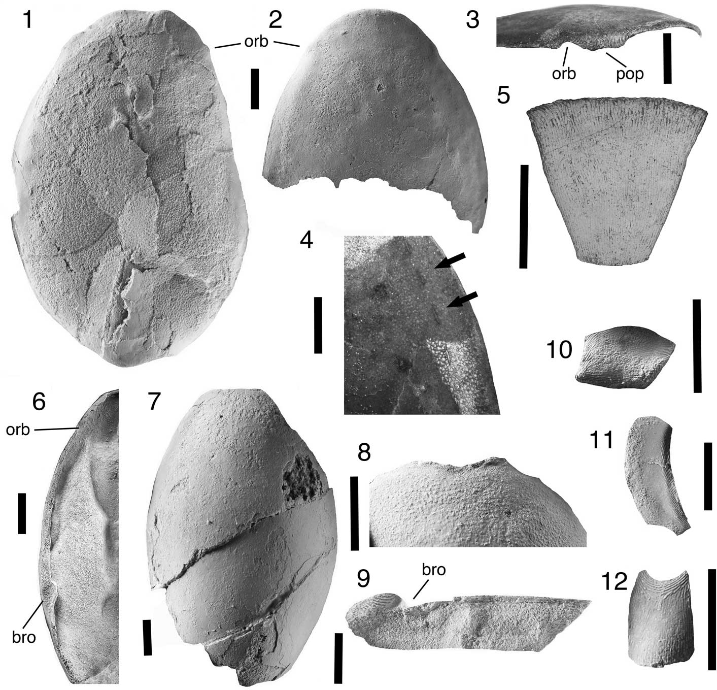
Phyllonaspis laevis n. gen. n. sp. (all specimens dusted with ammonium chloride). Abbreviations: bro, branchial opening; orb, orbit; pop, preorbital process. All scale bars represent 5.0 mm: (1) FMNH-PF14494, holotype dorsal shield in dorsal view; (2–4) FMNH-PF14495; (2) anterior part of dorsal shield in dorsal view; (3) oblique anterior view; (4) anterior margin showing sensory canal system (arrows); (5) FMNH-PF14518, median oral plate; (6) FMNH-PF14503, lateral view of reversed dorsal shield showing the branchial opening; (7) FMNH-PF14505, ventral shield; (8) FMNH-PF14509, ventral shield showing anterior margin and notches for the reception of oral plates; (9) FMNH-PF14511, incomplete right branchial plate; (10) FMNH-PF14517, ventrolateral scale; (11) FMNH-PF14514, dorso-lateral scale; (12) FMNH-PF14515, median ridge scale.
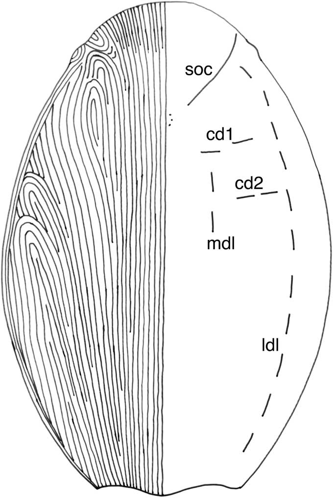
Phyllonaspis laevis n. gen. n. sp., restoration of dorsal shield in dorsal view showing the ridge pattern diagrammatically (ridges actually finer than drawn) and the sensory canal system. Abbreviations: cd1, cd2, first and second transverse commissures; ldl, lateral dorsal canal; mdl, median dorsal canal; soc, supraorbital canal.

Diagnosis
Medium-sized Phyllonaspis with an oval dorsal shield and smooth lateral margins to the lateral brim.
Occurrence
From the late Emsian Sandy Member of the Sevy Dolomite in the Northern Egan Range, near Ely, east-central Nevada.
Description
Medium-sized cyathaspid in which the dorsal shield is oval in outline, broadly rounded anteriorly, the lateral margins are convex, and the posterior margin has a broad, rounded, median lobe flanked by shallow concavities (Fig. 2.1, 2.2). The dorsal surface of the shield is gently convex except in the posterior median area where a slight posterior median crest is developed. There is no median rostral lobe (Fig. 2.3), but there is a narrow (1.9 mm), unornamented, maxillary brim. The orbits are anteriorly placed and the orbital notch and preorbital process are well developed (Fig. 2.2, 2.3). The branchial opening is placed in the posterior third of the shield (Fig. 2.6) and its posterior margin is formed by the anterior edge of a narrow (1.0 mm deep) ventrally directed lateral lamina that extends to the posterior margin of the shield (Fig. 2.6) forming a postorbital lobe. The posterior margin of the branchial opening is beveled to direct flow laterally and posteriorly (Fig. 2.6). A lateral dorsal brim is present, originating behind the orbit and continuing over the branchial opening before dying out posteriorly into a faint carina at the posterior margin, separating the dorsal face of the shield from the vertical lateral lamina. The outer margin of the brim is smooth (Fig. 2.1).
The ornamentation consists of very fine, continuous, flat-topped ridges (9–10 per mm) that are longitudinal over most of the shield and meet the lateral margins at a low angle (Fig. 3). No epitega are demarcated. The lateral margin is formed of a band of 7–8 ridges, parallel to the margin and coarser (6–7 per mm), and more round-topped than those in the central part of the shield. Anterior to the branchial opening the ridges of the central part of the shield merge with those of the lateral margin, however, the lateral ridges remain coarser and spaced more widely apart than those on the more interior part of the shield. The coarse ornament continues in the more marginal part of the rostrum. The longitudinal ridges form whorls against the lateral ridges in the area between the branchial opening and the orbit and appear to be related to the position of the lateral longitudinal canal. The ridges diverge slightly round the pineal area but there is no pineal macula.
The sensory canal system is visible in some areas as small pores that open on the surface of the shield and are demarcated by short ridge sections. In some areas, the system consists of short segments (~1.5 mm long) consisting of two pores with a length of canal between them (Fig. 2.4). In other areas, such as the supraorbital canal, the canal system appears to be continuous. One specimen (FMNH-PF14495, Fig. 2.2, 2.4) exhibits the system for much of the anterior part of the shield. This follows a typical cyathaspid pattern with supraorbital canals extending from anterior to the orbits to the pineal area, and medial and lateral longitudinal canals connected by transverse commissures (Fig. 3). The first two commissures can be seen, but no specimen shows the most posterior ones. No infraorbital canal can be seen, but the lateral longitudinal canal does appear to connect to the supraorbital canal (Fig. 3).
The ventral shield is oval in outline with a bluntly rounded anterior and a posterior margin that is straight or has a slight posterior median lobe (Fig. 2.7). The anterior margin has a shallow median depression that is very convex posteriorly, but shallow anteriorly, and is flanked on each side by slight lateral concavities (Fig. 2.8). The ornament consists of fine (9–10 per mm), continuous, flat-topped ridges. The pattern is generally longitudinal, but the ridges meet the posterolateral margins of the shield at a fairly steep angle. Anterior to that they form whorls in association with pores of the lateral longitudinal canal system. Approaching the anterior margin there is a central band of slightly diverging ridges that is about 3.0 mm wide where it meets the anterior margin before flaring laterally. This area corresponds to the shallow depression in the anterior margin that is probably for reception of the median oral plate (Fig. 2.8). The pores of the lateral line canal system are only visible on a small number of specimens and indicate the presence of the lateral longitudinal canals only.
Several partial branchial plates are present in the collection. Of these, the most complete is FMNH-PF14511 (Fig. 2.9), which is a single right branchial plate missing the anterior and posterior terminations but including the branchial notch. The plate is elongated and parallel sided, 21.5 mm long, and 4.9–5.5 mm wide. It is broken just behind the branchial notch; dorsally and posterior to the notch a series of coarse, sharp-crested ridges curve into the branchial opening. Anteriorly a narrow band of ridges form a dorsal border to the plate below which dorso-ventrally directed ridges cover the plate.
A single oral plate is available for this species (Fig. 2.5). In outline it is a symmetrical keystone-shaped trapezoid. The longer of the two parallel sides is 7.9 mm long, the shorter 3.1 mm long. The two diverging sides are each 7.0 mm long and the antero-posterior length of the plate is 6.7 mm. The plate is flat over most of its surface and slightly convex in each antero-lateral corner. The ornament is of fine (9–10 per mm), flat-topped ridges, similar to those found elsewhere on the shield, that are arranged in a longitudinal pattern and flare into the antero-lateral corners, additional ridges being added periodically at the lateral margins. The lateral margins are bordered by a band of 2–3 ridges. It is assumed that the narrower side forms the base and that this is a central oral plate, similar in position, although not in shape, to those already recorded for several species of Poraspis (Elliott and Petriello, Reference Elliott and Petriello2011).
There are only a few body scales recognized for this species. FMNH-PF14514 (Fig. 2.11) is a dorsolateral scale 12.0 mm long and 3.4 mm wide with Phyllonaspis-type ornament that is oriented at right angles to the long axis. The ventral margin shows two straight edges that would have articulated with the ventrolateral scales. The scale forms a narrow anterior projection dorsally, with a concave embayment behind it that would have articulated with the median dorsal scales. A small polygonal scale that may be a ventrolateral scale (Fig. 2.10), is 5.4 mm long and 3.5 mm wide, with straight anterior margins, and a rounded posterior. A small symmetrical convex scale, 4.2 mm long and 2.7 mm wide, is probably a median ventral scale (Fig. 2.12). It is concave posteriorly and convex anteriorly with a narrow anterior border formed of six ridges, covered by antero-posteriorly directed ridges over the rest of the scale. One scale, that may be a median dorsal scale, is symmetrical and convex with a straight anterior margin and a slightly convex posterior margin. It differs from other scales in having longitudinal ornament that diverges from the mid line as it approaches the anterior edge, forming a narrow band parallel to the anterior border.
Etymology
From the Latin laevis, smooth or polished, in recognition of the smooth surface of the shield.
Other material
Ten dorsal shields FMNH-PF14495–14504; six ventral shields FMNH-PF14505–14510; three branchial plates FMNH-PF14511–14513; four scales FMNH-PF14514–14517; and one oral plate FMNH-PF14518.
Remarks
This species can be separated from the other two in the genus by its more oval shape and the lack of any serrations along the margin of the lateral brim.
Phyllonaspis serratus new species
Figures 4.1–4.7, 5.5, 6.3, Table 2
Phyllonaspis serratus n. gen. n. sp. (all specimens dusted with ammonium chloride). Abbreviations: bro, branchial opening; orb, orbit. All scale bars represent 5.0 mm: (1) FMNH-PF14519, holotype dorsal shield in dorsal view; (2) FMNH-PF14520, dorsal shield in dorsal view; (3) FMNH-PF14528, internal mold of ventral shield; (4) FMNH-PF14530, right branchial plate; (5) FMNH-PF14533, median ridge scale; (6) FMNH-PF14534, lateral scale; (7) FMNH-PF14539, lateral oral plate with arrows showing the course of the pore line.

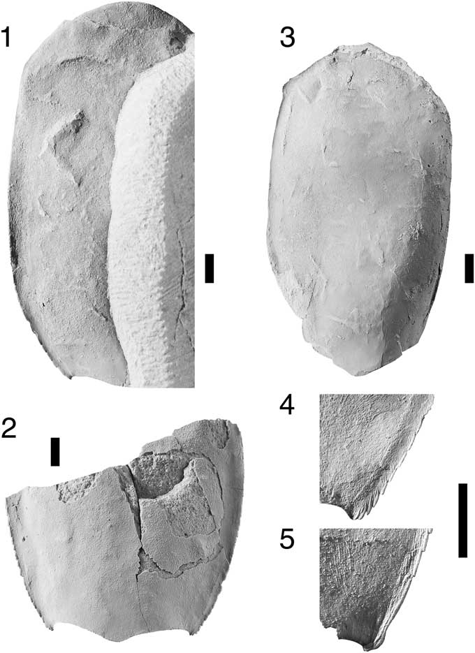
Phyllonaspis taphensis n. gen. n. sp. (all specimens dusted with ammonium chloride). All scale bars represent 5.0 mm: (1) FMNH-PF14540, holotype dorsal shield in dorsal view; (2) FMNH-PF14541, posterior part of dorsal shield in dorsal view; (3) ventral shield; (4, 5) details of the posterior lateral margins of the dorsal shield in (4) Phyllonaspis taphensis, FMNH-PF14541, and (5) Phyllonaspis serratus, FMNH-PF14520, to show characteristic differences.

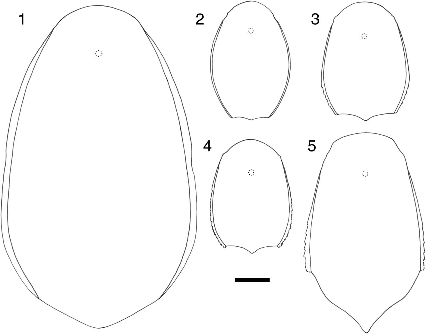
Representative members of the subfamily Boothiaspidinae drawn to the same scale. Scale bar represents 20 mm. (1) Boothiaspis ovata (redrawn from Broad, Reference Broad1973); (2) Phyllonaspis laevis; (3) P. serratus; (4) P. taphensis; (5) Alainaspis platyrhina (redrawn from Elliott and Dineley, Reference Elliott and Dineley1985)

1999 Undescribed cyathaspidid Reference Elliott, Reed and JohnsonElliott et al., fig. 4E.
Diagnosis
Medium-sized Phyllonaspis with a quadrangular outline to the dorsal shield; lateral margins of the lateral brim are serrated over the posterior third, but cease before the posterior margin.
Occurrence
From the late Emsian Sandy Member of the Sevy Formation in the Pavant Range, near Kanosh, west-central Utah (Davis, Reference Davis1983).
Description
Medium-sized cyathaspid with a slightly convex dorsal shield that is bluntly rounded anteriorly, convex laterally, and broadest at about 2/3 of the length of the shield (Fig. 4.1, 4.2). The posterior margin is slightly convex laterally, flanking a small, rounded, central lobe. No median lobe is present on the rostrum, the orbits are anteriorly placed and shallow, and the antorbital process is weakly developed. The post-branchial lobe cannot be seen and a slight convexity on the lateral margin of the dorsal shield marks the position of the branchial opening. A projecting lateral brim is present, originating behind the orbit and continuing posteriorly over the branchial opening. The brim is about 3.0 mm wide behind the orbit and narrows posteriorly to 1.0 mm at the posterior margin, becoming laterally serrated towards the posterior border (Fig. 4.1, 4.2).
The ornament consists of continuous, fine (10 per mm), flat-topped ridges. These are longitudinal over the central part of the shield and diverge laterally over the posterior part of the shield. No epitega are present. In the rostral area, the ridge pattern is longitudinal centrally but divergent laterally, with a concave-forward whorl in the center of the anterior margin. The position of the pineal organ is marked by the presence of complementary whorls so that ridges appear to radiate from a common center. Over a marginal border about 2.5 mm wide the ridges become coarser (7 per mm), round-topped, and separated from each other. This border of coarser ridges continues over the orbit where it becomes parallel to the plate margin and narrows posteriorly to about 1.0 mm wide in the branchial area. From just behind the orbit, one very coarse ridge forms the lateral margin. About 12.0 mm from the posterolateral corner this breaks up into a series of serrations, each formed by a short (1.5 mm) offset length of coarse ridge. The serrations do not continue to the posterolateral corner of the shield because over the last 6.0 mm a single continuous marginal ridge follows a slightly sinuous course before forming a convex curve at the posterolateral margin (Fig. 4.1, 4.2). The main pattern of ridges on the shield is longitudinal, however, laterally the ridges flare and meet the lateral border of coarse ridges almost perpendicularly. Closer to the orbit, the ridges of the central field grade into those of the lateral border. In this area also a series of two or three large whorls is formed.
The sensory canal system is only present in a few specimens and is incomplete in those cases. Only the more anterior pores of the system are distinct. A supraorbital canal, an infraorbital canal, and part of the lateral longitudinal canal can be recognized. These appear to be continuous where seen, but are not connected to each other. The lateral longitudinal canal can be traced in part by the disruption in the ridge pattern along the anterior part of its length.
The ventral shield is represented by only one complete example (Fig. 4.3) and some fragments. It is slightly convex anteriorly and more deeply so posteriorly. The anterior margin is bluntly rounded, convex laterally, with the posterior margin gently concave to either side of a blunt central projection. Sensory canals are visible in only one specimen and show parts of the central and lateral dorsal canals. There is an insufficient amount of the shield preserved to show the overall pattern, but it is noticeable that the system is not continuous because the pores occur in pairs connected by short canals.
One almost complete right branchial plate is present in the collections together with two partial specimens (Fig. 4.4). The plate is elongated (34.0 mm long as preserved) and narrows posteriorly to a blunt termination. The dorsal margin is slightly convex, interrupted 8.5 mm from the posterior end by the branchial notch, and the ventral margin is slightly concave. The branchial notch is delineated by a visceral surface that is a continuation of the posterodorsal part of the plate, and the posterior part of the anterior portion of the plate, which overlaps it. Both surfaces have convex dorsal margins and between them is the branchial duct, which is thus directed posteriorly. The anterior part of the plate has parallel dorsal and ventral margins and is about 6.0 mm deep. The plate is ornamented by fine (14 per mm), flat-topped, dentine ridges over much of its surface (Fig. 4.4), which are directed dorsally from the posterior and posteroventral margins and become very coarse (7 per mm) and triangular in section as they reach the dorsal margin. Anteriorly a narrow band of slightly coarser (10 per mm), round-topped, ridges forms a dorsal border to the plate. Over much of the plate the ridges are oriented dorsoventrally and form a series of whorls as they reach the dorsal margin. Although the anterior margin is not present in the main specimen, in one incomplete specimen it is abrupt and straight.
One small incomplete plate is identified as a possible lateral plate from the oral cover. It is elongated, 8.5 mm long and 4.0 mm wide at its widest point (Fig. 4.7). Natural edges are present along one long margin and three short margins, and the other edge is curving and apparently broken. A significant feature of the plate is the presence of a row of ten pores that form a curving line in the mid-line of the plate, but parallel to its natural long edge. The ornament is mainly longitudinal; parallel to the long edge, but curving across the plate to meet the broken edge of the plate at a steep angle. The pores are situated for the most part in a central loop in the ornament, which in part of the plate becomes a junction at which ridges meet at a steep angle. Identification of the plate is based on its shape and on the presence of the pore row. Pores are present on the dorsal and ventral shields, but not as close to the plate margins as shown here, also the plate margins are usually demarcated by coarser ridges that are not seen here. A lateral plate is indicated because this is the only small plate positioned where a sensory canal connects the dorsal and ventral shields. There are two possibilities because both the infraorbital and supraorbital canals cross from the dorsal to the ventral plates. The incomplete nature of this plate makes it difficult to decide its orientation or complete shape, but it seems most likely that it is the lateral plate bearing the supraorbital canal.
A number of scales are present and consist mostly of dorsolateral scales that are very similar in size and appearance to the scale from P. laevis and average 12.7 mm in length and 4.4 mm in width (Fig. 4.6). One scale appears to be a median ridge scale (Fig. 4.5). It is oval, convex, and 5.5 mm long by 3.2 mm wide with longitudinal ornament.
Etymology
From the Latin serratus, toothed like a saw, in recognition of the serrated margin of the shield.
Other material
Eight incomplete dorsal shields FMNH-PF14520–14527; two incomplete ventral shields FMNH-PF14528, 14529; three branchial plates FMNH-PF14530–14532; six scales FMNH-PF14533–14538; and one lateral oral plate FMNH-PF14539.
Remarks
This species can be separated from Phyllonaspis laevis by the more quadrangular shape of the dorsal shield and by the fact that the lateral brim is serrated. The serrations continue to the posterior margin of the shield in P. taphensis, but in P. serratus they cease about 6.0mm before the posterior border and the margin continues smoothly to terminate in a convex curve.
Phyllonaspis taphensis new species
Diagnosis
Medium-sized Phyllonaspis with a quadrangular outline to the dorsal shield; lateral margins of the lateral brim are serrated to the posterior margin where they form a small cornu.
Occurrence
From the late Emsian Lippincott Member of the Lost Burro Formation, at Trail Canyon in the Panamint Mts., Death Valley, California.
Description
Medium-sized cyathaspid with a slightly convex dorsal shield, the convexity becoming more pronounced posteriorly. The shield is bluntly rounded anteriorly and convex laterally; the posterior margin is slightly convex laterally, flanking a small, rounded, central lobe (Fig. 5.1, 5.2). No median lobe is present on the rostrum; the orbits are anteriorly placed and shallow, and the antorbital process is weakly developed. The post-branchial lobe cannot be seen. A slight marginal convexity on the lateral margin of the dorsal shield marks the position of the branchial opening. A projecting lateral brim is present, originating behind the orbit and continuing posteriorly over the branchial opening to the posterolateral corner of the shield. The brim is about 2.3 mm wide behind the orbit and narrows posteriorly to 1.3 mm at the posterior margin. It becomes laterally serrated shortly after its origination and continues to the posterior border where a small cornu is formed (Fig. 5.2, 5.4).
The ornament consists of continuous, fine (10 per mm), flat-topped ridges. These are longitudinal over the central part of the shield and diverge laterally and anteriorly. No epitega are present. In the rostral area, the ridge pattern is longitudinal centrally but divergent laterally, and a concave-forward whorl is formed in the center of the anterior margin. Over a marginal border about 2.0 mm wide the ridges become coarser (8 per mm), round-topped, and separated from each other. This border of coarser ridges continues over the orbit where it becomes parallel to the plate margin and narrows posteriorly to about 1.0 mm wide in the branchial area. From just behind the orbit one very coarse ridge forms the lateral margin and almost immediately this breaks up into a series of serrations, each formed by a short (2.5–3.0 mm) offset length of coarse ridge. At the posterior margin these become very short (0.8–1.0 mm) and the last section forms a small, serrated, cornu (Fig. 5.4). The main pattern of ridges on the shield is longitudinal, however, the ridges flare laterally and meet the lateral border of coarse ridges almost perpendicularly. Closer to the orbit the ridges of the central field grade into those of the lateral border. Whorls are formed along the lateral margins where ridges of the central field meet those of the lateral margin.
A few sections of the sensory canal system can be seen in some specimens. These indicate a system similar to that in the other two species.
There is one complete ventral shield for this species associated with the holotype dorsal shield (Fig. 5.3). It has slightly convex sides and a broadly convex anterior margin with a central facet 4.2 mm wide. Posteriorly it has a broad median lobe and is highly convex, but it becomes shallower anteriorly.
Etymology
From the Latin taphos, a tomb, and Latin ensis, a suffix denoting locality, in recognition of Death Valley as the collecting locality for this species.
Other material
One incomplete dorsal shield and seven fragments FMNH-PF14541–14548; one ventral shield FMNH-PF14549; and one incomplete branchial plate FMNH-PF14550.
Remarks
This species is most similar to Phyllonaspis serratus and shares with it a serrated margin to the lateral brim. However, in P. taphensis the serrations continue to the posterolateral margin of the shield while in P. serratus the serrations cease well before that (Fig. 5.4, 5.5). It is differentiated from P. laevis by the more quadrangular shape of the dorsal shield and the fact that P. laevis has a smooth margin to the lateral brim.
Discussion
The oral cover in the Cyathaspididae
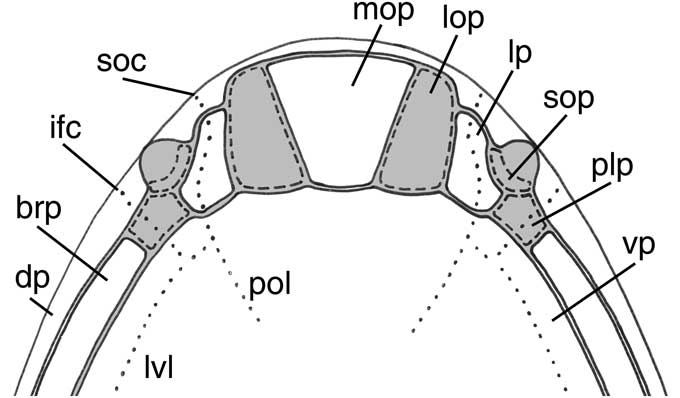
No articulated material of the oral cover is available for Phyllonaspis, however two isolated plates are known. The first is a median oral plate of P. laevis (Fig. 2.5), and the second a lateral plate of P. serratus (Fig. 4.7), which together allow a partial reconstruction of the oral cover (Fig. 7). The median oral plate is a symmetrical, keystone-shaped trapezoid with a length of 6.7 mm. The longer of the two parallel sides is 7.9 mm long, the shorter is 3.1 mm long. The symmetrical shape suggests that this is a median plate, the narrower side forming the base and articulating with the anterior margin of the ventral shield. The second plate is elongated and irregular in shape and is characterized by the presence of a series of ten pores of the sensory canal system forming a curving line through the center. Although incomplete, this plate has one long natural edge and three short natural edges forming one end of the plate. The other side is curving and broken. The presence of the pores identifies it as one of the lateral plates because these are the only small, pore-bearing plates in the carapace of a cyathaspid, situated as they are between the dorsal and ventral plates at the point where the infraorbital and supraorbital canals continue from the dorsal surface to the ventral where they connect with the postoral and lateral ventral canals (Fig. 7). The asymmetric shape also supports this identification because it indicates that the plate must be a lateral rather than a median oral plate. The only other lateral plate reported for a cyathaspid is by Kiaer and Heintz (Reference Kiaer and Heintz1935; fig. XXXIII.1), who illustrate a small polygonal lateral plate with two pores attributed to Homalaspidella nitida (Homalaspidella nitidus Strand, Reference Strand1934). Based on its shape and size, this would probably have been a posterior lateral plate bearing pores of the infraorbital canal. Denison (Reference Denison1960, fig. 121) illustrated several lateral plates for the heterostracan Allocryptaspis laticostatus. Although Denison’s view that Allocryptaspis was a cyathaspid has been challenged (Elliott et al., Reference Elliott, Reed and Loeffler2004), the general arrangement of plates within the oral cover can be taken to provide an indication of the arrangement in the Cyathaspididae. The illustrated lateral plates are mostly small polygonal plates with lines of pores between the suborbital plate and the anterolateral margin of the ventral shield in Denison’s reconstruction (Denison, Reference Denison1960, figs. 121C, D, E, 122), and included the junction of the lateral ventral canal with the ventral termination of the infraorbital and supraorbital canals. One plate, however, is elongated with a line of pores along its axis and therefore shows similarity to the plate described here. Denison identified this as a lateral plate and placed it lateral to the oral plates with the supraorbital canal running through it in his reconstruction of the oral cover (Denison Reference Denison1960, figs. 121G, 122).
Reconstruction of the oral cover in Phyllonaspis. Known plates are shown in white. Abbreviations: brp, branchial plate; dp, dorsal plate; ifc, infraorbital canal; lop, lateral oral plate; lp, lateral plate; lvl, lateral ventral canal; mop, median oral plate; plp, posterior lateral plate; pol, postoral canal; soc, supraorbital canal; sop, suborbital plate; vp, ventral plate.

The reconstruction of the oral cover provided here (Fig. 7) is based on the proportions of P. laevis because this is the species for which the complete median oral plate is known. Superimposing an outline of the dorsal and ventral shields leaves a space between the anterior margins that presumably equates to the area occupied by the oral cover in life (Fig. 7). The large median oral plate occupies a central position and fits in the space between the anterior margin of the ventral shield and the maxillary brim of the dorsal shield. However, it is not wide enough to completely fill the space, so there must have been at least one lateral oral plate on each side (lop in Fig. 7). The lateral plate can be fitted next to the lateral oral plate and curving under the assumed position of the suborbital plate (sop in Fig. 7). In that position the line of pores that run across it can be seen to represent the continuation of the supraorbital canal from the dorsal shield and onto the ventral shield where it joins the postoral canal. The infraorbital canal also probably passed through a small posterior lateral plate on its path to the ventral plate. This plate would have abutted the branchial plate, which has an abrupt anterior termination in Phyllonaspis, and probably would have been an oblong plate similar to that illustrated by Kiaer and Heintz (Reference Kiaer and Heintz1935, plate XXXIII.1) for Homalaspidella nitida or those illustrated by Denison (Reference Denison1960, fig. 121) for Allocryptaspis laticostatus.
The oral cover in heterostracans is constructed of a complex of small plates, therefore they commonly disarticulated before the preservation of the rest of the carapace. For this reason there is a paucity of information about the construction of the oral cover in heterostracan taxa. The earliest heterostracan for which the oral cover is known is Athenaegis chattertoni (Soehn and Wilson, Reference Soehn and Wilson1990), in which a series of small rod-like oral plates are arranged in a V-shaped pattern on either side of a central postoral plate. A pair of lateral oral plates flanked this arrangement on each side. Janvier (Reference Janvier1996, fig. 4.8) placed Athenaegis as the sister-group to higher heterostracans, therefore the structure of its oral cover is taken as being primitive.
Different arrangements of the plates of the oral cover are now known in cyathaspids. In the Poraspidinae, two different species of Poraspis show a simple arrangement in which there is a large, oblong oral plate that fills most of the oral area and may have been flanked by small lateral or lateral oral plates (Dineley and Loeffler, Reference Dineley and Loeffler1976; Elliott and Petriello, Reference Elliott and Petriello2011). Anglaspis is also in the Poraspidinae and appears to have an arrangement in which two fairly large oblong plates on each side flank a smaller central plate (Bendix-Almgreen, Reference Bendix-Almgreen1986). The Boothiaspidinae, which are here considered to be closely related to the Poraspidinae, are now shown to have a large central plate that was probably flanked by a pair of lateral oral plates, which is similar to that seen in Poraspis. The only other published cyathaspid oral area is from Capitaspis giblingi (Elliott, Reference Elliott2013), a cyathaspidinid, in which there are clearly two rows of plates in the oral area, including a large median plate in the oral row. For a discussion of possible feeding habits in the Cyathaspididae see Elliott (Reference Elliott2013).
The best documented heterostracan oral covers are known from the Pteraspididae (Blieck, Reference Blieck1984) and show an increasing complexity through the evolution of the group with narrow elongated plates attached to the anterior margin of the ventral shield in the most primitive taxa being followed by the addition of one and then two rows of postoral plates in the more advanced taxa. At present it is not possible to recognize similar trends within the Cyathaspididae because the relationships within the family are poorly understood. The most complete treatment of the family was carried out by Denison (Reference Denison1964) who published a comprehensive review in which he recognized the cyathaspids as a family, the Cyathaspididae. Within the family he recognized a series of sub-families and proposed a phylogeny in which the Tolypelepidinae were recognized as the ancestral stock, with the Cyathaspidinae being derived from them by the loss of scale-like components of the shield. The Irregulareaspidinae and the Poraspidinae were thought to have developed from early Cyathaspidinae, the poraspids probably representing an artificial assemblage of genera that had independently lost distinct epitega. No further attempts to develop an understanding of the relationships of the Cyathaspididae were made until 1996 when Janvier (Reference Janvier1996) showed in cladistic form (although without a cladistic analysis), as part of a phylogeny of the Heterostraci, a simple cyathaspid phylogeny in which Nahanniaspis represented basal forms and Anglaspis and Torpedaspis more advanced forms, while the ctenaspids and amphiaspids formed a sister-group (Janvier, Reference Janvier1996; fig. 4.8). Most recently a first attempt at a cladistic analysis of the family (Lundgren and Blom, Reference Lundgren and Blom2013) is unfortunately flawed by the inclusion of groups that have been shown not to be cyathaspids: the ctenaspids (Elliott and Blieck, Reference Elliott and Blieck2010); Allocryptaspis (Elliott et al., Reference Elliott, Reed and Loeffler2004); and Ariaspis (Elliott and Swift, Reference Elliott and Swift2010).
Age and distribution of the Boothiaspidinae
Temporal control has been placed on the Early Devonian faunas from the western United States by Elliott and Johnson (Reference Elliott and Johnson1997) who showed that the base of the Grassy Flat Member of the Water Canyon Formation in northern Utah, the basal Lippincot Member of the Lost Burro Formation of Death Valley, California, the Sandy Member of the Sevy Dolomite in east-central Nevada and western Utah (Davis, Reference Davis1983), and the Beartooth Butte Formation of Beartooth Butte in northern Wyoming (Dehler, Reference Dehler1995) could be correlated based on their vertebrate faunas. Although age-diagnostic invertebrates are rare, the Beartooth Butte Formation at Beartooth Butte has a spore determination of middle to late Emsian (Tanner, Reference Tanner1983), an age that is supported by dating of the vertebrate-bearing channel of the Lippincott Member. Although the channel sediments lack diagnostic invertebrates and spores, the upper part of the Hidden Valley Dolomite into which it is cut is considered to be middle Emsian in age (Johnson et al., Reference Johnson, Sandberg and Poole1988), and above lies the Lippincott Member (the basal part of which is separated as the cherty argillaceous unit), which contains conodonts of late Emsian age at Mesquite Springs in the Panamint Range (Johnson et al., Reference Johnson, Sandberg and Poole1988). Although the upper age limit of the vertebrate fauna cannot be constrained, the lower limit is clearly the top of the Hidden Valley Dolomite into which the channel is cut, and hence it cannot be older than the middle Emsian. Thus, on balance, the age of the western United States fauna is considered to be late Emsian (inversus-serotinus Zones).
Alainaspis and Boothiaspis are widely distributed in late Silurian and Early Devonian rocks on the central Arctic islands, Nunavut, Arctic Canada (Broad, Reference Broad1973; Elliott and Dineley, Reference Elliott and Dineley1985, Reference Elliott and Dineley1991). Alainaspis is reported from the Somerset Island Formation on Somerset Island and the lower member of the Peel Sound Formation on Prince of Wales Island (Elliott and Dineley, Reference Elliott and Dineley1985). Boothiaspis is also known from those localities and from the upper part of the Douro Formation on Somerset Island and the Devon Island Formation on Ellesmere Island (Broad, Reference Broad1973). The Somerset Island Formation is considered to be an age equivalent of the lower member of the Peel Sound Formation on Prince of Wales Island (Miall et al., Reference Miall, Kerr and Gibling1978), which was dated to the late Ludlow to early Pridoli (Uyeno, Reference Uyeno1980) based on conodonts and graptolites. However, a study by Mayr et al. (Reference Mayr, Brent, de Freitas, Frisch, Nowlan and Okulitch2004) correlated the Somerset Island Formation and the lower member of the Peel Sound Formation with the lower member of the Peel Sound Formation as defined by Thorsteinsson (Reference Thorsteinsson1980) and Uyeno (Reference Uyeno1980) and dated this part of the section to the Lochkovian (eurekaensis Zone). The Douro Formation is Ludlow in age and almost entirely in the siluricus Zone, while the upper part is dated to the lowermost latialata Zone (Mayr et al., Reference Mayr, Brent, de Freitas, Frisch, Nowlan and Okulitch2004). The Devon Island locality was dated to the Pridoli by Thorsteinsson (Broad, Reference Broad1973), based on the presence of a monograptid dated as Pridoli on Cornwallis Island. Thus the Boothiaspis and Alainaspis localities are late Ludlow to early Lochkovian in age, which places them somewhat older than the Emsian age of the Phyllonaspis localities.
Elliott and Blieck (Reference Elliott and Blieck2010) and Elliott and Petriello (Reference Elliott and Petriello2011) noted the discrepancy in age between the Arctic and western US faunas, and suggested that this temporal distribution represented the time needed for dispersal of the organisms from a center in the Arctic to the western United States. The Old Red Sandstone Continent was divided by shallow seaways in the late Silurian–Early Devonian (see e.g., Ziegler Reference Ziegler1988, Scotese Reference Scotese1997), which may have facilitated dispersal of early vertebrates that seem to have inhabited proximal marine environments at this time because they are typically associated with an impoverished marine fauna composed of euryhaline forms (Cruziana, leperditiid ostracodes, ceratiocarid phyllocarids, and lingulids) (Blieck and Janvier Reference Blieck and Janvier1999; Elliott et al., Reference Elliott, Schultze and Blieck2015). Johnson et al. (Reference Johnson, Elliott and Wittke2000) hypothesized a series of pathways for the radiation of actinolepid arthrodires based on the presence of these open seaways. However, throughout the Early Devonian, these seaways were reduced or closed, eliminating easy pathways of dispersal. Thus dispersal of the Arctic fauna would probably have been confined to coastal routes around the Old Red Sandstone Continent.
If dispersal took place from the Canadian Arctic to the western United States, evidence should be available in pre-Emsian age (Lochkovian or Pragian) sediments along the probable pathway around the western coast of the Old Red Sandstone Continent. There is a paucity of such information, however, as no Pragian localities and only two of Lochkovian age are known from the western United States (Turner and Murphy, Reference Turner and Murphy1988; Elliott and Johnson, Reference Elliott and Johnson1997). The Mackenzie Mt. fauna (Adrain and Wilson, Reference Adrain and Wilson1994), which contains an extensive fauna of Lochkovian age, includes poraspids, which also are found in the western US (Elliott and Petriello, Reference Elliott and Petriello2011), and is now known to contain ctenaspids (personal communication, K. Soehn, 2011), which was not thought to be the case when the western US ctenaspid was described (Elliott and Blieck, Reference Elliott and Blieck2010). Although highly speculative, the preponderance of evidence supports the view that dispersal followed a coastal route from the central Arctic to the western US.
Acknowledgments
This research was supported by Northern Arizona University Organized Research grants, which are gratefully acknowledged. Death Valley National Monument provided collecting permits and T. Elliott, J. Evans, R. Ilyes, H. Johnson, and R. Thomas are thanked for collecting assistance; J. Tucker assisted with the initial part of this study; S. Swift carried out some of the photography; and J. Wittke provided the SEM image. I benefitted greatly from the helpful reviews of A. Blieck and an anonymous reviewer.