I. INTRODUCTION
Donepezil hydrochloride (C24H29NO3⋅HCl, or donepezil HCl), one of the first acetylcholinesterase (AChE) inhibitors designed specifically for the treatment of mild to moderate Alzheimer's disease (Sugimoto et al., Reference Sugimoto, Yamanishi, Iimura and Kawakami2000), was developed by Eisai Co. Ltd. and is marketed using the tradename Aricept. The systematic IUPAC name for donepezil HCl is 2-[(1-Benzyl-4-piperidinyl)methyl]-5,6-dimethoxy-1-indanone hydrochloride (1:1). A two-dimensional (2D) drawing of the molecular structure is illustrated in Figure 1.
A 2D drawing of the molecular structure of donepezil HCl.

Donepezil salts and free base exhibit numerous different polymorphs (Imai et al., Reference Imai, Wanatabe, Kajima, Ishihama, Ohtsuka and Tanaka2000; Park et al., Reference Park, Lee, Lee, Choi, Mao, Kang, Choi and Lee2013). Powder X-ray diffraction (PXRD) data for the donepezil HCl polymorph examined in this work, known as Form III, has been observed previously (Imai et al., Reference Imai, Wanatabe, Kajima, Ishihama, Ohtsuka, Tanaka and Narabu1997, Reference Imai, Wanatabe, Kajima, Ishihama, Ohtsuka and Tanaka2000; Vrbinc et al., Reference Vrbinc, Jordan-Kotar, Smrkolj and Vrecer2007). The structure does not appear to be present in the Cambridge Structural Database (CSD; Groom et al., Reference Groom, Bruno, Lightfoot and Ward2016) or the open literature, although ORTEP plots of a single-crystal structure attributed to donepezil hydrochloride were published in a review paper without providing the atomic coordinates (Sugimoto et al., Reference Sugimoto, Yamanishi, Iimura and Kawakami2000). Raw data from the Powder Diffraction File (PDF, ICDD 2020) entry 00-060-1199 for donepezil hydrochloride, collected at the University College London (Vickers, 2005), was used to obtain a crystal structure which was subsequently verified with a density functional theory (DFT) optimization.
II. EXPERIMENTAL
Laboratory PXRD data were previously obtained by Martin Vickers at the University College London (UCL) using a Stoe StadiP diffractometer in transmission mode (Vickers, 2005). The diffractometer was equipped with a copper anode operated at 40 kV and 30 mA, and an incident beam germanium monochromator (λ = 1.54059 Å). The sample was mounted in a 0.5 mm diameter glass capillary and using a 6° linear position-sensitive detector, data were collected between 5° and 35° 2θ in 0.01° steps, re-binned to give a data-step of 0.02°. The raw data were published online (Vickers, 2005) and in the PDF (Gates-Rector and Blanton, Reference Gates-Rector and Blanton2019) as part of PDF entry 00-060-1199.
Pattern indexing with DICVOL06 (Boultif and Louër, Reference Boultif and Louer2004) suggested a monoclinic unit cell with lattice parameters a = 14.3669 Å, b = 11.8394 Å, c = 13.5554 Å, β = 107.803°, and a cell volume of 2195.3 Å3 (M20 = 21.0, F20 = 59.6), in strong agreement with the initial assessment made at UCL (Vickers, 2005) and tabulated in the PDF entry. Space group determination with ChekCell (Laugier and Bochu, Reference Laugier and Bochu2000) suggested space group P21/c as the most plausible option. Searches of the CSD using the formula “C24 H30 Cl N O3” (0 hits), name “donepezil” (12 hits) and a reduced cell search with 2.5% tolerances on the cell parameters (19 hits) did not yield a match for donepezil HCl.
A donepezil molecule was extracted from the CSD entry SUBTIS04 (Park et al., Reference Park, Lee, Lee, Choi, Mao, Kang, Choi and Lee2013). A hydrogen atom was added to the molecule on the nitrogen site using Avogadro (Hanwell et al., Reference Hanwell, Curtis, Lonie, Vandermeersch, Zurek and Hutchison2012). The molecule was converted to a Fenske-Hall Z-matrix with Open Babel (O'Boyle et al., Reference O'Boyle, Banck, James, Morley, Vandermeersch and Hutchison2011) and combined with a chlorine atom to solve the structure with FOX (Favre-Nicolin and Černý, Reference Favre-Nicolin and Černý2002), using 40 sets of parallel tempering with 2 × 10⁶ trials/set. These sets yielded one solution with a cost function of ~3000 that was significantly lower than the other sets.
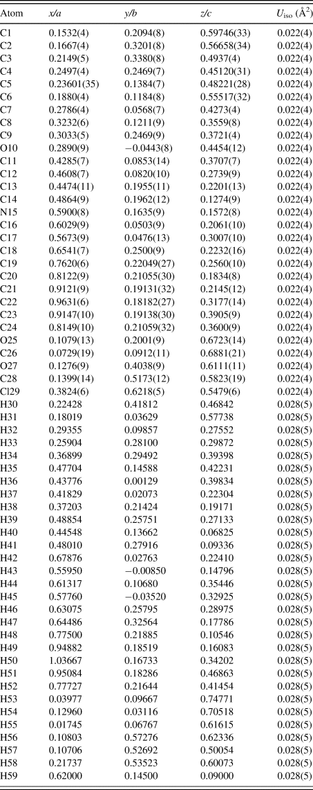
Rietveld refinement of the crystal structure was performed with the GSAS/EXPGUI program (Toby, Reference Toby2001; Larson and Von Dreele, Reference Larson and Von Dreele2004). Restraints on the bonds, angles, and planar restraints on the rings were applied using values determined by the Mogul 1.7 module of the CSD (Bruno et al., Reference Bruno, Cole, Kessler, Luo, Motherwell, Purkis, Smith, Taylor, Cooper, Harris and Orpen2004). The background was refined using a Chebyshev polynomial with 12 terms. The positions of the C, N, O, and Cl atoms were refined, while the positions of the H atoms remained fixed but were periodically optimized using Avogadro. An overall isotropic displacement parameter was refined for the C, N, O, and Cl atoms, with the H atoms constrained to 1.3 times this value.
The crystal data, data collection, and refinement details are summarized in Table I.
The crystal data, data collection, and refinement parameters obtained for donepezil HCl.

Density function theory (DFT) geometry optimization was performed on donepezil HCl using CRYSTAL17 (Dovesi et al., Reference Dovesi, Erba, Orlando, Zicovich-Wilson, Civalleri, Maschio, Rerat, Casassa, Baima, Salustro and Kirtman2018). Basis sets were obtained from the literature for the C, H, N, O (Gatti et al., Reference Gatti, Saunders and Roetti1994), and Cl atoms (Vilela Oliveira et al., Reference Vilela Oliverira, Peintinger, Laun and Bredow2019). The calculations used the B3LYP functional (Becke, Reference Becke1993) and D3 dispersion correction (Grimme et al., Reference Grimme, Antony, Ehrlich and Krieg2010) with eight k-points.
III. RESULTS AND DISCUSSION
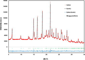
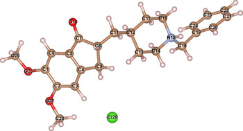
The final Rietveld refinement for donepezil HCl is illustrated in Figure 2, and the refined atomic coordinates are given in Table II, while the labeling of the non hydrogen atoms is illustrated in Figure 3. While the data range is limited, visually the Rietveld refinement looks reasonable. A structural check of the bond lengths and angles with Mogul (Bruno et al., Reference Bruno, Cole, Kessler, Luo, Motherwell, Purkis, Smith, Taylor, Cooper, Harris and Orpen2004) yields only one bond length and two bond angles with z-scores over 1, the largest being the bond length C18–N15 (z-score = 1.70), suggesting that the refined structure is reasonable. Similar to a previous structure solution for trandolapril (Reid et al., Reference Reid, Kaduk and Vickers2016), the limited PXRD data range absolutely necessitates a comparison of the structure with a DFT optimization.
A plot illustrating the final Rietveld refinement for donepezil HCl obtained with GSAS.

The atom labels used for the structure description of donepezil HCl. The figure was prepared with VESTA (Momma and Izumi, Reference Momma and Izumi2011).

The Rietveld-refined crystal structure of donepezil HCl with lattice parameters a = 14.3662(9) Å, b = 11.8384(6) Å, c = 13.5572(7) Å, and β = 107.7560(26)°. All atoms were refined with fixed site occupancies of 1 (on Wyckoff 4e sites).

The DFT-optimized atomic coordinates for donepezil HCl are shown in Table III, and select bond lengths and angles from the Rietveld-refined and DFT-optimized structures are compared in Table IV. The root-mean-square (RMS) Cartesian difference between the Rietveld-refined and DFT-optimized structures is 0.14 Å; the excellent agreement strongly suggests that the crystal structure is correct (van de Streek and Neumann, Reference van de Streek and Neumann2014). The RMS displacement calculated using the molecular overlay function of Mercury (Macrae et al., Reference Macrae, Bruno, Chisholm, Edgington, McCabe, Pidcock, Rodriquez-Monge, Taylor, van de Streek and Wood2008) is 0.08 Å, also suggesting excellent agreement. A molecular overlay of the Rietveld-refined and DFT-optimized structures is illustrated in Figure 4.
A molecular overlay of the Rietveld-refined structure of donepezil HCl, in red, and the DFT-optimized structure, in blue. The RMS Cartesian displacement of the nonhydrogen atoms is 0.14 Å. The figure was prepared with Mercury (Macrae et al., Reference Macrae, Bruno, Chisholm, Edgington, McCabe, Pidcock, Rodriquez-Monge, Taylor, van de Streek and Wood2008).

The DFT-optimized crystal structure of donepezil HCl with fixed lattice parameters a = 14.36617 Å, b = 11.83837 Å, c = 13.55723 Å, and β = 107.756°.

A comparison of the Rietveld-refined and DFT-optimized values of select bond lengths and angles from the donepezil HCl crystal structure.

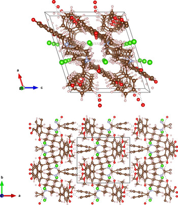
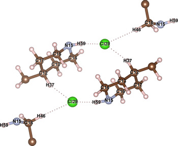
The hydrogen bonds observed in donepezil HCl via the DFT optimization are shown in Table V, and views of the structure along the b- and c-axes are illustrated in Figure 5. The structure is held together by columns of chloride anions parallel to the b-axis, where each anion is hydrogen-bonded to three individual donepezil molecules. This creates zigzag columns of donepezil and chloride along the b-axis, as viewed down the c-axis (Figure 5, bottom). The donepezil molecules form layers parallel to the (101) planes, with donepezil molecules in adjacent layers connected by a ring of hydrogen bonds, with graph set R2,4(14) (Etter, Reference Etter1990; Bernstein et al., Reference Bernstein, Davis, Shimoni and Chang1995) through two chloride anions, as illustrated in Figure 6. Each chloride anion has another hydrogen bond, via H46, to a third donepezil molecule. Weaker hydrogen bonds with oxygen acceptors connect molecules both within layers and between adjacent layers.
The DFT-optimized crystal structure of donepezil HCl, viewed along the b-axis (top) and c-axis (bottom). The atom types can be identified by color including carbon (brown), oxygen (red), nitrogen (blue), chlorine (green), and hydrogen (pink). Hydrogen bonds are represented by the dotted lines. The unit cell is outlined in black. The figure was prepared with VESTA (Momma and Izumi, Reference Momma and Izumi2011).

A close-up view illustrating the hydrogen bonds around the chloride anions. The figure was prepared with VESTA (Momma and Izumi, Reference Momma and Izumi2011).

The most prominent hydrogen bonds observed in the donepezil HCl structure and their parameters obtained from the DFT modeling.

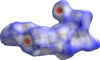
The Hirshfeld surface (Hirshfeld, Reference Hirshfeld1977; Spackman and Byrom, Reference Spackman and Byrom1997; Turner et al., Reference Turner, McKinnon, Wolff, Grimwood, Spackman, Jayatilaka and Spackman2017) of donepezil HCl is illustrated in Figure 7. The volume enclosed within the Hirshfeld surface (539.29 Å) represents 98.2% of one-fourth of the unit cell volume, suggesting that the molecules are not tightly packed. The close contacts, illustrated in red in Figure 7, generally involve the hydrogen bonds.
The Hirshfeld surface of donepezil HCl. Regions in red represent intermolecular contacts shorter than the sum of the van der Waals radii, while regions in blue represent longer contacts and regions in white represent contacts equal to the sum of the van der Waal radii.

CIF files for the final Rietveld refinement and the DFT-optimized structure of donepezil HCl have been included in the Supplementary material.
IV. DEPOSITED DATA
The Crystallographic Information Framework (CIF) files containing the results of the Rietveld refinement (including the raw data) and the DFT geometry optimization were deposited with the ICDD. The data can be requested at info@icdd.com.
ACKNOWLEDGEMENTS
Much thanks to Martin Vickers for the deposition of his excellent PXRD pharmaceutical data in the PDF, and his support for this work. JR dedicates this work to Ada Phyllis Louise Reid (1910–1998), who lost her battle with Alzheimer's disease.
FUNDING
The Canadian Light Source is supported by the Canadian Foundation for Innovation, the Natural Sciences and Engineering Research Council of Canada, the National Research Council Canada, the Canadian Institutes of Health Research, the Government of Saskatchewan, Western Economic Diversification Canada, and the University of Saskatchewan.
CONFLICTS OF INTEREST
The authors have no conflicts of interest to declare.