INTRODUCTION
Besides the well-known Dirofilaria immitis (Spirurida, Onchocercidae), canine infestations by filarioids are due to nematodes whose adults colonize predominantly subcutaneous tissues, muscular fasciae, retropharyngeal and axillary lymphatics (genera Acanthocheilonema, Brugia, Cercopithifilaria and Dirofilaria), and ocular tissues (Onchocerca lupi) (Sréter and Széll, Reference Sréter and Széll2008). With the exception of D. immitis, Dirofilaria repens and Acanthocheilonema reconditum, all other filarioids of dogs have been little studied and the information on their aetiology, life history as well as their actual impact on human and veterinary medicine is meagre (Otranto and Eberhard, Reference Otranto and Eberhard2011; Brianti et al. Reference Brianti, Gaglio, Napoli, Giannetto, Dantas-Torres, Bain and Otranto2012a).
Since the elevation of Cercopithifilaria Eberhard, Reference Eberhard1980 to the genus rank by Bain et al. (Reference Bain, Baker and Chabaud1982a), 28 species parasitizing primates, ungulates, rodents, carnivores and marsupials have been classified or described within this genus (Bain et al. Reference Bain, Uni and Takaoka2002). Cercopithifilaria adults are usually localized beneath cutaneous tissues, whereas microfilariae are always in the dermis (Bain et al. Reference Bain, Uni and Takaoka2002), the infestation being transmitted by ixodid ticks (Ixodida, Ixodidae) (Winkhardt, Reference Winkhardt1980; Bain et al. Reference Bain, Petit and Chabaud1986; Spratt and Haycock, Reference Spratt and Haycock1988; Petit et al. Reference Petit, Bain, Carrat and de Marval1988).
Cercopithifilaria grassii (=Filaria grassii) was described more than a century ago by Noè (Reference Noè1907) from a dog from Rome. This little-known filarioid presents typical ‘gigantesche’ (from Italian, giant, measuring about 650 μm in length) microfilariae (Noè, Reference Noè1907), which are completely distinct from those of Cercopithifilaria bainae presenting short microfilariae (about 180 μm) described, only once, in a dog from south-eastern Brazil (Almeida and Vicente, Reference Almeida and Vicente1984). Since their original description, both species have never been reported again, with the exception of occasional findings of developing larvae of Cercopithifilaria sp. in ixodid ticks in Switzerland (Bain et al. Reference Bain, Aeschlimann and Chatelanat1982b) and northern Italy (Pampiglione et al. Reference Pampiglione, Canestri Trotti and Marchetti1983).
Following the recent morphological description and molecular characterization of microfilariae of Cercopithifilaria sp. sensu Otranto et al. Reference Otranto, Brianti, Dantas-Torres, Weigl, Latrofa, Gaglio, Cauquil, Giannetto and Bain2011 (herein after referred to as Cercopithifilaria sp. I) from a dog from Sicily, Italy, this filarioid was retrieved in dogs from Spain, Greece and southern Italy (i.e., Apulia, Basilicata and Sicily regions), with prevalence rates reaching up to 21·6% (Otranto et al. Reference Otranto, Brianti, Latrofa, Annoscia, Weigl, Lia, Gaglio, Napoli, Giannetto, Papadopoulos, Mirò, Dantas-Torres and Bain2012a). The occurrence of Cercopithifilaria sp. I in dogs overlapped with the distribution of its vector, the brown dog tick Rhipicephalus sanguineus (Brianti et al. Reference Brianti, Otranto, Dantas-Torres, Weigl, Latrofa, Gaglio, Napoli, Brucato, Cauquil, Giannetto and Bain2012b). In addition, the wide distribution of Cercopithifilaria sp. I paralleled the high intraspecific nucleotide variation (mean = 1·2%) detected among mitochondrial cytochrome c oxidase subunit 1 (cox1) sequences of examined nematodes (n = 2287) from skin samples and ticks, with up to 16 haplotypes characterized to date (Otranto et al. Reference Otranto, Latrofa, Brianti, Annoscia, Parisi, Dantas-Torres, Bain and Gasser2012b). Although scientific knowledge on Cercopithifilaria sp. I infesting dogs has recently been refined, several questions on its aetiology still need to be addressed since no adults are available for proper taxonomical identification.
Following a comprehensive coupled morphological and molecular analysis of numerous microfilariae collected from dogs and ticks from the Mediterranean area, this paper reports the presence of at least 3 different species of Cercopithifilaria in dogs. In particular, a new microfilaria of Cercopithifilaria (referred to as Cercopithifilaria sp. II) is morphologically identified and molecularly characterized herein, and compared with morphological and molecular features of microfilariae of Cercopithifilaria sp. I and C. grassii. Sequence data for the latter species have been herein reported for the first time after more than a century since its original description. Morphological and molecular analyses were congruent and proved the coexistence of at least 3 different species of Cercopithifilaria in dogs from Mediterranean Europe.
MATERIALS AND METHODS
Sample collection and identification procedures
Microfilariae were collected and examined from the skin of dogs (n = 329) living in southern Italy i.e., Putignano (Apulia) (n = 115), Matera (Basilicata) (n = 50), Valledolmo (Sicily) (n = 113) and Spain (La Vera) (n = 51). From February 2011 to February 2012, samples were collected under the context of a previous study aimed to estimate the prevalence of Cercopithifilaria sp. I in dogs and ticks (Otranto et al. Reference Otranto, Brianti, Latrofa, Annoscia, Weigl, Lia, Gaglio, Napoli, Giannetto, Papadopoulos, Mirò, Dantas-Torres and Bain2012a). Briefly, sampled dogs were naturally exposed to R. sanguineus infestation (Brianti et al. Reference Brianti, Pennisi, Brucato, Risitano, Gaglio, Lombardo, Malara, Fogliazza and Giannetto2010; Lorusso et al. Reference Lorusso, Dantas-Torres, Lia, Tarallo, Mencke, Capelli and Otranto2010; Dantas-Torres and Otranto, Reference Dantas-Torres and Otranto2011; G. Mirò, personal communication). Skin samples were taken using disposable scalpels from the right shoulder, inter-scapular or temporal region (about 0·5 × 0·5 × 0·6 cm) and soaked in saline solution for 10 min at 37 °C. A drop of sediment of soaked skin sample was placed on a glass slide and observed under light microscopy (2 fields of 10 × 10 mm cover slip each) after adding a drop of methylene blue (1%). Dermal microfilariae were processed and measured using the software AxioVision rel. 4·8 (Carl Zeiss Germany) and microscopic images were acquired using a digital camera (Zeiss Axiocam MRc, Carl Zeiss Germany) mounted directly on the microscope (Zeiss Axioscop 2 plus, Carl Zeiss Germany). In addition, 6 ticks from Spain were dissected and microfilariae from digestive caeca were prepared for the morphological description. For a detailed morphological analysis, several sealed preparations were available of skin microfilariae and microfilariae from ticks. The cover slips of a few slides were unsealed with a scalpel and the microfilariae removed and placed in a drop of lactophenol in order to re-orient the microfilariae. Drawings were made with an optic microscope equipped with a camera lucida and measurements were made on drawings.
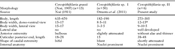
Microfilariae were morphologically identified based on key characters and compared to those of Cercopithifilaria sp. I (Otranto et al. Reference Otranto, Brianti, Dantas-Torres, Weigl, Latrofa, Gaglio, Cauquil, Giannetto and Bain2011) and C. grassii (Noè, Reference Noè1907), as reported in Table 1, as well as with other Cercopithifilaria spp. (data not shown).
Measurements (in micrometers) and morphological features of dermal microfilariae of Cercopithifilaria spp. found in dogs

* Lateral alae included.
A skin sample was taken from the injured skin of a dog from Spain suffering from pruritic, diffuse, erythaematous and dermatitis but not presenting any flea infestation or any other apparent causes for the dermatological affection. Biopsies were fixed in 4% buffered formalin solution (pH 7·4), paraffin embedded and routinely processed for light microscopy. Sections (thickness, 5 μm) were stained with haematoxylin and eosin.
All procedures of this study were performed in accordance with ethical principles of animal experimentation and approved by the ethical commission of the Faculties of Veterinary Medicine of Bari, Messina and Madrid.
Molecular amplification and phylogenetic analyses
Following morphological identification, microfilariae were isolated using an eyelash mounted on a stick and genomic DNA extracted from individual specimens using a commercial kit (Dneasy Blood & Tissue Kit, Qiagen GmbH, Hilden, Germany) in accordance with the manufacturer's instructions.
The cox1 (∼689 bp) and 12S (∼330 bp) gene fragments, which are usually employed for barcoding of filarioids (Ferri et al. Reference Ferri, Barbuto, Bain, Galimberti, Uni, Guerriero, Ferté, Bandi, Martin and Casiraghi2009) were amplified as previously reported (Otranto et al. Reference Otranto, Brianti, Dantas-Torres, Weigl, Latrofa, Gaglio, Cauquil, Giannetto and Bain2011). For phylogenetic analysis a partial cox1 (pcox1) fragment of Cercopithifilaria spp. (304 bp) was amplified using specific primers and procedures as described elsewhere (Otranto et al. Reference Otranto, Brianti, Dantas-Torres, Weigl, Latrofa, Gaglio, Cauquil, Giannetto and Bain2011).
Amplicons were purified using Ultrafree-DA columns (Amicon, Millipore; Bedford, USA) and then sequenced directly using the Taq DyeDeoxyTerminator Cycle Sequencing Kit (v.2, Applied Biosystems) in an automated sequencer (ABI-PRISM 377). Sequences were determined from both strands (using the same primers individually as for the PCR) and the electropherograms verified by eye. Pairwise comparisons of sequence differences (D) were made using the formula D = 1- (M/L), where M is the number of alignment positions at which the 2 sequences have a base in common, and L is the total number of alignment positions over which the 2 sequences are compared (Chilton et al. Reference Chilton, Gasser and Beveridge1995). In order to ensure open reading frames, all nucleotide sequences of the cox1 fragment were conceptually translated into amino acid sequences using the invertebrate mitochondrial code by MEGA5.0 (Tamura et al. Reference Tamura, Peterson, Peterson, Stecher, Nei and Kumar2011). Sequences were aligned using ClustalW program (Larkin et al. Reference Larkin, Blackshields, Brown, Chenna, McGettigan, McWilliam, Valentin, Wallace, Wilm, Lopez, Thompson, Gibson and Higgins2007) and compared with those available in the GenBank dataset by BLAST analysis. To investigate the relationships among filarioids of the Onchocercidae family, sequences of both genes were analysed with those available in GenBank. All the pcox1 haplotypes of the Cercopithifilaria sp. I previously sequenced (AN: JF461457; JF925148–JF925151; JQ305155–JQ305163; JQ753309; JQ753310) were included in the analysis. The evolutionary history was inferred by MEGA4.0, using cox1 and 12S sequences, under Neighbor-Joining methods using 8000 replicates bootstrap values. Thelazia callipaeda (Spirurida, Thelaziidae) was chosen as an out-group. A bootstrap support of = 0·70 was considered significant. The nucleotide sequences analysed in this study are available in the GenBank (Accession numbers: JQ837809–JQ837812).
RESULTS
Morphological identification
Cercopithifilaria sp. I sensu Otranto et al.Reference Otranto, Brianti, Dantas-Torres, Weigl, Latrofa, Gaglio, Cauquil, Giannetto and Bain2011
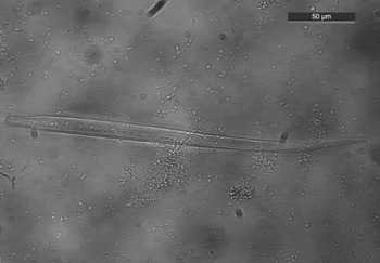
Description: Microfilariae bodies were short and constant in width for the main part, mean length of 185·7 ± 7·2 μm and a mean width (in dorso-ventral view) of 6·9 ± 1 μm (Table 1, Fig. 1). The body cuticle was thick bearing transverse striations, with no sheath. For a detailed morphological description of species type of microfilariae see Otranto et al. (Reference Otranto, Brianti, Dantas-Torres, Weigl, Latrofa, Gaglio, Cauquil, Giannetto and Bain2011).
Light microscopy of microfilariae of Cercopithifilaria sp. I from subcutaneous tissue.

Cercopithifilaria grassii (Noè, Reference Noè1907)
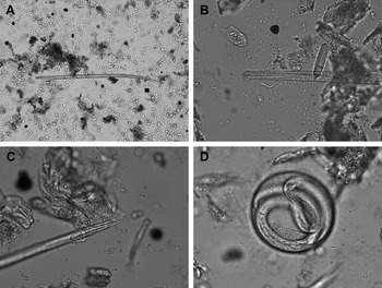
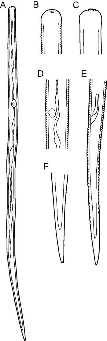
Description: (based on 4 microfilariae recovered from a dog from Sicily, Italy, and 2 from a dog from La Vera, Spain): Microfilariae 645–670 μm long and 15–17 μm wide (Figs 2A and 3A). Head rounded, slightly bulbous; sclerotized convex formation and tiny left hook at apex (Figs 2B and 3B,C); body cylindrical with thick cuticle and striae, which are interrupted in the lateral plane (Fig. 3C); no lateral alae; tail long, thick, conical, with a blunt extremity that may appear bifid depending on the orientation; the last 20 μm of the tail are exclusively cuticular and striae are no longer visible (Figs 2C and 3E,F). Internally, no nuclei visible, except that of the excretory cell near the excretory pore; presence of an axial cordon not well defined and irregular in diameter; anus identified in 1 microfilaria. For a microfilaria 645 μm long, width 17 μm, excretory cell at 185 μm from anterior extremity (Fig. 3D), anus at 118 μm from posterior extremity, cuticular posterior part 28 μm long. Some eggs were found to contain similar microfilariae (Fig. 2D).
Light microscopy of microfilariae of Cercopithifilaria grassii from subcutaneous tissue. (A) General view; (B) rounded bulbous head; (C) posterior cuticular extremity of larvae without striae; (D) microfilaria in an unshaded egg.

Microfilaria of Cercopithifilaria grassii. (A) General microfilaria; (B) head, left sinistral view; (C) head, dorso-ventral view; (D) excretory pore, sinistral view; (E) posterior region, sinistral view; (F) caudal extremity, dorso-ventral view. Scale bars: A = 100 μm; B–D = 10 μm; E, F = 25 μm.

Remarks: Noè (Reference Noè1911) provided a precise morphometrical description of the microfilariae of C. grassii in dogs from Italy. The metrical and morphological characters of the microfilaria described above correspond well to those of C. grassii as described by Noè (Reference Noè1911) (see Table 1). Based on their similarities, we identified these larvae collected from dogs in Italy and Spain as C. grassii.
Cercopithifilaria sp. II
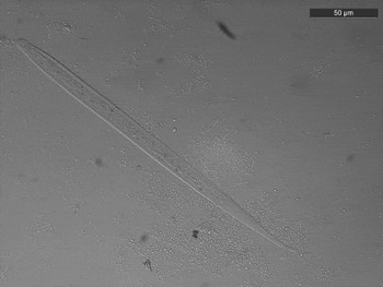
Description: (based on microfilariae from skin snips from 2 dogs and from the digestive caeca of ticks from La Vera, Spain; Fig. 4). In microfilariae from ticks, nuclei were most often slightly altered. Body navicular in dorso-ventral view, due to the wide lateral alae (Fig. 5A), filiform in lateral view till the long robust conical posterior extremity (Fig. 5B). Transverse striae conspicuous (well identified in lateral and sublateral views), interrupted on lateral lines (as observed in dorso-ventral view) and striae closer in the conical posterior part. Lateral alae beginning about 10 μm from anterior extremity (Fig. 5C); posteriorly fused and forming a thick cuticular cone (Fig. 5A,G). Anterior end rounded with very tiny left subterminal cephalic hook (Fig. 5D). Cephalic space about 10 μm long (Fig. 5C,D). At mid-body, excluding the lateral alae, body transverse section slightly elongated dorso-ventrally (9–7 μm in diameter, excluding the lateral alae); number of nuclei per transverse line 3–4 in dorso-ventral view and 4–5 in lateral view (Fig. 5E,F). The excretory pore was not visible in Fig. 3E because the microfilaria was not cleared in lactophenol (Fig. 5E). The last caudal nuclei are 58–70 μm from tip; the non-cuticular part of the body is regularly attenuated in dorso-ventral view but its beginning is marked by an abrupt narrowing in lateral view (Fig. 5G,H); tail extremity attenuated. For a microfilaria 280 μm long, body width in dorso-ventral view 12 μm (7 μm, lateral alae excluded); in lateral view 9 μm; beginning of lateral alae at 11 μm from anterior extremity, excretory pore at 90 μm from anterior end, last nucleus 60 μm from tail tip, conical cuticular posterior extremity 40 μm long.
Light microscopy of microfilaria of Cercopithifilaria sp. II from subcutaneous tissue.

Microfilaria of Cercopithifilaria sp. II. (A) General dorso-ventral view (Pertu, tick 432, mf 2); (B) general lateral view; (C) anterior end, dorso-ventral view; (D) anterior end, sinistral view; (E) mid-body, dorso-ventral view; (F) mid-body, lateral view; (G) posterior part, dorso-ventral view; (H) posterior part lateral view. The excretory pore was not visible because the microfilaria was not cleared in lactophenol. Scale bars: A, B = 50 μm, C–F = 10 μm, G, H = 25 μm.

Remarks: With the large lateral alae, the body size and the shape of the caudal region the present microfilaria is distinct from those described in the genus Cercopithifilaria, from dogs in Europe (see above) and Brazil (Almeida and Vicente, Reference Almeida and Vicente1984), viverids (Bain et al. Reference Bain, Wamae and Reid1989), cercopithecids (Eberhard, Reference Eberhard1980; Bain et al. Reference Bain, Baker and Chabaud1982a, Reference Bain, Wamae and Reid1988), lagomorphs (Bartlett, Reference Bartlett1983), ursids (Uni, Reference Uni1984), murids (Spratt and Varughese, Reference Spratt and Varughese1975), porcupines (Bain et al. Reference Bain, Baker and Chabaud1982a, Reference Bain, Petit and Chabaud1986), ungulates (Fain, Reference Fain1977; Chabaud et al. Reference Chabaud, Landau and Petit1978; Winkhardt, Reference Winkhardt1980; Bain et al. Reference Bain, Baker and Chabaud1982a; Uni et al. Reference Uni, Suzuki and Katsumi1998, Reference Uni, Suzuki, Baba, Mitani, Takaoka, Katsumi and Bain2001, Reference Uni, Bain, Takaoka, Fujita and Suzuki2002), didelphids (Esslinger and Smith, Reference Esslinger and Smith1979; Bain et al. Reference Bain, Baker and Chabaud1982a) (see references in Bain et al. Reference Bain, Baker and Chabaud1982a and Uni et al. Reference Uni, Suzuki, Baba, Mitani, Takaoka, Katsumi and Bain2001, Reference Uni, Bain, Takaoka, Fujita and Suzuki2002). We consider these microfilariae as belonging to an unknown species, Cercopithifilaria sp. II.
Material examined: Slide-mounted microfilariae were deposited in the collection of the Muséum National d'Histoire Naturelle, Paris, France (MNHM), under Accession numbers reported in the following: Cercopithifilaria sp. I (194YU, 284YU, 407YU, 408YU, Apulia, Italy; 2YT, 3YT from Sicily, Italy), Cercopithifilaria sp. II (367YU, 368YU, 372YU from La Vera, Spain) and C. grassii (395YU from Sicily, Italy; 409YU from Spain).
Molecular analyses
The morphological differentiation among the 3 dermal kinds of microfilariae was confirmed by differences in cox1 and 12S sequences examined and those available in the GenBank database. Indeed, the mean level of interspecific pairwise (Pwc) distance (%) for cox1 gene was 11·4%, ranging from 10·1% to 12·8% in Cercopithifilaria sp. II vs C. grassii and Cercopithifilaria sp. I vs Cercopithifilaria sp. II, respectively. By comparing the nucleotide sequences of cox1 and 12S of Cercopithifilaria spp. with the most frequent species of filarioids affecting dogs (i.e., A. reconditum, D. immitis, and D. repens) the mean interspecific Pwc distance (%) was of 11·9% and 19% for cox1 and 12S, respectively. Analogously, the overall 12S interspecific difference (mean = 17·7%) ranged from 14·3% to 19·5% in Cercopithifilaria sp. I vs Cercopithifilaria sp. II and Cercopithifilaria sp. I vs C. grassii, respectively. The cox1 mitochondrial sequences from dermal microfilariae of Cercopithifilaria sp. I, Cercopithifilaria sp. II, and C. grassii had a typical average base composition of AT content (64·6%) and a bias at the third codon position to AT (75·4%) compared with the first and second positions (59·3%). The cox1 sequences included 489 conserved and 97 variable and singleton sites, none being parsimony-informative. Most variable sites (n = 78; 80·4%) were at the third codon position, whereas the others (n = 19; 19·6%) were at the first and second codon positions. Nucleotide variations were represented by transitions (Ts) rather than transversions (Tv), with a Ts/Tv ratio of 2·2. The conceptual translation at third codon position of cox1 sequence led to 194 amino acids without stop codons. Most (80·2%) nucleotide alterations were synonymous, with the exception of non-synonymous nucleotide substitutions, which resulted in amino acid alterations (not shown). The 12S sequence obtained from specimens examined differed from one another by transitions and transversions (Ts/Tv = 0·5) and insertion/deletion events. The alignment of the 12S sequences resulted in a total of 284 characters including gaps (not shown), with an overall AT bias of 77·6%. Sequences included 216 conserved and 63 variable sites, of which 60 were singletons and none were parsimony-informative. In particular, cox1 interspecific difference ranged from 9·6% to 14·7% in D. immitis vs D. repens and Cercopithifilaria sp. I vs A. reconditum, respectively. Analogously, 12S interspecific difference ranged from 10·8% to 24·2% in D. immitis vs D. repens and A. reconditum vs Cercopithifilaria sp. II, respectively.
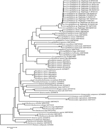
Phylogenetic analyses of the sequence dataset here produced with those of other Onchocercidae available in GenBank for both genes were concordant in clustering these with other sequences of Cercopithifilaria spp. to the exclusion of Dirofilaria spp., Onchocerca spp. and Acanthocheilonema spp. In particular, Onchocerca spp., Setaria spp., and Brugia spp. were clustered together and differentiated by a second group including Cercopithifilaria spp. and Acanthocheilonema spp. The branches were supported by high bootstrap values in their main nodal points. The phylogenetic reconstruction obtained from the alignment of cox1 sequence data is shown in Fig. 6.
Phylogenetic tree based on regions of cox1 sequence data, compared with those representative filaroids of the family Onchocercidae available in GenBank™. The tree was constructed using the Neighbor-Joining (NJ) method and rooted against Thelazia callipaeda (Spirurida, Thelaziidae) out-group.

Distribution and prevalence of the three Cercopithifilaria spp. and histology of Cercopithifilaria sp. II
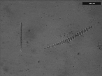
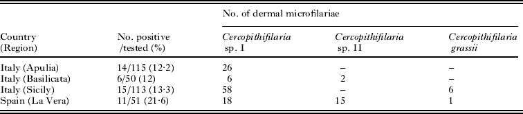
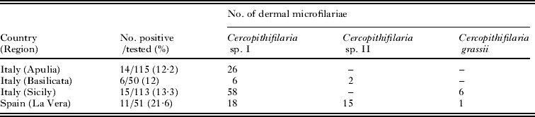
Larvae of Cercopithifilaria spp. examined were collected from 46 (14%) out of 329 dogs examined with overall prevalence rates of 12% and 21·6%, in Basilicata and La Vera, respectively (Table 2). A total of 132 microfilariae of Cercopithifilaria spp. were detected in skin samples from 46 positive dogs (Table 2). Almost all the dermal microfilariae (n = 108, 81·8%) collected from Apulia (n = 26, from 14 positive dogs), Basilicata (n = 6, from 6 positive dogs), Sicily (n = 58, from 15 positive dogs) and La Vera (n = 18, from 5 positive dogs) were morphologically identified as Cercopithifilaria sp. I. Seventeen (12·9%) out of 132 microfilariae (2 from 2 dogs from Basilicata and 15 from 3 dogs from La Vera) were morphologically identified as Cercopithifilaria sp. II. Finally, 7 out of 132 (5·3%) dermal microfilariae (6 from 1 Sicilian dog and 1 from 1 Spanish dog) were identified as C. grassii. Cercopithifilaria sp. I and Cercopithifilaria sp. II mixed infestations were detected in Basilicata (Fig. 7) whereas Cercopithifilaria sp. I and C. grassii were detected in Sicily. Finally, co-occurrence of Cercopithifilaria sp. I, Cercopithifilaria sp. II and C. grassii mixed infestations was detected in dogs from La Vera.
Microfilariae of Cercopithifilaria sp. I, and Cercopithifilaria sp. II simultaneously detected in a dog from Basilicata (southern Italy).

Number of dogs positive for the three species of Cercopithifilaria herein considered broken down according to country and region of sampling and microfilariae isolate from skin samples and their specific identification based on morphological characters
The Sicilian and the Spanish dogs from which C. grassii was re-described were previously shown to be infested with another species of Cercopithifilaria, the former being the original source of Cercopithifilaria sp. I (Otranto et al. Reference Otranto, Brianti, Dantas-Torres, Weigl, Latrofa, Gaglio, Cauquil, Giannetto and Bain2011; Brianti et al. Reference Brianti, Otranto, Dantas-Torres, Weigl, Latrofa, Gaglio, Napoli, Brucato, Cauquil, Giannetto and Bain2012b). A year later, when the dog died naturally, no Cercopithifilaria sp. I microfilariae were detected, but instead those of C. grassii. The Spanish dog had Cercopithifilaria sp. II in July 2011, but in February 2012 the skin snip performed contained C. grassii.
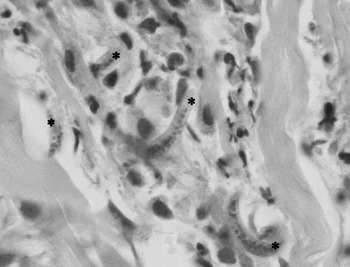
The histological examination showed multifocal interstitial dermatitis characterized by the presence of neutrophils, eosinophils and lymphocytes in association with microfilariae. These were located outside the vessel lumina (Fig. 8).
Histopathology from skin. In the interstitium of the dermis there are neutrophils, a few eosinophils and microfilariae (asterisk). Haematoxylin-eosin stain.

DISCUSSION
From their dermal localization, the microfilariae studied herein could belong to either the genus Onchocerca or Cercopithifilaria. Both genera include filarioids of dogs little known to parasitologists and veterinarians due to the difficulties in detecting dermal microfilariae in skin biopsy tissues (most dog owners are unwilling to authorize this procedure). Morphological and molecular evidence were concordant in indicating that the microfilariae studied belong to 3 different species of Cercopithifilaria. Besides Cercopithifilaria sp. I recently identified (Otranto et al. Reference Otranto, Brianti, Dantas-Torres, Weigl, Latrofa, Gaglio, Cauquil, Giannetto and Bain2011), C. grassii is here re-described for the first time. This finding is of relevance to parasitology, because after more than a century since its first description (Noè, Reference Noè1907), bona fide evidence for its existence has finally been provided. Indeed, over the last decade, there was a great confusion on the taxonomical identity of this species, which is still reported in the literature as ‘Dipetalonema grassii’ presenting blood-circulating instead of skin microfilariae (Vakalis and Himonas Reference Vakalis and Himonas1997; Tarello, Reference Tarello2004). Finally, in the present study, based on a morphological appraisal and comparison of microfilariae within the genus, a new species has been defined as Cercopithifilaria sp. II.
Additionally, comparing the 3 types of microfilariae it becomes evident that the exceptionally large C. grassii microfilaria presents with an unusual internal anatomy. Indeed, instead of the many nuclei close to each other, which are those of the cells constituting the anlagen of the diverse organs (Bain, Reference Bain1970a,Reference Bainb, Reference Bain1972; McLaren, Reference McLaren1972), the digestive cordon seems organized. This might suggest that the first-stage larva of C. grassii would be laid at a later state of organization than in all other onchocercids, or it might be a different style of diapause without miniaturization of the microfilaria, most likely a consequence of the small number of microfilariae produced by the tiny females of C. grassii (Noè, Reference Noè1907).
A previous study showed that Cercopithifilaria sp. I is widespread among dogs from urban, wooded and rural Mediterranean areas (Otranto et al. Reference Otranto, Brianti, Latrofa, Annoscia, Weigl, Lia, Gaglio, Napoli, Giannetto, Papadopoulos, Mirò, Dantas-Torres and Bain2012a) and presents a quite structured genetic population with up to 16 haplotypes identified so far (Otranto et al. Reference Otranto, Latrofa, Brianti, Annoscia, Parisi, Dantas-Torres, Bain and Gasser2012b). Accordingly, while microfilariae of Cercopithifilaria sp. I were found at a very high prevalence in animal populations from Italy (Otranto et al. Reference Otranto, Brianti, Latrofa, Annoscia, Weigl, Lia, Gaglio, Napoli, Giannetto, Papadopoulos, Mirò, Dantas-Torres and Bain2012a), C. grassii and Cercopithifilaria sp. II seem to be less prevalent. Indeed, Cercopithifilaria sp. II was only detected in rural areas of Basilicata and La Vera, which possibly suggests the participation of tick species linked to wooded environment (e.g., Ixodes ricinus) in its biological life cycle. Interestingly, the finding of 3 Cercopithifilaria spp. in dogs from a wooded environment in Spain suggests a high level of filarioid circulation in this geographical area, in spite of the small number of animals examined.
The occurrence of 3 different Cercopithifilaria spp. in sampled dogs confirms the high degree of diversity of this genus. Indeed, 3 Cercopithifilaria spp. were identified in the same monkey species (Bain et al. Reference Bain, Wamae and Reid1988) and 5 in the Japanese serow (Uni et al. Reference Uni, Suzuki, Baba, Mitani, Takaoka, Katsumi and Bain2001). The coexistence of congeneric species in the same host has been explained by 2 hypotheses: (i) persistence of different species acquired a long time ago at the time of expansion from common hosts (reflecting ancient evolution of these species in the host) (Inglis, Reference Inglis and Dawes1971) or, (ii) a single species diversified over time of host evolution (Chabaud and Durrete-Desset, Reference Chabaud and Durette-Desset1978). Definitively, more information on the morphology of stages of other species of Cercopithifilaria stages, their localization in the host and natural history could provide further data to support the above hypotheses. Without any doubt the participation of ticks in the biological life cycle of these filarioids and their role in the transmission of different strains/populations might have played a role in the speciation of these 3 microfilariae considering the high intraspecific variability detected by the genetic characterization of cox1 within Cercopithifilaria sp. I.
Whether the presence of these 3 species of filarioids is linked to a different pathogenicity in the animal is still unknown and would deserve further investigation. In this study, we demonstrated that gross skin lesions occurred as well as histological lesions in a dog from Spain infested by Cercopithifilaria sp. II. Further studies need to address the pathogenicity of these filarioids and their participation in the occurrence of skin lesions in undiagnosed conditions.
Adults of Cercopithifilaria spp. are very tiny (e.g., in C. grassii males are from 7 to 8 mm long and 46 μm wide; females are from 17 to 21 mm long and 110 μm wide and C. bainae males are from 7·28 to 9·10 mm long and 44 μm wide; females are from 13·6 to 17·85 mm long and 100 μm wide), and are in the skin or in the subcutaneous tissues of dogs, making their retrieval challenging. In this study the retrieval of a few ovoid eggs containing microfilariae of C. grassii suggests that they may have escaped from a female accidentally cut during skin biopsy and further supports the cutaneous or subcutaneous localization of the adult worms of C. grassii as already reported by Noè (Reference Noè1907). In addition, although adult worms of Cercopithifilaria sp. I were searched for, at least in 1 dog from Sicily, only 1 female and 1 male of A. reconditum were recovered.
In contrast to adults, microfilariae are easier to detect (by soaking skin samples). Xenodiagnosis (detection of developing larvae in their arthropod vector) may be useful for the detection of skin-dwelling microfilariae and used for identification on different species after confirming that similar microfilariae are found in dog skin. Undoubtedly, a reliable diagnosis using PCR primers specific for the 3 species might be useful to gain more information on dog filarioids. The great diversity of these neglected filarioids in dogs was unexpected and it probably presents aspects of great biological interest considering the complex interactions occurring among hosts, ticks and Cercopithifilaria spp. in different environments.
ACKNOWLEDGMENTS
The authors thank Dr Giada Annoscia, Tina Lacasella, Giancarlo Dipaola and Viviana Tarallo (University of Bari, Italy), Gabriella Gaglio (University of Messina, Italy), Ana Montoya, Leticia Hernández, Rocío Checa and Alba Bello (University Complutense of Madrid, Spain) for some laboratory and field work, Francesca Abramo (University of Pisa, Italy) for the histological examination and Lénaïg Halos (Merial, France) for partially supporting this research.