Introduction
Oocytes cryopreservation could facilitate the preservation of genetic resources in farm and laboratory animals. Freezing of immature oocytes could enlarge the sources of oocytes for studies on the mechanism of oocyte maturation and animal biotechnology such as cloning. Moreover, in a clinical context, it would allow the preservation of reproductive potential of cancer patients who are at risk of losing their ovarian function after chemotherapy and irradiation. Nevertheless, poor development of the oocytes after cryopreservation greatly inhibited its application in these fields (Katayama et al., Reference Katayama, Stehlik, Kuwayama, Kato and Stehlik2003; Paynter et al., Reference Paynter and Fuller2004).
In oocytes, freezing and thawing damage differ according to meiosis stages (Barnes et al., Reference Barnes, Daniani, Looney and Duby1997; Hochi et al., Reference Hochi, Ito, Hirabayashi, Ueda, Kimura and Hanada1997). Freezing of matured oocytes has low developmental potential due to the disorganization of the spindle (Aman & Parks, Reference Aman and Parks1994), disruption of chromosomes (Aman & Parks, Reference Aman and Parks1994; Men et al., Reference Men, Monson, Parrish and Rutledge2003), and altered distribution of cortical granules (Fuku et al., Reference Fuku, Xia and Downey1995). Unlike matured oocytes, immature oocytes do not possess a spindle apparatus and have a period of maturation to recover cryoinjury.
Immature oocytes at germinal vesicle (GV) stage have a lower membrane permeability and stability than matured ones (Agca et al., Reference Agca, Liu, Peter, Critser and Critser1998; Hong et al., Reference Hong, Chung, Lim, Ko, Yoon, Yee and Cha1999). Cryopreservation of oocytes at this stage could induce membrane injury of oocyte or cumulus cells (Goud et al., Reference Goud, Goud, Qian, Van Der, Maele and Dhont2000; Ruppert-Lingham et al., Reference Ruppert-Lingham, Paynter, Godfrey, Fuller and Shaw2003), which plays an important role in the maturation of the oocyte via gap junction (Anderson & Albertini, Reference Anderson and Albertini1976; Fagbohun & Downs, Reference Fagbohun and Downs1991; Eppig, Reference Eppig1994; Mori et al., Reference Mori, Amano and Shimizu2000; Gilchrist et al., Reference Gilchrist, Ritter and Armstrong2004). The germinal vesicle breakdown (GVBD) oocyte is at the intermediate stage between the GV and matured oocytes and its membrane is more permeable and stable than that of GV oocytes. Therefore, oocytes at GVBD might be a good alternative for cryopreservation of immature oocytes. GVBD oocytes were more resistant to cooling than GV oocytes in porcine (Huang & Holtz, Reference Huang and Holtz2002), bovine (Luna et al., Reference Luna, Ferrari and Rumpf2001) and buffalo (Sharma & Loganathasamy, Reference Sharma and Loganathasamy2007), possibly be due to the different incidence of diploid oocytes after freezing (Luna et al., Reference Luna, Ferrari and Rumpf2001). However, little is known about the effect of meiosis on the membrane cryoinjury in cumulus cells after oocytes freezing. Moreover, what stage was suitable for immature oocytes cryopreservation in mice was not clear.
In the present study, immature oocytes at GV or GVBD stage were vitrified using OPS method. The effects of the meiosis stage on cumulus cell damage and the subsequent development of the oocytes after vitrification were evaluated in order to choose the suitable stage for cryopreservation in mouse immature oocytes and offer a technical reference for the oocyte cryopreservation in human and farm animals.
Materials and Methods
All chemicals and media were purchased from Sigma Chemical Co., unless otherwise indicated.
Source of oocytes
Kunming (KM) mice (Academy of Military Medical Sciences) were maintained in the room at 20–22 °C under a 14 h (6:00–20:00) light and 10 h (20:00–6:00) dark schedule. All experimental protocols concerning the handling of mice were in accordance with the requirements of the Institutional Animal Care and Use Committee at China Agricultural University. Female mice aged 6–8 weeks were induced to superovulate by i.p. injection of 10 IU of pregnant mare's gonadotrophin (PMSG) (Ningbo Hormone Products Co.). Oocytes at the GV and GVBD stages were collected at 46 h and 54 h post PMSG injection respectively. The ovaries were removed immediately into 4 ml M2 medium supplemented with 4 mg/ml bovine serum albumin (BSA) (Albumin fraction V powder, roche diagnostics GmbH Mannheim, Germany). The GV or GVBD oocytes were released into 1 ml of M2 medium containing 0.1 mg/ml dcAMP by puncturing ovaries with a 28 g micro-inject needle. Then the oocytes were washed three times in M2 medium and only those with normal morphologies were used in this experiment.
Cryopreservation of oocytes
Vitrification solutions
EDFS30: ethylene glycol (EG) and dimethyl sulphoxide (DMSO) were diluted to 15% and 15% (v/v) in M2 medium containing 30% (w/v) Ficoll (FW:70000) plus 0.5M sucrose.
10% EG + 10% DMSO: EG and DMSO were diluted to 10% and 10% (v/v) in M2 medium.
Vitrification and warming of oocytes
Oocyte handling was performed at ambient temperature (25 ± 0.5 ℃); all vitrification media and oocytes were maintained at 37 ℃ on a hot plate (Wenesco, Inc. Chicago, USA). Oocytes were vitrified in EDFS30 by OPS method. Firstly, oocytes were pretreated in 10% EG + 10% DMSO for 30 s, then transferred to the EDFS30 in the narrow end of the pulled straw, and held for 25 s. Then the straws were immediately plunged into liquid nitrogen (LN2). Fifteen oocytes were loaded into each OPS. After storage for at least 24 h in LN2, oocytes were taken out for thawing. The tip of OPS was put into 0.5 mol/l sucrose, and the oocytes were released and kept in 0.5mol/l sucrose for 5 min. Finally, the oocytes were placed into 100 μl droplets of M2 medium in a petri dish (35 mm × 10 mm, Corning Incorporated, Corning) for the experimental use.
Assessment of cumulus cells of oocytes
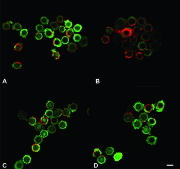
Fresh or thawed immature oocytes were incubated in dark at 37 °C in M2 medium + 4 mg/ml BSA +0.1 mg/ml dcAMP + 0.1 mg/ml fluorescence diacetate (FDA) and 0.1 mg/ml propidium iodide for 10 min. The oocytes were washed twice in M2 medium containing 4 mg/ml BSA and 0.1 mg/ml dcAMP, then placed into a droplet of the same solution in a petri dish covered with mineral oil and viewed using a confocal microscope (Nikon). Cumulus cells with an intact cell membrane fluoresced green whereas those with a damaged cell membrane fluoresced red. Each oocyte was double-blinded scored for membrane integrity of the cumulus cells using the Ruppert-Lingham's scoring system (Ruppert-Lingham et al., Reference Ruppert-Lingham, Paynter, Godfrey, Fuller and Shaw2003). An oocyte was scored as 1 for 71–100% of the cumulus cells with intact membranes, 2 for 51–70%, 3 for 21–50% and 4 for 0–20%.
In vitro maturation (IVM)
Fresh or thawed immature oocytes were cultured in 30 μl droplets of Waymouth's MB752/1 medium (Invitrogen) supplemented with 5% foetal bovine serum (FBS) (Invitrogen), 75 μg/ml penicillin G-K, 50 μg/ml streptomycin, 10 ng/ml epidermal growth factor, 27.5 μg/ml sodium pyruvate (Invitrogen), 5 μg/ml insulin, 5 μg/ml transferrin, 10 μg/ml follicle stimulation hormone (FSH) and 10 μg/ml luteinizing hormone (LH) (Bioniche Inc.) under mineral oil at 37 °C in an atmosphere of 5% CO2 in air for 10 h (GVBD) or 18 h (GV) until maturation stage.
In vitro fertilization and development of embryos
In vitro matured oocytes were placed into 75 μl of human tubal fluid (HTF) medium (Quinn et al., Reference Quinn, Kerin and Warnes1985) supplemented with 4 mg/ml BSA and then 10 μl of capacitated sperm from Kunming mice (sperm which had been incubated for 1–1.5 h in HTF medium supplemented with 4 mg/ml BSA at 37°C) was added to the oocytes. The mixture was incubated at 37 °C, in an atmosphere of 5% CO2 in air for 4–6 h. Oocytes were then washed in HTF medium and transferred into 75 μl drops of HTF medium. At this point, the oocytes were assessed for survival. Oocytes having dark or granular cytoplasm, or being non-spherical or shrunken were defined as abnormal. The normal oocytes were cultured in HTF at 37°C, in an atmosphere of 5% CO2 in air. The numbers of 2-cell and blastocyst embryos were counted 24 h and 4 days post insemination respectively.
Statistical analysis
All experiments were repeated at least three times. Pairwise comparisons were made between the experimental and control groups using the chi-squred test with Statistical Analysis System (SAS) software (SAS Institute). The p value of less than 0.05 was considered statistically significant.
Results
Membrane integrity of cumulus cells after vitrification of immature oocytes at different stages
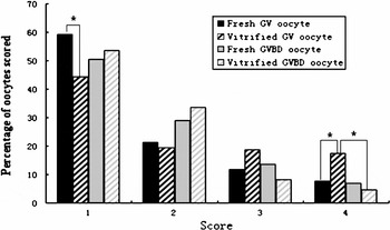
Staining of vitrified oocytes revealed that GV oocytes had a higher incidence of cumulus cell damage than GVBD oocytes; lots of damage of the former occurred in the inner layers of cumulus, but most of damage in the latter occured only in outer layers of cumulus cells (Fig. 1). Minority of GV oocytes were scored 1 and most of them scored 2, 3, and 4. However, a majority of GVBD oocytes were scored 1 and 2 and a few of them scored 4 (Fig. 2).
Representative photos of cumulus membrane damage of immature oocytes. (a) Fresh GV oocytes; (b) vitrified GV oocytes; (c) fresh GVBD oocytes; (d) vitrified GVBD oocytes. Scale bar represents 25 μm.

Distribution of scores assigned following staining. Fresh GV oocytes (n = 103), vitrified GV oocytes (n = 117), fresh GVBD oocytes (n = 103) and vitrified GVBD oocytes (n = 110). Scoring system: percentage of cumulus cells with intact membranes 100–71% as 1, 70–51% as 2, 50–21% as 3 and 20–0% as 4. *indicates that each two groups differed significantly (p < 0.05).

Survival and maturation rate of frozen–thawed immature oocytes at different stages
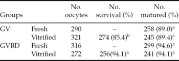
As shown in Table 1, the survival rate of vitrified GVBD oocytes (94.1%) were significantly higher (p < 0.05) than those of GV oocytes (85.4%). The maturation rates were similar among the experimental and control groups.
Survival and maturation of frozen–thawed immature oocytes at different stage.

a, b Values with different superscripts within each column are significantly different (p < 0.05).
In vitro fertilization and subsequent development of immature oocytes vitrified at different stage
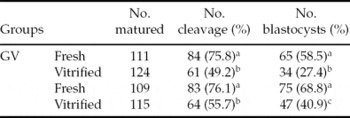
The data were shown in Table 2. Both frozen GV and GVBD oocytes showed significantly lower cleavage and blastocyst rates (p < 0.05) than those of corresponding control groups after IVF. However, the blastocyst rate of vitrified GVBD oocytes was significantly higher (p < 0.05) than that of vitrified GV oocytes (40.9% vs. 27.4%), though there was no difference in cleavage rate (55.7% vs. 49.2%).
In vitro fertilization and subsequent development of immature oocytes vitrified at different stages.
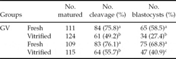
a–c Values with different superscript within same column are significantly different (p < 0.05).
Discussion
The efficiency of oocyte freezing was affected by many factors, such as cryoprotectant type, the freezing method and cooling or thawing rates, each of which may be responsible for the cryodamage of the oocyte. The present study investigated the effect of the meiosis stage on efficiency of oocyte freezing. Mouse GV or GVBD oocytes were vitrified using the OPS method, the cumulus cell cryodamage and subsequent development of oocytes were evaluated after vitrification.
In the present study, the cumulus cells stained red following fluorescence staining, indicating that the cumulus cell was damaged. The oocytes were scored for different grades according to the proportion of intact cumulus cells (100–71% as 1, 70–51% as 2, 50–21% as 3, 20–0% as 4). From to the result, vitrified GV oocytes revealed a high incidence of cumulus cell damage and a great many of the oocytes scored grade 4. However, oocytes vitrified at the GVBD stage revealed higher membrane integrity in cumulus cells. This may be due to the cytological changes in the cumulus cell during the development of the oocyte (Alworth & Albertini, Reference Alworth and Albertini1993). Another study also demonstrated that vitrification of GV oocytes produced lysis in the cumulus cells and significantly disrupted communication between the cumulus cells and oocyte. However, when the oocytes were vitrified at the matured stage, cumulus cells did not show any alterations and looked similar to fresh oocytes (Carmen et al., Reference Carmen, Paloma, Enrique, Carlos, Carolina, Aida, Lina, Santiago de la, Alba, Nieves and Maite2005).
Unlike cumulus cells, the injury of oocytes immediately after vitrification was difficult to assess, due to presence of cumulus cells; therefore, the survival rate of oocytes was assessed when cumulus cells were dispersed. At this time point, survival rate of oocytes at the GVBD stage was significantly higher than those cells vitrified at the GV stage. Similar results were obtained in porcine (Huang & Holtz, Reference Huang and Holtz2002), bovine (Barnes et al., Reference Barnes, Daniani, Looney and Duby1997) and buffalo (Sharma & Loganathasamy, Reference Sharma and Loganathasamy2007). The low permeability to water and cryoprotectant (Men et al., Reference Men, Monson and Rutledge2002) and low stability of plasma membrane (Hong et al., Reference Hong, Chung, Lim, Ko, Yoon, Yee and Cha1999) of the GV oocytes have been considered responsible for a higher incidence of damage and a lower survival in the oocytes vitrified at this stage.
In the present study, the developmental impairment of vitrified GV oocyte was most extensive, although the maturation rate of the GV oocyte did not decrease after vitrification. Previous studies in mouse have reported similar results. In one study, GV oocytes were frozen using a vitrification method and had a blastocyst rate of 26.2 compared with 48.5% of control groups (Aono et al., Reference Aono, Abe, Hara, Sasada, Sato and Yoshida2005). In another study, in which GV oocytes were cooled at 10 ℃/min to –150℃ and plunged into LN2, blastocyst rate had significantly decreased (61.9% vs.20.8%) after thawing (Ruppert-Lingham et al., Reference Ruppert-Lingham, Paynter, Godfrey, Fuller and Shaw2006), which was lower than the result found (27.4%) using the OPS vitrification method in the present study. This difference might be due to the different methods used for oocyte cryopreservation. Previous reports showed that the OPS method could dehydrate the oocytes more adequately and avoid ice crystal formation when compared with the slow freezing method (Vajta et al., Reference Vajta, Holm, Kuwayama, Booth, Jacobsen, Greve and Callesen1998; Chen et al., Reference Chen, Lien, Chen, Chao, Ho and Yang2001), which would result a the higher blastocyst rate than that of slow freezing method.
Our results showed that vitrification of oocytes at GVBD stage revealed higher membrane integrity for cumulus cells, survival rate and developmental potential of oocytes when compared at the GV stage, indicating that GVBD oocytes are more suitable stage for cryopreservation than GV oocytes. Normal cumulus cells could transform some factors, such as choline, uridine and inositol into oocytes through gap junctions (Gilchrist et al., Reference Gilchrist, Ritter and Armstrong2004; Byskov et al., Reference Byskov, Andersen, Nordholm, Thogersen, Xia, Wassmann, Andersen, Guddal and Roed1995; Chian et al., Reference Chian, Niwa and Sirard1994; Stephen, Reference Stephen2001), which had an important effect on maturation and subsequent development of oocytes (Fagbohun & Downs, Reference Fagbohun and Downs1991; Rodriguez & Farin, Reference Rodriguez and Farin2004). Therefore, the better maintenance of cumulus cells in vitrified GVBD oocytes would highly associated with the higher survival and subsequent development potential of oocytes.
On the other hand, the difference in cytology could also be associated with the different developmental potentials of oocytes vitrified at the GV or GVBD stages. The chromosomes in GV oocytes are not condensed but distributed dispersedly within the nuclear envelope. Cryopreservation of oocytes at the GV stage resulted nuclear abnormality (Liu et al., Reference Liu, Sun, Li, Jiao and Wang2003) or aneuploidy (Eroglu et al., Reference Eroglu, Toner, Leykin and Toth1998), which could be responsible for the poor development of GV oocytes after in vitro fertilization (Akiyama et al., Reference Akiyama, Nagata and Aoki2006). GVBD oocytes, howver, have resumed meiosis and show suitable conditions for nuclear and cytoplasmic changes. The use of oocytes vitrified at this stage would efficiently decrease the effect of freezing on the chromosomal structure and consequent development. Another cryopreservation study in bovine also demonstrated that oocytes at the GVBD stage produced higher cleavage and blastocyst rates than those at the GV stage (Barnes et al., Reference Barnes, Daniani, Looney and Duby1997), which was in agreement with the present study.
In conclusion, differences in cumulus cell damage or developmental potential appeared to be dependent on the meiosis stage of the vitrified oocytes. Vitrified GVBD oocytes have a better developmental potential than that of GV oocytes, partially due to the low injury of cumulus cells, and this was the suitable stage for cryopreservation of immature oocytes in mice.
Acknowledgements
The present work was supported by State ‘863’ Planning (project no. 2004AA213071) and National Key Technology Research and Development Program Foundation (No. 2006BAD14B08).