Introduction
Fear acquisition and extinction have become the focus of much scientific interest in recent years. In the laboratory, fear is acquired when a neutral stimulus (conditioned stimulus) elicits a fear response (conditioned response) after being repeatedly paired with an aversive event (unconditioned stimulus). Fear extinction occurs when the conditioned stimulus is presented alone and the conditioned response is eventually diminished. Research on how fears are extinguished is especially relevant, since it may inform the treatment of fear/anxiety-related disorders (Graham & Milad, Reference Graham and Milad2011). For example, exposure therapy is the main component of cognitive-behavioural therapy (CBT) for anxiety disorders and relies upon the basic principles of fear extinction (Hofmann, Reference Hofmann2007). In exposure therapy, an individual is repeatedly exposed to an event that has previously resulted in aversive consequences until his/her fear diminishes.
Data from the animal literature and recent human neuroimaging studies have delineated the brain areas involved in fear extinction, namely the amygdala, the ventromedial prefrontal cortex (vmPFC) and the hippocampus (Milad & Quirk, Reference Milad and Quirk2012). Each of these areas could be related to different extinction mechanisms. While initial extinction learning seems to depend on the amygdala, the consolidation and retention of extinction learning (extinction recall) would seem to depend on the vmPFC (Phelps et al. Reference Phelps, Delgado, Nearing and LeDoux2004; Kalisch et al. Reference Kalisch, Korenfeld, Stephan, Weiskopf, Seymour and Dolan2006; Sotres-Bayon et al. Reference Sotres-Bayon, Cain and LeDoux2006; Quirk & Mueller, Reference Quirk and Mueller2008). In turn, the hippocampus has been associated with the contextual modulation of fear extinction (Bouton, Reference Bouton2004; Milad et al. Reference Milad, Wright, Orr, Pitman, Quirk and Rauch2007).
Two recent studies have suggested that structural differences in the vmPFC may be associated with differences in fear extinction in humans. Milad et al. (Reference Milad, Quinn, Pitman, Orr, Fischl and Rauch2005) were the first to report cortical thickness in one sub-region of the vmPFC, the medial orbital frontal cortex (mOFC), to be associated with extinction recall in a sample of healthy individuals. This finding was recently replicated by an independent research group (Hartley et al. Reference Hartley, Fischl and Phelps2011). Given that exposure therapy relies on fear extinction mechanisms, it is plausible that the results obtained in fear extinction studies (specifically, those involving extinction recall) translate into the clinical world, i.e. that size variability, in particular vmPFC sub-regions, predicts – at least partially – exposure therapy outcomes. Identifying such predictors could eventually guide treatment planning in anxiety disorders (Rauch et al. Reference Rauch, Shin and Phelps2006).
Nevertheless, to date, there have been very few studies on this topic in clinical samples. Bryant et al. (Reference Bryant, Felmingham, Whitford, Kemp, Hughes, Peduto and Williams2008) used voxel-based morphometry (VBM) to test whether the volume of another vmPFC sub-region, the rostral anterior cingulate cortex (rACC), predicted response to CBT in post-traumatic stress disorder (PTSD) patients. In this study, treatment responders and healthy controls had larger rACC volume than non-responders, and rACC volume was significantly associated with pre- to post-treatment symptom reduction. The study, however, did have a number of limitations, including a small sample (n = 13), a relatively arbitrary criterion of treatment response (failing to fulfil PTSD criteria after treatment), and a lack of examination of other brain regions known to be involved in fear extinction (e.g. mOFC). Very recently, Hoexter et al. (Reference Hoexter, Dougherty, Shavitt, D'Alcante, Duran, Lopes, Diniz, Batistuzzo, Evans, Bressan, Busatto and Miguel2012), also using VBM techniques, found that the volume of the subgenual ACC (overlapping with the subcallosal cortex; SC) predicted pre- to post-treatment symptom reduction in a sample of treatment-naive obsessive-compulsive disorder (OCD) patients receiving group CBT as part of a randomized trial. Again, the sample size of this study was relatively small (n = 15). Furthermore, there was no control group to test for pre-treatment differences in the brain areas studied. This is important, given previous reports of volume reductions in areas such as the mOFC or the ACC in OCD patients in comparison with healthy controls (Pujol et al. Reference Pujol, Soriano-Mas, Alonso, Cardoner, Menchon, Deus and Vallejo2004; Rotge et al. Reference Rotge, Guehl, Dilharreguy, Tignol, Bioulac, Allard, Burbaud and Aouizerate2009).
Therefore, although previous studies suggest that morphometric variability in different sub-regions of the vmPFC may predict the outcome of exposure therapy (or more generally CBT) in anxiety disorders, there are indeed some conflicting results. Thus, while the volumes of the rACC and the SC were not associated with extinction recall in healthy subjects (Milad et al. Reference Milad, Quinn, Pitman, Orr, Fischl and Rauch2005), they predicted CBT outcome in PTSD and OCD patients (Bryant et al. Reference Bryant, Felmingham, Whitford, Kemp, Hughes, Peduto and Williams2008; Hoexter et al. Reference Hoexter, Dougherty, Shavitt, D'Alcante, Duran, Lopes, Diniz, Batistuzzo, Evans, Bressan, Busatto and Miguel2012). Conversely, to date, morphometry of the mOFC has not been related to CBT outcome in any anxiety disorder.
Methodological differences between studies may explain such divergences, since Milad et al. (Reference Milad, Quinn, Pitman, Orr, Fischl and Rauch2005) focused on cortical thickness measurements whereas Bryant et al. (Reference Bryant, Felmingham, Whitford, Kemp, Hughes, Peduto and Williams2008) and Hoexter et al. (Reference Hoexter, Dougherty, Shavitt, D'Alcante, Duran, Lopes, Diniz, Batistuzzo, Evans, Bressan, Busatto and Miguel2012) assessed regional volumes. Although both cortical thickness and regional volumetry have been widely used as valid measurements of brain morphometry, they refer to partially different aspects of brain structure, since regional volumetry is also influenced by cortical surface area and cortical folding (Hutton et al. Reference Hutton, Draganski, Ashburner and Weiskopf2009). Consequently, combining both measurements may be useful to fully characterize brain anatomy (Hutton et al. Reference Hutton, Draganski, Ashburner and Weiskopf2009; Labate et al. Reference Labate, Cerasa, Mula, Mumoli, Gioia, Aguglia, Quattrone and Gambardella2012).
In the present study we tested whether morphometric variations in the different sub-regions of the vmPFC were related to exposure therapy outcome in a sample of OCD patients. In addition, baseline morphometric differences in relation to healthy controls were also assessed for these brain areas. Importantly, with the aim of overcoming previous methodological differences between studies and to comprehensively characterize such differences and the correlations between vmPFC sub-regions and exposure therapy outcome, we used regional measurements of both cortical thickness and volume. We focused on OCD in view of the recent interest in ‘new’ OCD models based on fear extinction deficits (Milad & Rauch, Reference Milad and Rauch2012). Furthermore, exposure therapy (using response prevention) is one of the treatments of choice for OCD. From a behavioural perspective, OCD is characterized by the presence of conditioned fear responses (which could be established through Pavlovian aversive conditioning), and exposure (using response prevention, which blocks escape/avoidance) is effective as it provides an opportunity for the extinction of such responses (see Abramowitz, Reference Abramowitz2006). Exposure is therefore an analogue of extinction training. However, although exposure is a highly effective treatment for OCD, there is significant across-subject variability in treatment response.
On the basis of previous reports (Pujol et al. Reference Pujol, Soriano-Mas, Alonso, Cardoner, Menchon, Deus and Vallejo2004; Venkatasubramanian et al. Reference Venkatasubramanian, Zutshi, Jindal, Srikanth, Kovoor, Kumar and Janardhan Reddy2012), we predicted that OCD patients would show morphometric alterations in the mOFC and the rACC in comparison with healthy controls. Also based on previous clinical work (Bryant et al. Reference Bryant, Felmingham, Whitford, Kemp, Hughes, Peduto and Williams2008; Hoexter et al. Reference Hoexter, Dougherty, Shavitt, D'Alcante, Duran, Lopes, Diniz, Batistuzzo, Evans, Bressan, Busatto and Miguel2012), we hypothesized that the size of the rACC and SC would be associated with the outcome of exposure therapy for OCD.
Method
Participants
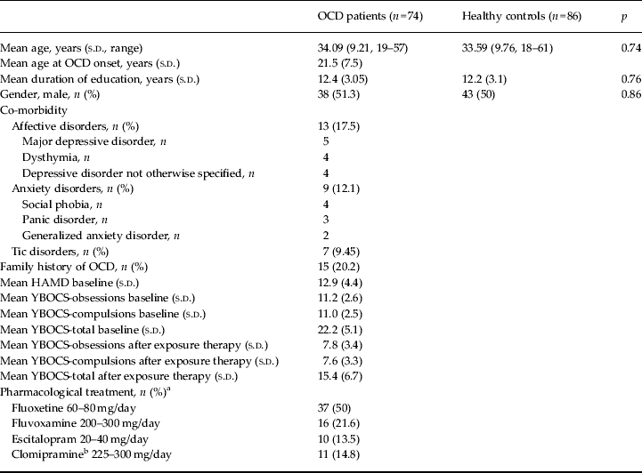
A total of 74 OCD patients and 86 healthy controls were included in the study. Sociodemographic and clinical characteristics of both groups are presented in Table 1. Patients were recruited from the OCD Unit at Bellvitge University Hospital (Barcelona, Spain) between 2009 and 2011. All patients met Diagnostic and Statistical Manual of Mental Disorders, fourth edition (DSM-IV) criteria for OCD (APA, 1994) and had shown OCD symptoms for at least 1 year. Diagnoses were independently assigned by two psychiatrists with extensive clinical experience in OCD, who separately interviewed the patients using the Structured Clinical Interview for DSM-IV Axis I Disorders – Clinician Version (SCID-IV-CV; First et al. Reference First, Spitzer, Gibbon and Williams1996). Patients were eligible for the study when both clinicians agreed on all diagnostic criteria. Exclusion criteria for the patients were as follows: age under 18 or over 65 years, current or 6-month past history of psychoactive substance abuse/dependence, co-morbid psychotic or bipolar disorders, mental retardation, presence or past history of serious organic or neurological disorder (except tic disorder), and any contraindication to magnetic resonance imaging (MRI) scanning. Co-morbid depressive and anxiety disorder symptoms were not considered to be exclusion criteria provided that OCD was the main diagnosis and the primary reason for seeking assistance. The presence of lifetime depressive symptoms (including lifetime major depressive disorder, MDD) was also assessed with the SCID-IV-CV.
Sociodemographic and clinical characteristics of study participants

OCD, Obsessive-compulsive disorder; s.d. standard deviation; HAMD, Hamilton Depression Rating Scale; YBOCS, Yale–Brown Obsessive-Compulsive Scale.
a A benzodiazepine tapering protocol was initiated at the beginning of the pharmacological trial.
b Patients under clomipramine treatment should have reported a previous history of failure with selective serotonin reuptake inhibitors.
Healthy controls were recruited from the same sociodemographic environment as patients. Each control underwent the SCID-IV non-patient version (First et al. Reference First, Spitzer, Gibbon and Williams2002) to exclude any current or past Axis I disorder. The other exclusion criteria were the same as for the OCD group.
Participants in both groups gave written informed consent after receiving a complete description of the study, which was approved by the research and ethics committee of Bellvitge University Hospital. The investigation was carried out in accordance with the Declaration of Helsinki.
Study procedures
OCD patients received an open trial of standard exposure therapy (including response prevention) after partial or non-response to a 12-week pharmacological trial with a selective serotonin reuptake inhibitor or clomipramine following recommended guidelines (Koran et al. Reference Koran, Hanna, Hollander, Nestadt and Simpson2007). After achieving the maximum recommended and tolerated doses of medication, patients initiated exposure therapy. The time lapse between the end of the pharmacological trial and the initiation of exposure therapy ranged between 1 and 2 weeks. During this interval, a structural MRI examination was performed (see below). Importantly, medication was kept stable during exposure therapy (pharmacological profiles are shown in Table 1). Of an initial sample of 83 patients who fulfilled the inclusion criteria, three subjects refusing to initiate exposure therapy and six who dropped out before completing the first five sessions were duly classified as non-completers.
MRI acquisition
Participants in both groups were scanned with a 1.5-T scanner (Signa Excite system; General Electric, USA) equipped with an eight-channel phased-array head coil. A high-resolution T1-weighted anatomical image was obtained for each subject using a three-dimensional fast spoiled gradient inversion-recovery prepared sequence with 130 contiguous slices in the axial plane (repetition time = 11.8 ms, echo time = 4.2 ms and flip angle = 90°, within a field of view of 30 cm, with a 256 × 256 pixel matrix and a slice thickness of 1.2 mm).
Exposure therapy
Exposure therapy was manualized (Kozak & Foa, Reference Kozak and Foa1997) and applied by an experienced therapist who was blind to the study's hypotheses. The study involved 20 individual weekly sessions lasting approximately 45 min. The first two sessions were devoted to psychoeducation, the introduction to the behavioural model of OCD and the development of an exposure hierarchy; sessions 3–18 consisted of gradual exposure to items of the hierarchy, with instructions for strict response prevention from compulsions. Between sessions, homework consisting of exposure to stimuli similar to those addressed in the sessions was assigned (60 min daily). The final two sessions were devoted to relapse prevention.
Assessment and treatment response
The Yale–Brown Obsessive-Compulsive Scale (YBOCS; Goodman et al. Reference Goodman, Price, Rasmussen, Mazure, Fleischmann, Hill, Heninger and Charney1989) was administered by experienced clinicians to the OCD group to assess illness severity both before and after exposure therapy. Co-morbid depressive symptoms were measured by means of the Hamilton Depression Rating Scale (HAMD; Hamilton, Reference Hamilton1960).
Treatment response was defined as a 35% or greater decrease in the YBOCS total score according to operational criteria (Pallanti et al. Reference Pallanti, Hollander, Bienstock, Koran, Leckman, Marazziti, Pato, Stein and Zohar2002), according to which 35 patients (47.2%) were regarded as responders to exposure therapy [mean YBOCS reduction: 49.3 (s.d. = 11.0)%] and 39 (52.7%) as non-responders [mean YBOCS reduction: 17.8 (s.d. = 10.6)%].
Image processing
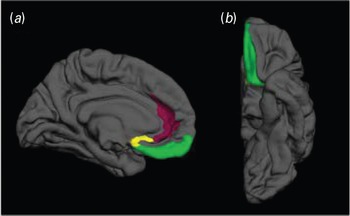
After inspection for imaging artefacts, subcortical and cortical parcelation was performed using the publicly available FreeSurfer software (version 5.1; http://ftp.nmr.mgh.harvard.edu/). The standard pipeline was used, including a volume- and a surface-based processing stream. Volume-based stream involved the following stages: affine registration to the MNI305 template, initial volumetric labelling, intensity correction for field inhomogeneities, intensity normalization, removal of non-brain tissue and linear and non-linear transformations to a probabilistic brain atlas (Fischl et al. Reference Fischl, Salat, Busa, Albert, Dieterich, Haselgrove, van der Kouwe, Killiany, Kennedy, Klaveness, Montillo, Makris, Rosen and Dale2002, Reference Fischl, Salat, van der Kouwe, Makris, Segonne, Quinn and Dale2004). The surface stream was focused on modelling the boundary between white matter and cortical grey matter (GM; denoted as white surface), in addition to the pial surface. The white surface was parcelled into 74 cortical regions of interest (ROIs) through the assignment of a neuroanatomical label for each surface location (vertex) according to the Destrieux et al. (Reference Destrieux, Fischl, Dale and Halgren2010) parcelation scheme. Cortical thickness and GM volumes values were then calculated for the 74 cortical ROIs. Specifically, the distance between the pial and the white surface was calculated for each brain vertex and averaged across all the vertices of each ROI to obtain regional cortical thickness, while regional cortical volumes were calculated by multiplying the number of voxels in each parcelation unit by the voxel volume (1.17 × 1.17 × 1.2 mm). However, on the basis of our hypotheses, in the present study we only considered three ROIs per hemisphere: (1) mOFC (corresponding to the gyrus rectus plus the medial orbital sulcus); (2) rACC (corresponding to the anterior part of the cingulate gyrus and sulcus); and (3) the SC (corresponding to Brodmann area 25 and the most ventral part of Brodmann area 32). The selected ROIs are displayed in Fig. 1.
Representation of the regions of interest used in this study, which are overlaid on mid-sagittal (a) and ventral (b) sections of a left hemisphere brain template. The medial orbitofrontal cortex is depicted in green, the rostral anterior cingulate cortex in violet, and the subcallosal cortex in yellow.

Statistical analyses
An analysis of covariance (ANCOVA) was used to compare regional measurements of cortical thickness and volume in the selected ROIs between patients and controls using age, gender, lifetime MDD and total GM volume as covariates. Total GM volume was calculated for each subject by multiplying the number of voxels classified as GM by the voxel volume. In the patient group, partial correlations were calculated between ROI measurements and exposure therapy outcome (percentage of total YBOCS reduction between pre- and post-exposure therapy assessments). In addition, we compared ROI measurements between treatment responders and non-responders also by means of an ANCOVA test. In both analyses related to treatment response, in addition to age, gender, lifetime MDD and total GM volume, we also controlled for pre-exposure therapy HAMD scores and YBOCS reduction after the pharmacological trial. The Bonferroni correction for multiple comparisons was used, and α was set at p < 0.0083 (0.05/6 ROIs, three per hemisphere) in each analysis. These analyses were conducted using the Statistical Package for Social Sciences software (SPSS version 20; IBM, USA).
Results
Baseline morphometric differences between patients and controls
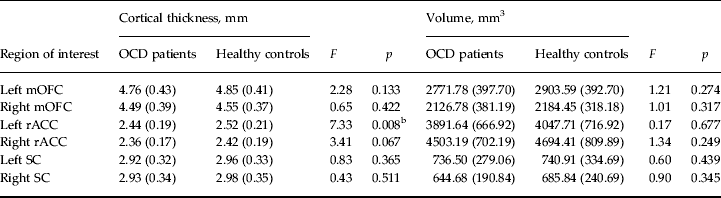
As presented in Table 2, cortical thickness analyses revealed that the left rACC was thinner in OCD patients in comparison with healthy controls. Patients and controls did not differ in the cortical thickness of the right rACC, the mOFC or the SC. Results from the volumetric analyses are also shown in Table 2. OCD patients and healthy controls did not differ in any of the volumetric measurements.
Differences between OCD patients (n = 74) and healthy controls (n = 86) in our regions of interest a

Data are given as mean (standard deviation).
OCD, Obsessive-compulsive disorder; mOFC, medial orbitofrontal cortex; rACC, rostral anterior cingulate cortex; SC, subcallosal cortex.
a Controlling for age, gender, lifetime major depressive disorder and total grey matter volume.
b Significant difference after Bonferroni correction.
Morphometric correlates of exposure therapy outcome
YBOCS scores
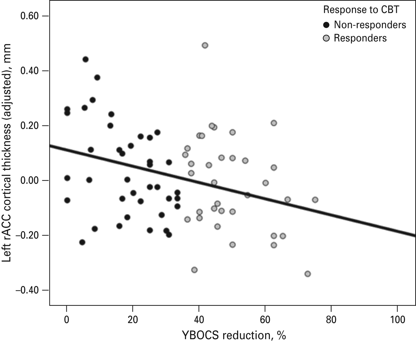
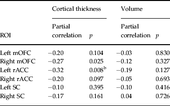
Significant negative partial correlations were observed between the cortical thickness measurements of the right mOFC and the left rACC and exposure therapy outcome assessed by the percentage of reduction in YBOCS scores. However, following the Bonferroni correction, only the association with the left rACC (r–0.32, p = 0.008) remained statistically significant (see Fig. 2). Regarding volumetric measures, none of the ROIs assessed was significantly associated with exposure therapy results (see Table 3). Given the age range of our sample and the possible effects of menopausal status on brain measures (e.g. Goto et al. Reference Goto, Abe, Miyati, Inano, Hayashi, Aoki, Mori, Kabasawa, Ino, Yano, Iida, Mima and Ohtomo2011) and fear extinction (see Zeidan et al. Reference Zeidan, Igoe, Linnman, Vitalo, Levine, Klibanski, Goldstein and Milad2011), we repeated this analysis controlling for such a status in female participants, and the results remained unchanged.
Cortical thickness of the left rostral anterior cingulate cortex (rACC) shows an inverse relationship with exposure therapy outcome [assessed as the percentage of reduction in Yale–Brown Obsessive-Compulsive Scale (YBOCS) scores, r – 0.32, p = 0.008], after controlling for age, gender, lifetime major depressive disorder, total grey matter volume, pre-treatment depression score and YBOCS reduction after the pharmacological trial. Obsessive-compulsive disorder patients are labelled as responders and non-responders. CBT, Cognitive-behavioural therapy.

Correlational analyses of ROI measurements and exposure therapy results in OCD patients (n = 74) a

ROI, Region of interest; OCD, obsessive-compulsive disorder; mOFC, medial orbitofrontal cortex; rACC, rostral anterior cingulate cortex; SC, subcallosal cortex; YBOCS, Yale–Brown Obsessive-Compulsive Scale.
a Controlling for age, gender, lifetime major depressive disorder, total grey matter volume, pre-treatment depression score and YBOCS reduction after the pharmacological trial.
b Significant correlation after Bonferroni correction.
To confirm the above results, a post-hoc hierarchical multiple regression analysis was conducted with the pre–post YBOCS percentage reduction as the dependent variable and the cortical thickness values of the six ROIs as predictor variables (stepwise method). Age, gender, lifetime MDD, total GM volume, pre-treatment depression (HAMD score) and YBOCS reduction after pharmacological trial were initially forced into the model to control for their possible influence. The results of this analysis showed that cortical thickness of the left rACC was the only significant predictor of exposure therapy outcome, explaining 8.3% of additional variance over and above the effects of the confounding covariates (full-model R 2 = 0.27, F 7,66 = 3.52, p = 0.003; R 2 change after including cortical thickness of the left rACC = 0.083, F 1,66 change = 7.51, p = 0.008).
Treatment responders versus non-responders
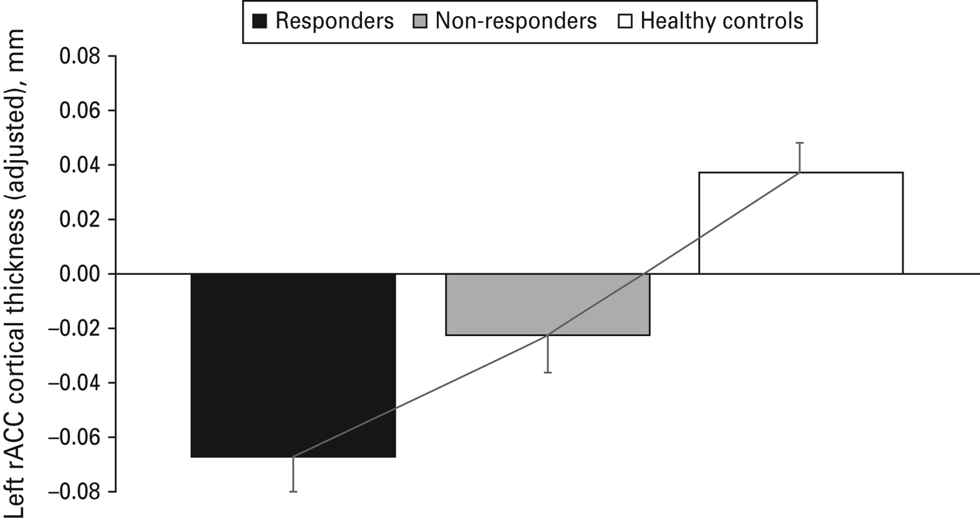
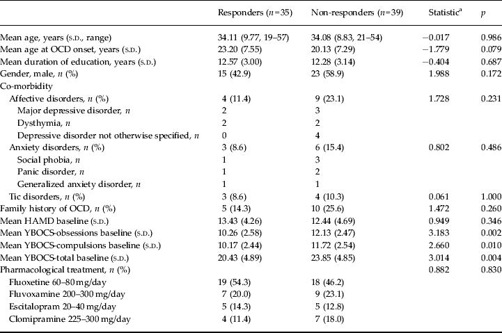
Finally, we divided our patient sample into two groups according to their response to exposure therapy treatment. A total of 39 subjects were considered treatment non-responders (YBOCS reduction <35%), while 35 patients did respond to exposure therapy (YBOCS reduction ⩾35%). These groups did not differ in any sociodemographic or clinical variable with the exception of the YBOCS score prior to exposure therapy initiation, which was lower in treatment responders (see Table 4). The inclusion of this variable as a further covariate in the above analyses, however, did not alter their significance. Regarding morphometric measurements, we observed that the thickness of the left rACC was significantly different across the three study groups (F 2,153 = 4.25, p = 0.008), with treatment responders showing a thinner left rACC than non-responders and healthy controls. Indeed, left rACC thickness showed a significant linear increase across the study groups (polynomial contrast, linear trend, p = 0.006). The results are shown in Fig. 3.
Cortical thickness of the left rostral anterior cingulate cortex (rACC, adjusted for confounding covariates) in exposure therapy responders and non-responders and healthy controls. Values are means, with standard deviations represented by vertical lines. The significant linear increase observed across groups is represented by a solid line.

Sociodemographic and clinical variables in exposure therapy responders and non-responders

s.d., Standard deviation; OCD, obsessive-compulsive disorder; HAMD, Hamilton Depression Rating Scale; YBOCS, Yale–Brown Obsessive-Compulsive Scale.
a For quantitative variables, t test; for qualitative variables, χ 2.
Discussion
In the present study we aimed to investigate the relationship between the vmPFC, a brain area typically associated with fear extinction in animal and human studies (Milad & Quirk, Reference Milad and Quirk2012), and exposure therapy outcomes in a relatively large sample of OCD patients. Importantly, the alleged role of the vmPFC in fear extinction seems to translate into clinical practice, as the region has been associated to exposure therapy outcome in anxiety disorders (Bryant et al. Reference Bryant, Felmingham, Whitford, Kemp, Hughes, Peduto and Williams2008; Hoexter et al. Reference Hoexter, Dougherty, Shavitt, D'Alcante, Duran, Lopes, Diniz, Batistuzzo, Evans, Bressan, Busatto and Miguel2012). Of the three sub-regions within the vmPFC assessed here, we found that the left rACC showed cortical thickness reductions in OCD patients. Also, this region was significantly associated to exposure therapy outcome. Specifically, our analyses showed that, after adjusting for unspecific confounding factors, the thickness of the left rACC explained 8% of inter-subject variability in response to exposure therapy.
Morphometric differences between OCD patients and healthy controls
In comparison with healthy controls, OCD patients showed cortical thinning in the left rACC. Conversely, none of the vmPFC sub-regions showed significant between-group differences in volume. Our results diverge from those of other studies reporting cortical thickness reductions in OCD involving the left mOFC (Shin et al. Reference Shin, Yoo, Lee, Ha, Lee, Lee, Kim, Kim and Kwon2007) and the right rACC (Venkatasubramanian et al. Reference Venkatasubramanian, Zutshi, Jindal, Srikanth, Kovoor, Kumar and Janardhan Reddy2012). Likewise, the lack of significant between-group differences in volume measurements is at odds with previous reports of mOBF and rACC volume reductions (Pujol et al. Reference Pujol, Soriano-Mas, Alonso, Cardoner, Menchon, Deus and Vallejo2004; Rotge et al. Reference Rotge, Guehl, Dilharreguy, Tignol, Bioulac, Allard, Burbaud and Aouizerate2009). These discrepant findings may be partially attributed to methodological factors, such as the methods used for volumetric quantification and the precise clinical features of the OCD samples assessed. Thus, rACC volume reductions have mainly been detected using semi-automated or manual approaches (Rotge et al. Reference Rotge, Guehl, Dilharreguy, Tignol, Bioulac, Allard, Burbaud and Aouizerate2009), but not with automated approaches (Radua & Mataix-Cols, Reference Radua and Mataix-Cols2009; Rotge et al. Reference Rotge, Langbour, Guehl, Bioulac, Jaafari, Allard, Aouizerate and Burbaud2010), while mOBF volume reductions have particularly been observed in subsamples of OCD patients with co-morbid major depressive disorder (Cardoner et al. Reference Cardoner, Soriano-Mas, Pujol, Alonso, Harrison, Deus, Hernandez-Ribas, Menchon and Vallejo2007). Indeed, the thorough control of possible confounding factors (i.e. lifetime MDD) performed here must certainly have contributed to circumscribe our findings to those regions more specifically related to OCD. All in all, comparisons between different methodological approaches and OCD samples differing in particular clinical features would need to be conducted in order to fully characterize the nature of such alterations. Be that as it may, the existence of structural abnormalities in brain regions relevant to the consolidation and retention of fear extinction is in agreement with the idea that fear extinction deficits may partially account for the constellation of symptoms observed in OCD subjects (Milad & Rauch, Reference Milad and Rauch2012).
Prediction of exposure therapy outcome and the vmPFC
Cortical thickness of the left rACC was negatively associated with the response to exposure therapy. Although it might be argued that a correlation value of 0.32 is not particularly ‘strong’, it does indeed imply that 8% of the variability in exposure therapy response may be explained by morphometric factors. In addition, our correlational findings were further supported by the direct comparison between treatment responders and non-responders, showing a decreased cortical thickness in the left rACC of responder subjects. Although such data are broadly in agreement with the findings of Bryant et al. (Reference Bryant, Felmingham, Whitford, Kemp, Hughes, Peduto and Williams2008), in that they reported an association between regional volumes within the rACC and treatment response to exposure-based CBT in PTSD patients, there are significant differences between both studies. Apart from using a different patient population (PTSD versus OCD) and a different methodological approach (spatially constrained voxel-wise volumetric-only assessment versus the ROI approach used here to assess both cortical thickness and volume), Bryant's study showed a volumetric decrease in non-responders when compared with responders, whereas ours demonstrates the opposite pattern of results relating to cortical thickness. Indeed, although negative relationships between morphometric measurements and treatment response have been detected in other disorders (e.g. Arango et al. Reference Arango, Breier, McMahon, Carpenter and Buchanan2003), larger volumes or thicker cortices within the vmPFC (regardless of its precise location) have been typically associated with better fear extinction in both non-clinical (Milad et al. Reference Milad, Quinn, Pitman, Orr, Fischl and Rauch2005; Hartley et al. Reference Hartley, Fischl and Phelps2011) and clinical populations (Hoexter et al. Reference Hoexter, Dougherty, Shavitt, D'Alcante, Duran, Lopes, Diniz, Batistuzzo, Evans, Bressan, Busatto and Miguel2012).
Although the precise factors (methodological or others) leading to such contradictory results between studies remain elusive, it is noteworthy that, in a recent functional MRI study, a favourable response to CBT was associated with decreased activity in the rACC (Siegle et al. Reference Siegle, Thompson, Collier, Berman, Feldmiller, Thase and Friedman2012). Such a finding was interpreted in terms of the possibility that CBT provides or restores the necessary resources for an effective cortical inhibition of limbic activity, favouring top-down control strategies (Linden, Reference Linden2008). By contrast, drug therapy is supposed to directly down-regulate limbic activity, and indeed increased pre-treatment activity in the rACC predicts good response to antidepressant treatment in depressive patients (Chen et al. Reference Chen, Ridler, Suckling, Williams, Fu, Merlo-Pich and Bullmore2007). Further research would be needed to confirm whether such notions might also apply to morphometric measurements. Nevertheless, one possible explanation for our findings is that patients with smaller cortical thickness in the rACC show a greater fear extinction dysfunction and are therefore the best candidates for improvement after a therapy that is specifically tailored to enhance fear extinction capabilities. Alternatively, it may be also noted that the role of cortico-limbic interactions may differ depending on the brain hemisphere. Thus, while in the right hemisphere the interaction between the rACC and the amygdala seems to mediate behavioural adaptation to aversive events (for instance, in fear-conditioning paradigms), left rACC and amygdala interactions seem to mediate the effects of negative affect (Polli et al. Reference Polli, Wright, Milad, Dickerson, Vangel, Barton, Rauch and Manoach2009). In this sense, a left-lateralized reduction of rACC thickness may favour behavioural adaptations without interfering modulations from negative affect.
Beyond the above-mentioned correlation between rACC thickness and CBT response, we also observed a tendency to significance in terms of the correlation between the cortical thickness of the right mOFC and exposure therapy outcome. Again, however, it should be pointed out that our results differ from those reported by Milad et al. (Reference Milad, Quinn, Pitman, Orr, Fischl and Rauch2005), in which a thicker mOFC predicted a better retention of extinction learning. Nevertheless, such findings suggest that the mOFC may also play a relevant role in exposure therapy outcome, which should be researched further. Furthermore, contrary to what was recently reported by Hoexter et al. (Reference Hoexter, Dougherty, Shavitt, D'Alcante, Duran, Lopes, Diniz, Batistuzzo, Evans, Bressan, Busatto and Miguel2012), we did not observe any relation between the SC and treatment response. Nevertheless, contrary to what has been consistently reported for mood disorders (Fu et al. 2013), the SC has not been consistently related to OCD.
Strengths and limitations
The strengths of the present study include a clear hypothesis-driven approach based on solid translational research, a relatively large sample of patients and controls and the use of two different morphometric measurements. Its main limitation is that all the patients involved were undergoing pharmacological treatment and, therefore, morphometric assessments were subject to this potentially confounding factor. Although there is some concern as to the effects that psychotropic substances may have on brain anatomy, little evidence exists to suggest that OCD medication may induce morphometric brain alterations, such as neuronal loss or gliosis (Konradi & Heckers, Reference Konradi and Heckers2001). Furthermore, a meta-analysis of VBM studies performed with OCD samples concluded that medication has little effect on GM volume measurements (Radua & Mataix-Cols, Reference Radua and Mataix-Cols2009). Likewise, in another study assessing whole-brain cortical thickness, the measures of medication exposure were not correlated with the thickness of any cortical region showing a significant thinning in the sample of OCD patients (Shin et al. Reference Shin, Yoo, Lee, Ha, Lee, Lee, Kim, Kim and Kwon2007). Although the concurrent use of medication may limit the internal validity of our study, it nonetheless increases its clinical relevance, as, in everyday clinical practice, many OCD patients receive combined (i.e. medication plus exposure therapy) treatments (e.g. Boschen & Drummond, Reference Boschen and Drummond2012).
Furthermore, from a conceptual perspective, it might be argued that the evidence supporting the translation from fear extinction in the laboratory to exposure therapy in clinical practice is limited. Nevertheless, in at least one clinical study (with patients suffering from social anxiety disorder), a significant relationship was found between extinction retention and exposure therapy outcome (Berry et al. Reference Berry, Rosenfield and Smits2009). Likewise, certain molecules facilitating fear extinction (e.g. d-cycloserine) have been shown to improve exposure therapy outcome in clinical samples (see Norberg et al. Reference Norberg, Krystal and Tolin2008). Finally, although fear extinction (and retention) deficits may be characteristic of most anxiety disorders, such deficits have, to date, mainly been detected in PTSD patients (Milad et al. 2008, 2009) and, so far, only one study has shown them to characterize OCD (Milad et al. Reference Milad, Furtak, Greenberg, Keshaviah, Im, Falkenstein, Jenike, Rauch and Wilhelm2013). Nevertheless, fear extinction mechanisms have not been thoroughly investigated in OCD (Milad & Rauch, Reference Milad and Rauch2012).
Implications
First, from an academic point of view, the present research highlights the idea that the study of fear extinction and its brain correlates may provide new insight into the pathophysiology of OCD (Milad & Rauch, Reference Milad and Rauch2012). Second, from a methodological perspective, our findings indicate that the combined assessment of both volumetric and cortical thickness measurements may help in detecting the most relevant anatomical features associated with outcome variables. The results reported here suggest that cortical thickness measurements are more directly related to treatment outcome measurements. Indeed, integrating such morphometric measurements with other imaging (e.g. functional) and non-imaging (e.g. neuropsychological, genetic, or clinical) variables may increase our ability to predict treatment response.
Finally, the present study is also clinically significant insofar as it suggests that the assessment of the brain correlates of fear extinction may eventually lead to a better prediction of treatment response and/or the identification of new treatment targets. This latter aspect is especially relevant since current treatment options for severe OCD include neuromodulation strategies (Greenberg et al. Reference Greenberg, Rauch and Haber2010), and a better understanding of the brain areas involved in fear extinction may well guide such interventions. To this effect, for example, a recent study showed that repeated transcranial magnetic stimulation (rTMS) in combination with extinction training may enhance fear extinction in rats (Baek et al. Reference Baek, Chae and Jeong2012). In humans, a preliminary study in PTSD patients showed that adding rTMS to exposure therapy may also provide certain clinical benefits (Osuch et al. Reference Osuch, Benson, Luckenbaugh, Geraci, Post and McCann2009). Studies like the one presented here may contribute to the optimization of the different treatment strategies available for OCD and ultimately serve to improve patient management.
Acknowledgements
This study was supported in part by the Carlos III Health Institute (PI09/01331 PI10/01753, PI10/01003, CP10/00604, CIBER-CB06/03/0034) and by the Agencia de Gestió d'Ajuts Universitaris i de Recerca (AGAUR; 2009SGR1554). M.S. is funded by the Bellvitge Biomedical Research Institute (IDIBELL). C.L-S. is supported by the Spanish Ministry of Education, Culture and Sport (FPU12/01636). E.R. is supported by a ‘Rio Hortega’ contract from the Carlos III Health Institute (I.D. CM11/00077). C.S-M. is funded by a ‘Miguel Servet’ contract from the Carlos III Health Institute (CP10/00604). We also thank Gerald Fannon for revising the manuscript.
Declaration of Interest
None.