Introduction
The larval development of Fasciola hepatica, for example, within the body of Galba truncatula causes injury to the snail's viscera. In the weeks following miracidial penetration, the epithelium of four organs (albumin gland, digestive gland, gonad and kidney) progressively degenerates. An epithelial reconstitution subsequently develops after day 35 post-exposure (p.e.) at 20°C. Other cycles of epithelial necrosis–reconstitution subsequently occur up to the death of the infected snail (Rondelaud & Barthe, Reference Rondelaud and Barthe1983). Another injury is an amoebocytic proliferation in the snail's haemolymph, the intensity of which depends on the origin of snails used for infections (Rondelaud & Barthe, Reference Rondelaud and Barthe1981). This visceral response to F. hepatica infection was observed in most western European lymnaeids (Sindou et al., Reference Sindou, Cabaret and Rondelaud1991a); however, it is not specific to this helminth, as these visceral lesions were also found in snails infected by a protostrongylid nematode (Hourdin et al., Reference Hourdin, Rondelaud and Cabaret1990) or by any one of three digenean species other than F. hepatica (Moukrim & Rondelaud, Reference Moukrim and Rondelaud1992).
The fact that all the above observations were performed in snails, either infected with a single digenean species, or with two helminths developing one after the other, raises a problem. Indeed, little information is available on the parasite burden and tissue lesions developing in co-infected snails, i.e. snails able to sustain the simultaneous larval development of two digeneans. As G. truncatula has been known since the 1990s to be naturally co-infected with F. hepatica and Paramphistomum daubneyi (Rondelaud et al., Reference Rondelaud, Vignoles and Dreyfuss2004), it was of interest to study these parameters in these snails, as larval forms of both species often developed into separate compartments within the body of G. truncatula (Augot et al., Reference Augot, Abrous, Rondelaud and Dreyfuss1996). In view of this finding, the following three questions arise. How many rediae and cercariae can a snail harbour when it is experimentally co-infected with F. hepatica and P. daubneyi? What is the frequency of tissue lesions developing in the digestive gland and kidney of these infected snails? Are parasite burden and tissue lesions different from those found in naturally co-infected snails? To answer these questions, snails exposed experimentally first to P. daubneyi and second to F. hepatica were dissected between day 35 and day 49 p.e. to detect those that were co-infected; then they were put into Bouin's fixative. The same method was used for naturally co-infected snails. Serial sections were cut and stained to study parasite burden and the response of the snails' viscera to these co-infections.
Materials and methods
Experimentally infected snails
The population of G. truncatula was living in a road ditch located in the commune of Saint Michel de Veisse, department of Creuse, central France (45°79′87″N, 2°2′68″W). Six hundred snails, measuring 4 mm in height and belonging to the spring generation, were collected in May–June. The eggs of F. hepatica were collected from the gall bladders of heavily infected cattle at the slaughterhouse of Limoges, department of Haute Vienne, central France. To obtain eggs of P. daubneyi, adult worms were collected from the paunch of the same cattle and were placed in a saline solution (NaCl 0.9%, glucose 0.45%) for 4 h at 37 °C. All eggs were washed several times with spring water and were incubated at 20°C for 20 days in the dark (Ollerenshaw, Reference Ollerenshaw1971). Each snail was first subjected to a single miracidium of P. daubneyi for 4 h, and then to another miracidium of F. hepatica for the following 4 h (Augot et al., Reference Augot, Abrous, Rondelaud and Dreyfuss1996). After exposure, snails were subsequently reared for 30 days in polypropylene boxes (1 m × 55 cm, 15 cm deep) with 50 snails per box. The breeding method was already described by Abrous et al. (Reference Abrous, Roumieux, Dreyfuss, Rondelaud and Mage1998). Cos lettuce, originating from a private garden and grown without chemical treatment, was used to feed snails. These boxes were placed in an air-conditioned room under a constant temperature of 20 ± 1°C and a diurnal photoperiod of 12 h with a 3000–4000 lux light intensity over the boxes. Samples of 120 surviving snails each were collected from breeding boxes at days 35, 42 and 49 p.e., respectively. The choice of these dates for sampling G. truncatula was based on the fact that most snails infected either by F. hepatica or by P. daubneyi harboured cercariae-containing rediae and free cercariae within their bodies at that time (Abrous et al., Reference Abrous, Rondelaud and Dreyfuss2000). Snails were dissected under a stereomicroscope to detect those which harboured live forms of F. hepatica, P. daubneyi, or both. The 80 co-infected snails were dipped into Bouin's fixative after removal of shell fragments. Serial sections, 5 μm thick, were cut and stained with Harris’ haematoxylin and modified Gabe's trichrome (Moukrim et al., Reference Moukrim, Rondelaud and Barthe1988).
Naturally infected snails
From 1993 to 2002, 19,237 adult G. truncatula were collected in June–July and September–October from meadows surrounding 141 cattle-breeding farms of central France. These snails were dissected under a stereomicroscope to detect co-infections with F. hepatica and P. daubneyi. Only 111 snails were found harbouring live larval forms of both digeneans (Rondelaud et al., Reference Rondelaud, Vignoles and Dreyfuss2004). After dissection, shell fragments were removed and the bodies of co-infected snails were put into Bouin's fixative. Serial sections, 5 μm thick, were also stained with Harris’ haematoxylin and modified Gabe's trichrome (Moukrim et al., Reference Moukrim, Rondelaud and Barthe1988).
Snail groups
As the speed of larval development within co-infected G. truncatula was different for each digenean species (the growth of F. hepatica larvae might be quicker than that of P. daubneyi larvae, or vice versa), it is difficult to study tissue lesions occurring in snails in relation to the chronology of infections. For each type of snail infection (experimental or natural infections), two snail groups were considered (table 1). In the first group, the live rediae of F. hepatica were more numerous than those of P. daubneyi (F. hepatica > P. daubneyi group). In contrast, the number of P. daubneyi rediae was greater than those of F. hepatica in the second group (P. daubneyi > F. hepatica group). In each group, the species having the highest number of rediae was considered the dominating parasite (F. hepatica, for example, in the F. hepatica > P. daubneyi group).
Number of live rediae counted in G. truncatula in relation to the type of snail infections and species dominance.

Larval development of each digenean species
The F. hepatica rediae were easily recognizable by their appendages and their pharynx of great size. In contrast, those of P. daubneyi had only a small-sized pharynx. The rediae of both digeneans were counted on serial sections. The individual values recorded for each snail group and each digenean species were averaged and standard deviations were calculated. A one-way analysis of variance (Stat-Itcf, 1988) was used to establish levels of significance.
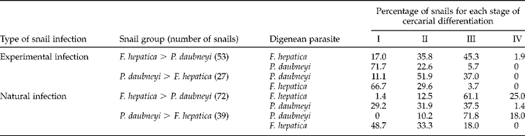
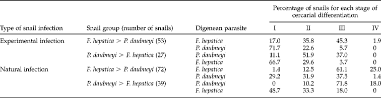
The few rediae developing for each digenean species (table 1) and the preferential growth of the first or the first two mother rediae (Augot et al., Reference Augot, Abrous, Rondelaud and Dreyfuss1996) did not allow us to follow precisely the larval development of each species over time. Owing to this difficulty, the growth of the first two mother rediae for each species was studied by considering the following four stages of cercarial differentiation (table 2): I, immature rediae without recognizable procercariae (immature cercariae); II, rediae containing short-tailed procercariae; III, rediae harbouring long-tailed cercariae (F. hepatica) or procercariae differentiating within the snail's body (P. daubneyi); IV, presence of free cercariae within the snail's body. The frequencies of snails for each stage of cercarial differentiation were determined for each digenean species and were subjected to a χ2 test (Stat-Itcf, 1988).
Percentage of G. truncatula in relation to the four stages of cercarial differentiation (I to IV) in the first or the two first free rediae of the first generation. Stages: I, immature rediae without recognizable procercariae; II, rediae containing short-tailed procercariae; III, rediae harbouring long-tailed cercariae (F. hepatica) or procercariae differentiating within the snail's body (P. daubneyi); IV, presence of free cercariae.
Tissue lesions
Tissue lesions were studied in the digestive gland and kidney. Indeed, most larvae of the dominating species developed within the intertubular spaces of the digestive gland, whereas the few larvae of the other digenean species were found in the mantle, the foot and/or other viscera such as the kidney. Three already-described lesions (Rondelaud & Barthe, Reference Rondelaud and Barthe1983; Moukrim & Rondelaud, Reference Moukrim and Rondelaud1992) were observed: multifocal epithelial necrosis, generalized epithelial necrosis, and epithelial reconstitution with cellular hyperplasia. The frequencies of tissue lesions are given in tables 3 and 4 in relation to the type of snail infections and snail group. A χ2 test (Stat-Itcf, 1988) was used to establish levels of significance.
Percentage of G. truncatula in relation to the occurrence of tissue lesions in their digestive gland.

Percentage of G. truncatula in relation to the occurrence of tissue lesions in their kidney.

Results
Parasite development
When the dominating species was F. hepatica or P. daubneyi, the number of free rediae per snail (table 1) was 3.5–4.2 and 2.4–3.2, respectively. In contrast, it was less than 2 rediae for the other species. Overall, the number of rediae noted for the dominating species was significantly higher (F = 5.37, P < 0.001) than that found for the other digenean species. A significant difference (F = 12.61, P < 0.001) between the numbers recorded for the dominating parasite and the other species was also found in the F. hepatica > P. daubneyi groups. The other differences were not significant.
Table 2 shows the frequencies of co-infected snails in relation to the four stages of cercarial differentiation. In each snail group, the quickest cercarial differentiation in the first two mother rediae was noted for the dominating species. However, all snails did not show the same stage of cercarial differentiation. In experimental co-infections, the highest percentages of snails were found for stages II (when P. daubneyi was the dominating species) and III (when F. hepatica rediae were the most numerous). In contrast, most snails harboured immature rediae (stage I) in the case of the non-dominating species. A significant difference (χ2 = 26.86, P < 0.001) between the frequencies of stage I was noted (when F. hepatica was dominating, the P. daubneyi larvae were present in more snails than expected, and vice versa). Another significant difference (χ2 = 22.11, P < 0.001) was also found for stage III (the P. daubneyi larvae were observed in more snails than expected when this species was dominating). In natural co-infections, most results were similar to those recorded in experimentally infected snails. Two significant differences between snail frequencies were found, for stage I (χ2 = 37.17, P < 0.001) and stage II (χ2 = 10.48, P < 0.01). Indeed, in each case, more snails than expected contained P. daubneyi larvae when F. hepatica was dominating, and vice versa. Two other significant differences were noted for stage III (χ2 = 16.54, P < 0.001) and stage IV (χ2 = 21.55, P < 0.001) stages. When F. hepatica was the dominating species, its larvae were present in more snails than expected. A similar finding was also noted for P. daubneyi.
Responses of the viscera of snails to parasite development
In the digestive gland of experimentally infected snails (table 3), the three types of tissue lesions were observed, whatever the species of dominating parasite. Most glands showed generalized epithelial necrosis. The other lesions were scarcer: 1.9% and 11.1% for multifocal necrosis, and 24.5% and 11.1% for reconstitution. For each type of lesion considered alone, the differences between frequencies were not significant. In naturally infected snails, generalized necrosis was significantly (χ2 = 18.24, P < 0.001) greater in the P. daubneyi > F. hepatica group than in the F. hepatica > P. daubneyi group. An inverse finding was found for reconstitution, as it was significantly (χ2 = 17.29, P < 0.001) more frequent in the F. hepatica > P. daubneyi group. Multifocal necrosis was scarce in both groups and showed non-significant differences.
In the kidney of experimentally infected snails (table 4), multifocal necrosis was significantly (χ2 = 12.69, P < 0.001) greater in the P. daubneyi > F. hepatica group, whereas generalized necrosis was significantly (χ2 = 4.19, P < 0.05) more frequent in the F. hepatica > P. daubneyi group. Similar findings were also noted for the frequencies of tissue lesions in naturally infected snails. Multifocal necrosis was more frequent (χ2 = 9.19, P < 0.05) in the P. daubneyi > F. hepatica group, whereas generalized necrosis was greater (χ2 = 6.28, P < 0.05) in the F. hepatica > P. daubneyi group.
Discussion
As for other digeneans having a redia stage in their life cycle, the most well-grown rediae of a species (F. hepatica for example) feed on the larvae of the other species (P. daubneyi in this case) when rediae of both species attempt to colonize the digestive gland of the snail. This fact explains why live rediae of F. hepatica and those of P. daubneyi developed into separate compartments within the body of these co-infected snails. Under these conditions, the differentiation of intraredial germinal masses might depend on the place of rediae. The first species to occupy the intertubular spaces of the digestive gland developed preferentially and was numerically dominating, as most nutrients were available for the growth of its larvae. In contrast, the other species only developed a few rediae (table 1), generally located in the haemolymphatic sinuses of renal lamellae. In our opinion, the location of larvae of each species might result from the speed of migration of each sporocyst through snail's tissues after penetration of the corresponding miracidium into G. truncatula. An argument in support of this approach described the different ways of migration followed by F. hepatica miracidia in relation to the point of their penetration (foot or mantle) into the snail (Préveraud-Sindou et al., Reference Préveraud-Sindou, Dreyfuss and Rondelaud1994). However, it is interesting to note that co-infected snails constituted a low percentage of G. truncatula exposed to both digeneans, as most of them only showed the larval development of one species, at least under experimental conditions (Abrous et al., Reference Abrous, Rondelaud and Dreyfuss1996; Augot et al., Reference Augot, Abrous, Rondelaud and Dreyfuss1996).
The response of the digestive gland to larval development of the dominating parasite agreed with reports of previous authors on the visceral pathology of snails mono-infected with F. hepatica (Barber, 1962; Rondelaud & Barthe, Reference Rondelaud and Barthe1978; Sindou et al., Reference Sindou, Cabaret and Rondelaud1991a, Reference Sindou, Rondelaud and Bartheb) or P. daubneyi (Moukrim & Rondelaud, Reference Moukrim and Rondelaud1992). Even if the number of free rediae was low for dominating species in the present study, the frequency of snails showing generalized necrosis in their digestive gland was similar to rates recorded in other snails experimentally infected with F. hepatica (Sindou et al., Reference Sindou, Rondelaud and Barthe1990a, Reference Sindou, Rondelaud and Bartheb, Reference Sindou, Rondelaud and Barthe1991b). The development of tissue lesions was thus independent of the number of larvae present within the intertubular spaces of this viscus. The destruction of digestive tubules by the movements of rediae and the secretion of their enzymes (Saint-Guillain, Reference Saint-Guillain1968) was the most frequently proposed hypothesis. However, it cannot completely explain the frequency of generalized necrosis in snails that only harboured a few rediae. In our opinion, the more likely hypothesis would be to admit the intervention of the snail's central nervous system in the development of this tissue lesion, via a specific agent, probably secreted by one or several neuroendocrine centres. An agent of that nature (schistosomin) had already been found by de Jong-Brink and her team in the haemolymph of Lymnaea stagnalis infected with Trichobilharzia ocellata (de Jong-Brink, Reference De Jong-Brink1990).
Compared to frequencies of tissue lesions in the digestive gland, those recorded in the kidney were more surprising, as most snails showed multifocal epithelial necrosis. Moreover, this lesion was significantly more frequent when P. daubneyi was the dominating species. This finding disagreed with reports by Rondelaud & Barthe (Reference Rondelaud and Barthe1983) and Moukrim & Rondelaud (Reference Moukrim and Rondelaud1992), as multifocal necrosis appeared in the kidney of mono-infected snails in the course of the first week p.e. and became generalized after day 21 p.e. at 20°C. Two hypotheses might be proposed to explain this difference. The first is to suppose that a sufficient number of free larvae within the snail would be necessary for the development of epithelial necrosis in the whole kidney. The second assumption is based on the presence of young and immature rediae, often located in the haemolymphatic sinuses of renal lamellae. As the structure of most rediae appeared normal during snail infection (Rondelaud & Barthe, Reference Rondelaud and Barthe1980), one may wonder about their pathogenic role, all the more so since the lamellar epithelium surrounding them was often normal. Finally, the greater frequency of multifocal necrosis noted in snails when P. daubneyi was the dominating species might be explained by proposing a less aggressive role of F. hepatica rediae towards the snail's kidney than that of P. daubneyi larvae.