Introduction
Foreign bodies in the cardiovascular system, such as prosthetic heart valves, carry risks of thrombosis and thromboembolic events. Prosthetic valve thrombosis leads to valve obstruction and dysfunction, which manifests as impaired leaflet coaptation, leaflet thickening, and valvular stenosis or insufficiency. Reference Dangas, Weitz, Giustino, Makkar and Mehran1 The incidence is higher in mechanical heart valves than bioprosthetic heart valves, with an annual rate ranging from 0.1 to 5.7%. Reference Lin, Tiong, Asher, Murphy, Thomas and Griffin2
Current standard-of-care for prosthetic valve thrombosis is with either systemic thrombolysis or surgical valve re-replacement, depending on patient factors such as New York Heart Association classification, thrombus size, and history of thromboembolism. Reference Das, Twomey, Khaddour and Dunning3 A case series by Hariram described five patients with prosthetic valve thrombosis who did not respond to thrombolytic therapy; each of these patients subsequently underwent percutaneous manipulation of their obstructed valve with an improvement of leaflet motion. Reference Hariram4 We present a case report describing successful transcatheter manipulation of the immobile leaflets of the mitral St. Jude valve in an infant presenting in extremis with pulmonary haemorrhage.
Case report
A 13-month-old male was followed closely from birth for progressive mixed mitral valve dysfunction secondary to congenital arcade mitral valve. At 9 months of age, he underwent surgical mitral valve replacement with a 17 mm St. Jude mechanical prosthesis. Four months after surgery, he presented with acute respiratory failure and haemoptysis, with bronchoscopy showing right pulmonary haemorrhage. Transthoracic echocardiography revealed severe right pulmonary vein stenosis. Furthermore, one of the mitral valve’s leaflets was completely immobile, and comparison to prior echocardiograms showed a rising transvalvular gradient over the past several months (up to 7 mmHg). Mitral valve inflow Doppler showed fused E-wave/A-wave indicating prosthetic valve dysfunction, which was suspected to be secondary to obstruction by adherent fibrinous and inflammatory material.
In the setting of the patient’s life-threatening pulmonary haemorrhage, all anticoagulation was held. After interdisciplinary discussions, the patient was deemed an extremely poor candidate for either systemic thrombolysis or cardiac surgery to address his dysfunctional prosthetic valve. He was therefore taken to the cardiac catheterisation laboratory for pulmonary vein angioplasty and percutaneous manipulation of the obstructed valve. Pre-catheterisation transoesophageal echocardiography showed no evidence of a left atrial clot, and the medial leaflet of the mechanical mitral valve was again noted to be immobile (Supplementary Video S1). With right femoral vein percutaneous access and echocardiographic guidance, a transseptal puncture was performed to access the left atrium. Balloon angioplasty of the severely stenotic right common pulmonary vein was successfully performed, with a reduction in the pulmonary vein gradient from 16 mmHg to 0 mmHg.
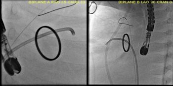
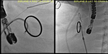
Attention was then focused on using catheters to manipulate the immobile leaflet of the mechanical mitral valve. Using echocardiographic and fluoroscopic guidance, the medial leaflet of the valve was struck repeatedly with multiple different catheters without significant improvement in leaflet excursion. A 5 Fr 4.0 curve Judkins right (JR) catheter was then reinforced with a 0.035” tip-deflecting wire, enabling the catheter to be advanced across the mechanical valve from the left atrium into the left ventricle (Fig 1). The catheter, with the tip-deflecting wire in situ, was then withdrawn from the left ventricle to position it across the medial hinge points of the valve leaflet and gentle force was repetitively applied to the immobile leaflet (Fig 2, Supplementary Video S2). Transcatheter manipulation of the medial leaflet with the 4.0 curve JR catheter, reshaped with the tip-deflecting wire, was repeated several times to push open the medial leaflet completely, leading to the return of normal leaflet excursion. Post intervention, there was no residual gradient across the mitral valve (left atrial pressure = 17 and left ventricular end-diastolic pressure = 18). A catheter was then placed in the left atrium for localised thrombolytic therapy.
After pushing against the obstructed medial leaflet of the prosthetic mitral valve, the catheter tip is advanced across the valve into the left ventricle as the additional curve from the tip-deflecting wire (not shown) is gently released.

A 5 Fr 4.0 curve JR catheter, reinforced with a tip-deflecting wire, is used to push open the immobile medial leaflet of the prosthetic valve. This procedure is repeated several times until the return of normal leaflet excursion is observed on fluoroscopy and echocardiography.

There were no complications during or immediately after the procedures. Afterwards, echocardiography showed the appropriate movement of the mechanical valve leaflets (Supplementary Video S1) and an improvement in the transvalvular gradient from 7 mmHg to 2 mmHg. In the subsequent weeks, the patient’s prosthetic valve continued to function well on echocardiographic evaluation. Unfortunately, due to severe illness and multiple co-morbidities, the patient had a complicated and tenuous course in the intensive care unit, and the family ultimately decided to withdraw care a few weeks after his catheterisation.
Discussion
Although uncommon, prosthetic valve thrombosis is a serious complication of prosthetic valve replacement. Lifelong warfarin is the standard anticoagulation therapy in patients with mechanical valves, but this can be difficult in infants because they require more frequent international normalised ratio (INR) testing and have fewer INR values within target range when compared to older children and adults. Reference Nishimura, Otto and Bonow5,Reference Streif, Andrew and Marzinotto6 Our patient, a 9-month-old infant at the time of surgical valve replacement, was therefore on enoxaparin rather than warfarin for anticoagulation and presented with valve thrombosis despite being at therapeutic levels for several months.
In a case series, Hariram described successful treatment of prosthetic mitral valve thrombosis in five adult patients via percutaneous manipulation and passage of a catheter across the valve. Reference Hariram4 We present a similar outcome in a 7.41 kg infant who, to our knowledge, is the youngest patient reported to undergo successful mechanical manipulation of an obstructed prosthetic valve. Smaller patients present a unique challenge with known limitations related to vascular access size and prograde catheter approach to the mitral valve. In our case, the acute angle needed to approach the immobile medial leaflet of the St. Jude valve in a relatively small left atrium made it challenging to propagate the necessary force onto the leaflet during mechanical thrusts to open the valve. Eventually, a tip-deflecting wire was used to reinforce the curve of the JR catheter so the bend of the catheter could prop open the valve. The catheter tip was gently advanced across the valve as the additional curve from the tip-deflecting wire was released.
Passage of a catheter across a prosthetic valve carries inherent risks, including catheter entrapment, valve disruption, and embolisation of a thrombus or pannus. During interdisciplinary discussions, our patient was deemed an extremely poor candidate for either surgery or systemic thrombolysis. Therefore, it was determined that percutaneous valve manipulation, despite its risks, provided the best opportunity to address the obstructed prosthetic valve. We used transoesophageal echocardiography to confirm the absence of a left atrial clot, perhaps mitigating the risk of thromboembolism. What requires further investigation is the risk of embolisation of pannus or inflammatory material during mechanical manipulation of a prosthetic valve.
Conclusion
Thrombosis of prosthetic valves leads to valve dysfunction and requires urgent management. Percutaneous manipulation of obstructed prosthetic valves is feasible and may be considered as a last resort in selective patients who have contraindications to surgery and systemic thrombolysis. Further evidence is necessary to demonstrate the efficacy of this intervention in patients with different risk profiles and different prosthetic valves.
Supplementary Material
To view supplementary material for this article, please visit https://doi.org/10.1017/S104795112000270X
Acknowledgements
We sincerely thank and acknowledge the help of Dr. Thomas Zellers, Professor of Pediatrics, UT Southwestern Medical Center, Dallas, Texas for his technical assistance during the procedure and for the appraisal of the manuscript.
Financial Support
This research received no specific grant from any funding agency, commercial or not-for-profit sectors.
Conflicts of Interest
None.