Introduction
Superior semicircular canal dehiscence syndrome is rare and was first described in 1998 by Minor et al.Reference Minor, Solomon, Zinreich and Zee1 They identified patients with a syndrome comprising vestibular symptoms and signs induced by sound or pressure changes in the middle ear. The symptoms were attributed to a thinning or complete absence of the part of the temporal bone overlying the superior semicircular canal. Vestibular symptoms and signs include vertigo, chronic disequilibrium and oscillopsia, with vertical–torsional eye movements in the plane of the superior canal evoked by sound or pressure changes. Audiometry often shows low-frequency conductive hearing loss, with normal or characteristically negative bone-conduction thresholds.Reference Sohmer, Freeman and Perez2 However, some patients experience conductive hyperacusis.Reference Stenfelt and Goode3 This presentation can be explained by the ‘third mobile window’ theory. According to this theory, in superior semicircular canal dehiscence the inner ear has three rather than two windows and is therefore more sensitive to sound and pressure changes.Reference Hiroven, Carey, Liang and Minor4 The aetiology of superior semicircular canal dehiscence has been heavily debated; it is still unclear whether the syndrome is congenital, acquired or a mixture of both. The incidence of superior semicircular canal dehiscence in the general population has been much investigated. In a study of 1000 cadaveric temporal bones, the incidence of superior semicircular canal dehiscence was 0.7 per cent, with an additional 1.3 per cent having a very thin bony layer (0.1 mm or less) covering the canal. Interestingly, the incidence of clinical superior semicircular canal dehiscence is much lower, although the reason why some patients present with symptoms and others do not is not fully understood.Reference Carey, Minor and Nager5
Diagnosing superior semicircular canal dehiscence requires a detailed medical history and the results of a medical examination, audiometry, high-resolution computed tomography (CT) and magnetic resonance imaging (MRI) of the temporal bones, and vestibular evoked myogenic potentials. Once the diagnosis has been confirmed, severe symptoms of superior semicircular canal dehiscence syndrome can be treated surgically via a middle cranial fossa, transmastoid or transcanal approach. The most common surgical techniques for managing this syndrome involve either plugging or resurfacing the superior canal.
Methods
A literature search using the key words ‘superior semicircular canal dehiscence’ was performed using the Allied and Complementary Medicine Database and the Embase, Health Management Information Consortium, Medline, PsycINFO, British Nursing Index, Cinahl and Health Business Elite databases for the search period January 2009 to May 2014. Systematic reviews, meta-analyses, randomised control trials, prospective and retrospective case series, case reports and observational studies were included. All abstracts and selected full text articles were reviewed.
In all, 205 papers and 4 key debated areas were identified from this search. The main questions were: What is the proposed aetiology of this syndrome? Does the dehiscence size correlate with the signs, symptoms and investigation results? What is the best way to investigate this syndrome? and What are the options for surgical management? In all, 35 papers were considered relevant to these questions. These four debated areas are discussed in detail.
Results
Aetiology
The aetiology of superior semicircular canal dehiscence is highly contentious, and has been much debated in the literature over the last five years. It is unclear whether it is a congenital, acquired or two-hit phenomenon.
Takahashi et al. macroscopically observed the middle cranial fossa of 42 adult cadavers and 4 fetal cadavers.Reference Takahashi, Tsunoda, Shirakura and Kitamura6 Six fetuses were also examined by CT; the volume data of the CT images obtained from four adults were used for comparison. The authors showed by macroscopic anatomy analysis and CT examination that in fetuses the superior semicircular canal and cochlea protrude into the cranium. In contrast, the superior semicircular canals of all adults were completely or almost entirely buried within the temporal bone. Therefore, they hypothesised that protrusion of the superior semicircular canal into the cranium during fetal development causes membranous labyrinth adhesion to the dura because the bony labyrinth develops much later than the membranous labyrinth. Such adhesion would in turn interfere with ossification and lead to a bony defect in the superior semicircular canal.
Several authors have explored the possibility that superior semicircular canal dehiscence is associated with a congenital morphological abnormality of the skull base. Patients with superior semicircular canal dehiscence frequently exhibit contralateral canal thinning and tegmen dehiscence or thinning.Reference Gracia-Tello, Cisneros, Crovetto, Martinez, Rodriguez and Lecumberri7–Reference El Hadi, Sorrentino, Calmels, Fraysse, Deguine and Marx10 These findings provide more supporting evidence for a congenital aetiology of superior semicircular canal dehiscence.
However, Nadgir et al. reported superior semicircular canal dehiscence to be more prevalent in older age groups, suggesting an acquired aetiology.Reference Nadgir, Ozonoff, Devaiah, Halderman and Sakai11 These authors assessed the prevalence of superior semicircular canal dehiscence in imaging data from multiple age groups. They retrospectively reviewed 306 consecutive temporal bone CT examinations performed between July 2005 and March 2007. Patients ranged in age from 7 months to 89 years and were subcategorised into 5 age groups. Twenty-four patients were identified with superior semicircular canal dehiscence and 137 had superior semicircular canal thinning. Each successive age category experienced a 93 per cent increase (95 per cent confidence interval (CI) 30–187 per cent) in the prevalence of superior semicircular canal dehiscence (p = 0.001). They concluded that the increased radiological prevalence of superior semicircular canal dehiscence in older age groups suggests that superior semicircular canal dehiscence is more commonly an acquired rather than a congenital condition. Despite this finding, Parlea et al. reported a patient with a solely developmental aetiology for superior semicircular canal dehiscence.Reference Parlea, Georgescu and Calarasu12 The case report described a 46-year-old woman presenting with Tullio phenomenon. Temporal bone CT showed that she had a defect of the left superior semicircular canal caused by an enlarged superior petrosal sinus receiving drainage from a large cerebellar developmental venous anomaly.
The two-hit aetiology of superior semicircular canal dehiscence has been much discussed since described by Minor in 2000.Reference Minor13 Whyte Orozco et al. performed a cadaveric study to test this theory.Reference Whyte Orozco, Martinez, Cisneros, Obon, Gracia-Tello and Angel Crovetto14 They demonstrated that a thinned temporal bone consists exclusively of the inner (or endosteal) layer separating the canal from the superior semicircular conduct. They concluded that the absence of the external and middle layers, besides reducing its thickness, makes the canal susceptible to suffering a second event leading to fracture and dehiscence.
There is a controversial suggestion that superior semicircular canal dehiscence may be related to osteopenia and osteoporosis. Crovetto et al. suggested that in superior semicircular canal dehiscence there is slight osteopenia of the roof of the superior semicircular canal associated with ageing, which is more pronounced in menopausal women.Reference Crovetto-De la Torre, Whyte-Orozco, Crovetto-Martinez, Whyte-Orozco, Obon-Nogues and Martinez-Rodriguez15 Moreover, Yu et al. argued that superior semicircular canal dehiscence may be associated with osteoporosis in elderly Asian women.Reference Yu, Teich, Moonis and Wong16 No investigations into whether vitamin D deficiency is associated with superior semicircular canal dehiscence have been reported.
Overall, the lack of agreement regarding the aetiology of superior semicircular canal dehiscence syndrome suggests that the cause may be multifactorial. Further research is therefore required to obtain an improved understanding of the aetiology.
Is dehiscence size important?
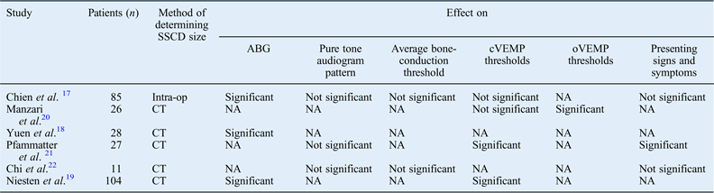
The question of whether the superior semicircular canal dehiscence size correlates with the signs and symptoms of this syndrome has been thoroughly investigated over the last five years. Six key papers have been published, involving a total of 281 patients; the superior semicircular canal dehiscence size was measured intra-operatively in 85 patients and via CT images in 196 patients. The studies investigated heterogeneous variables. However, there was consensus in three studies that the air–bone gap (ABG) size is significantly related to the superior semicircular canal dehiscence size.Reference Chien, Janky, Minor and Carey17–Reference Niesten, Hamberg, Silverman, Lou, McCall and Windsor19 Only one paper compared ocular vestibular evoked myogenic potential thresholds with superior semicircular canal dehiscence size, and found that they were significantly related.Reference Manzari, Burgess, McGarvie and Curthoys20 Several reports indicated that pure tone audiogram patterns and average bone-conduction thresholds were not significantly related to superior semicircular canal dehiscence size.Reference Chien, Janky, Minor and Carey17, Reference Pfammatter, Darrouzet, Gartner, Somers, Van Dinther and Trabalzini21, Reference Chi, Ren and Dai22 There has been disagreement about whether there is a significant relationship between both the superior semicircular canal dehiscence size and cervical vestibular evoked myogenic potential amplitudes and the patient's presenting signs and symptoms.Reference Chien, Janky, Minor and Carey17, Reference Manzari, Burgess, McGarvie and Curthoys20, Reference Pfammatter, Darrouzet, Gartner, Somers, Van Dinther and Trabalzini21 The findings of each report are summarised in Table I.
Key studies comparing sscd size with the clinical presentation and investigation results

SSCD = superior semicircular canal dehiscence; ABG = air–bone gap; cVEMP = cervical vestibular evoked myogenic potential; oVEMP = ocular vestibular evoked myogenic potential; intra-op = intra-operatively; NA = not applicable; CT = computed tomography
Investigations
Recently, there have been a few important findings about superior semicircular canal dehiscence, particularly regarding the use of imaging and vestibular evoked myogenic potentials for its diagnosis.
Multiple papers have reported that CT evaluation alone is unreliable when the clinical context of other investigations are not taken into consideration. There is a significant false positive risk when performing CT analysis of superior semicircular canal dehiscence.
Mondina et al. observed 3 dehiscences in 37 temporal bones by CT in the Poschl and Stenvers planes. However, no dehiscence was found by microscopic analysis.Reference Mondina, Bonnard, Barreau, Darrouzet and Franco-Vidal23 In a larger population, Tavassolie et al. compared the surgical and CT findings of 34 surgically confirmed superior semicircular canal dehiscence patients.Reference Tavassolie, Penninger, Zuniga, Minor and Carey24 They concluded that CT can overestimate the dehiscence size and can result in false positives for dehiscence detection. Crovetto et al. compared the CT and cadaveric temporal bone results of 604 ears.Reference Crovetto, Whyte, Rodriguez, Lecumberri, Martinez and Elexpuru25 They found that a higher incidence of superior semicircular canal dehiscence was obtained following CT scanning (3.6 per cent) compared with anatomical analysis (0.6 per cent). Masaki stressed the importance of diagnosing superior semicircular canal dehiscence based on both CT findings and clinical symptoms.Reference Masaki26 When a diagnosis of superior semicircular canal dehiscence syndrome in 164 ears was based solely on CT images, 80 per cent were false positives. As CT diagnosis of superior semicircular canal dehiscence can be unreliable, Eibenberger et al. investigated whether the limits of CT resolution allow the objective detection of bone structure defects as small as 0.1 mm in diameter.Reference Eibenberger, Carey, Ehtiati, Trevino, Dolberg and Haslwanter27 They used a bone cement phantom containing small, well-defined structural defects (diameter 0.1–0.4 mm) and tested both multislice and cone beam CT. The defects were reliably detected by comparing radiodensity variances between a region of interest (i.e. a region containing a hole) and a homogeneous region. Statistical noise analysis using the Fligner–Killeen test was used to predict the presence or absence of a hole (p ≥ 0.05). This analysis allowed the authors to distinguish real irregularities down to 0.1 mm (p ≤ 0.0001) from measured image noise or reconstruction errors.
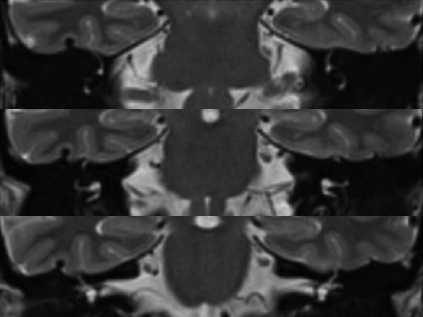
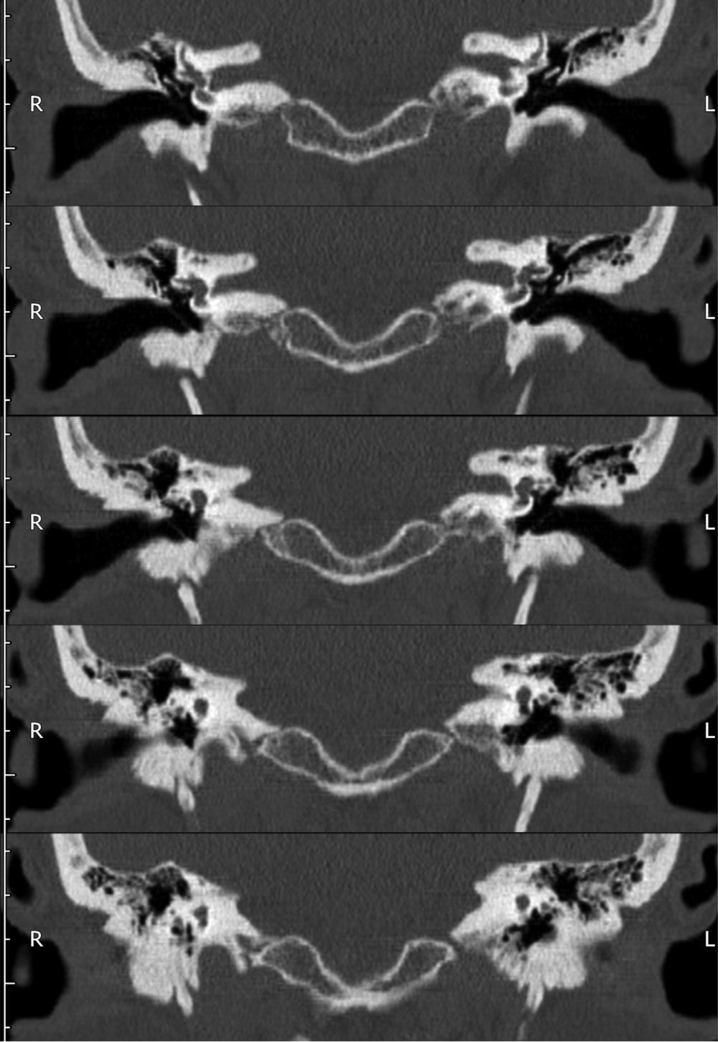
It is not unusual for patients with vertigo and symptoms of semicircular canal dehiscence to be investigated using both CT and MRI modalities. Browaeys et al. attempted to determine whether MRI alone is sufficient. They examined 112 consecutive patients (i.e. 224 ears) who underwent concurrent MRI and high-resolution CT of the temporal bones.Reference Browaeys, Larson, Wong and Patel28 They concluded that fast imaging employing steady-state acquisition (‘FIESTA’) MRI acquisition, with sensitivity and negative predictive values of 100 per cent, can conclusively exclude superior semicircular canal dehiscence. A negative MRI study therefore precludes the need for CT imaging to detect semicircular canal dehiscence; thus, only patients with a positive MRI study should have further CT evaluation. This approach may reduce costs and undue irradiation. To demonstrate the features seen using different imaging modalities, Figure 1 shows coronal T2-weighted MRI slices while Figure 2 shows coronal CT slices of the same patient with a right superior semicircular canal dehiscence.
Coronal T2-weighted magnetic resonance imaging scan, showing the superior semicircular canals lying close to the superior aspect of the temporal bone.
Consecutive coronal computed tomography images of the same patient as in Figure1, showing the right semicircular canal dehiscence. R = right; L = left
The importance of the vestibular evoked myogenic potential in diagnosing superior semicircular canal dehiscence syndrome has recently been investigated. Patients with this syndrome often show reduced vestibular evoked myogenic potential thresholds, thought to be due to the increased effectiveness of the transmission of sound energy to the sacculus caused by increased compliance of the inner ear.Reference Brantberg, Lofquist and Fransson29 The most common cervical myogenic response to vestibular stimuli is assessed; however, ocular and masseter response may also be evaluated. Cervical vestibular evoked myogenic potentials are obtained by first turning the patient's head away from the non-test ear. Electrodes are then placed on the forehead, anterior neck and over the sternocleidomastoid muscle of the test side. One hundred or so narrow band clicks of high intensity are then presented via inserts to the test ear to stimulate the ipsilateral saccule. The resultant myogenic activity is detected by the electrodes and averaged (Figure 3).
Graph showing the cervical vestibular evoked myogenic potential of a patient with superior semicircular canal dehiscence syndrome. The non-specific waveform from the abnormal ear is shown in blue, and the characteristic positive-negative-positive trace obtained from the normal side is shown in pink.
Milojcic et al. compared the audiometric and cervical vestibular evoked myogenic potential of ears with superior semicircular canal dehiscence with those of normal ears and of the unaffected ears of unilateral superior semicircular canal dehiscence patients.Reference Milojcic, Guinan, Rauch and Herrmann30 They found that patients with superior semicircular canal dehiscence syndrome had significantly larger ABGs at 0.25 and 0.5 kHz, and significantly lower cervical vestibular evoked myogenic potential thresholds at 0.5 kHz. They combined these two features to form an adjusted cervical vestibular evoked myogenic potential threshold. This measure allowed them to better differentiate superior semicircular canal dehiscence ears from normal or unaffected superior semicircular canal dehiscence ears, which they argued may be clinically applicable.
Other studies compared the use of ocular vestibular evoked myogenic potential versus cervical vestibular evoked myogenic potential for diagnosing superior semicircular canal dehiscence syndrome. Ocular vestibular evoked myogenic potentials represent extraocular myogenic activity in response to otolith stimulation, for example by air-conducted sound or bone-conducted vibration. Electromyographic activity of the extraocular muscles can be recorded using surface electrodes placed beneath the contralateral eye. Zuniga et al. demonstrated cervical vestibular evoked myogenic potential threshold sensitivity and specificity to range from 80 per cent to 100 per cent for diagnosing this syndrome; ocular vestibular evoked myogenic potential amplitudes demonstrated a sensitivity and specificity of greater than 90 per cent.Reference Zuniga, Janky, Nguyen, Welgampola and Carey31 Kantner and Gurkov argued that this was because ocular vestibular evoked myogenic potentials are likely to reflect predominantly utricular function, while cervical vestibular evoked myogenic potentials assess saccular function.Reference Kantner and Gurkov32 Vestibular evoked myogenic potentials are still not used regularly for diagnosing superior semicircular canal dehiscence. However, Zuniga et al. argued that ocular vestibular evoked myogenic potential testing in response to air-conducted sound provides an excellent screening test for superior semicircular canal dehiscence without radiation exposure.Reference Zuniga, Janky, Nguyen, Welgampola and Carey31 In addition, the ocular vestibular evoked myogenic potential amplitude measurement requires considerably less effort and time for patients compared with the cervical vestibular evoked myogenic potential threshold measurement.
Surgical management
In 2009, Vlastarakos et al. published a meta-analysis of the efficacy and complications of the various surgical techniques for treating superior semicircular canal dehiscence syndrome.Reference Vlastarakos, Proikas, Tavoulari, Kikidis, Maragoudakis and Nikolopoulos33 Their review identified a total of 64 primary operations for superior semicircular canal repair: 56 ears were operated on for vestibular complaints and 7 for auditory complaints. A total of 33 ears underwent canal plugging, 16 underwent resurfacing and 15 underwent capping. Success rates for these procedures were 97 per cent, 50 per cent and 93 per cent, respectively. The observed differences were statistically significant (p = 0.001). Resurfacing proved less effective than both plugging (p = 0.002) and capping (p = 0.01) techniques. Temporalis fascia combined with bone pâté and bone wax (for plugging), bone graft (for resurfacing) or hydroxyapatite cement (for capping) was commonly used as sealing material. Most operations were performed via a middle cranial fossa approach, with sensorineural hearing loss (SNHL) and disequilibrium being the most frequent complications.
There has been significant debate about which surgical approach is best for managing superior semicircular canal dehiscence syndrome, in particular when comparing a middle cranial fossa approach with a transmastoid approach. There have even been a few cases of superior semicircular canal dehiscence being managed via a transcanal approach. Ward et al. published hearing outcomes in 43 patients after surgical plugging of the superior semicircular canal by a middle cranial fossa approach.Reference Ward, Agrawal, Nguyen, Della Santina, Limb and Francis34 They concluded that low-frequency ABGs decrease after surgical plugging as a result of both increased bone-conduction thresholds and decreased air-conduction thresholds. In their study, surgical plugging via a middle cranial fossa approach in this syndrome was associated with mild, high-frequency SNHL that persisted in 25 per cent of patients with no change in speech discrimination.
There are several reports of the use of a transmastoid approach for treating superior semicircular canal dehiscence, all with promising outcomes. Beyea et al. performed transmastoid occlusion of the superior semicircular canal in 16 patients, 13 with vestibular symptoms, 2 with pulsatile tinnitus and 1 with hyperacusis.Reference Beyea, Agrawal and Parnes35 These symptoms were either greatly improved or completely resolved in 15 of the 16 patients who underwent transmastoid occlusion of the superior canal. Hearing was preserved in 14 patients and improved in 2 patients.
Both resurfacing the superior canal and the transmastoid approach for plugging the superior canal have drawbacks: plugging is invasive to the inner ear, while resurfacing requires a middle cranial fossa approach which is not without serious potential complications. Amoodi et al. therefore described their experience of treating superior semicircular canal dehiscence syndrome by resurfacing the canal defects of four patients via the transmastoid approach.Reference Amoodi, Makki, McNeil and Bance36 All patients reported symptom resolution, with audiograms showing some hearing improvement in three of them.
Fiorino et al. evaluated their results of treating superior semicircular canal dehiscence by plugging and resurfacing the defect via the transmastoid approach in six patients.Reference Fiorino, Barbieri, Pizzini and Beltramello37 There were no intra- or post-operative complications, and patients experienced the immediate relief of symptoms attributable to the dehiscence.
Lastly, Achar et al. performed a retrospective review of six patients with intractable symptoms due to superior semicircular canal dehiscence syndrome who were operated on via a transmastoid approach.Reference Achar, Nichani, Freeman and Lloyd38 One patient underwent canal plugging with fascia and five underwent resurfacing with cymba conchal cartilage. All patients had post-operative symptom resolution and sensorineural hearing preservation; none had major complications related to the approach or surgical technique used.
Overall, a literature analysis shows that canal plugging is associated with better symptomatic success rates compared with canal resurfacing techniques. However, the incidence of post-operative SNHL is higher with canal plugging than with resurfacing techniques. Recently, there has been a drive to surgically treat superior semicircular canal dehiscence syndrome via a transmastoid approach if the dehiscence anatomy and surgical expertise allow. Since 2009, 32 cases of the use of a transmastoid approach to treating this syndrome have been published in 4 papers. The reports concluded that the day-case procedure using a transmastoid approach is a safe, effective and less-invasive technique than the standard middle fossa approach, which has many potential complications and requires a longer hospital stay. However, the transmastoid approach is a new form of management for this syndrome and experience of this technique and its outcomes is limited.
In an attempt to improve the safety and efficacy of the middle cranial fossa approach, Peng and Shaia presented a case series of 10 patients with superior semicircular canal dehiscence syndrome that was surgically managed via an endoscopic middle cranial fossa approach (unpublished data). This provided better visualisation of the defect, a smaller preauricular incision, and shorter recovery and operative times. However, this technique limited the surgeons to performing a resurfacing technique to treat the dehiscence, the brain still required retracting and the patients were still subject to the risks associated with craniotomy. In all, 93 per cent of pre-operative symptoms showed post-operative resolution.
The transcanal approach was described only recently. Silverstein et al. reported a transcanal complete round window niche occlusion for treating superior semicircular canal dehiscence syndrome in 2009.Reference Silverstein and Van Ess39 This operation was performed on a 37-year-old man who presented with intermittent episodes of vertigo, left-sided hearing loss, bilateral tinnitus, left-sided aural fullness and hyperacusis, with imaging consistent with a superior semicircular canal dehiscence. These symptoms all resolved after the operation. The operation was again performed on a patient with a similar presentation; however, this patient's symptoms worsened post-operatively and so a transmastoid superior canal occlusion was performed. However, the result of the second operation was not published.
Banerjee et al. continued to treat superior semicircular canal dehiscence with round window occlusion using bone wax and fascia. He published his experience of treating two superior semicircular canal dehiscence syndrome patients with hyperacusis symptoms. Both patients reported a great post-operative reduction in symptoms, with cessation of the Tullio phenomenon. The authors concluded that this procedure is simple to perform and reversible, and that day-case surgery can be undertaken.Reference Nikkar-Esfahani and Banerjee40 However, much more evidence is required for this technique to be widely accepted.
More recently, Silverstein et al. published a retrospective, multicentre case series of 22 patients who were treated for symptomatic superior canal dehiscence using round window tissue reinforcement or complete round window occlusion.Reference Silverstein, Kartush, Parnes, Poe, Babu and Levenson41 Their analysis revealed a significant improvement in all symptoms except for hearing loss in the 19 patients who underwent round window reinforcement. In contrast, two of the three patients who underwent the alternative treatment of round window occlusion experienced symptom deterioration requiring revision surgery.
Discussion
Over the last four years, there has been much research aimed at gaining a better understanding of superior semicircular canal dehiscence syndrome. However, some areas remain unclear, such as its aetiology. Whether it is a congenital, acquired or a two-hit phenomenon remains uncertain. However, there have been major advancements in its investigation and surgical management. The importance of not relying solely on CT to diagnose superior semicircular canal dehiscence and of correlating CT finding to clinical findings and other investigations has been clarified.Reference Mondina, Bonnard, Barreau, Darrouzet and Franco-Vidal23–Reference Masaki26 This is due to the high risk of false positives when performing CT analysis for superior semicircular canal dehiscence. One paper reported that when a diagnosis was based solely on CT scans, 80 per cent of cases were false positives.Reference Masaki26 Browaeys et al. suggested that ordering both CT and MRI scans for a patient with suspected superior semicircular canal dehiscence is not always required, and that negative MRI precludes the need for CT imaging to detect semicircular canal dehiscence.Reference Browaeys, Larson, Wong and Patel28 Only patients with positive MRI should undergo further CT evaluation. Regarding the use of vestibular evoked myogenic potentials for diagnosing superior semicircular canal dehiscence syndrome, ocular vestibular evoked myogenic potential amplitudes demonstrated greater sensitivity and specificity compared with cervical vestibular evoked myogenic potential thresholds.Reference Zuniga, Janky, Nguyen, Welgampola and Carey31
The surgical management of superior semicircular canal dehiscence syndrome has shown much advancement in the last five years. A middle cranial fossa approach remains the surgical standard for treating this syndrome. However, there has been a move towards treatment via a transmastoid approach if the dehiscence anatomy and surgical expertise allow. This approach has been shown to be safe, effective and less invasive than the middle cranial fossa approach; it can be performed as a day-case operation.Reference Beyea, Agrawal and Parnes35–Reference Achar, Nichani, Freeman and Lloyd38 There have also been a few reports of using a transcanal approach to round window occlusion, particularly to treat hyperacusis.Reference Silverstein and Van Ess39–Reference Silverstein, Kartush, Parnes, Poe, Babu and Levenson41 This remains an experimental form of management of this syndrome. Resurfacing techniques have proven to be less effective than both plugging and capping techniques. However, the incidence of post-operative SNHL is higher with canal plugging than with resurfacing techniques.
• Superior semicircular canal dehiscence syndrome may be congenital, acquired or a two-hit phenomenon
• Air–bone gaps and ocular vestibular evoked myogenic potential thresholds are affected by dehiscence size
• Computed tomography diagnosis has false positive rates of up to 80 per cent; MRI diagnosis has sensitivity and negative predictive values of 100 per cent
• Ocular vestibular evoked myogenic potentials provide a specific and sensitive diagnosis
• Middle cranial fossa is the standard surgical approach, but there is a move towards a transmastoid approach
• Resurfacing techniques are less effective than plugging and capping techniques
Superior semicircular canal dehiscence syndrome is rare and relatively newly described. Much more research is required to fully understand its aetiology and pathophysiology, and the best surgical management.
Acknowledgements
We wish to thank A Wilkinson, Principal Clinical Scientist at the University Hospitals Bristol NHS Foundation Trust, Southmead Hospital, for providing the vestibular evoked myogenic potential data.