INTRODUCTION
Alexithymia is defined as a deficit in affect regulation, characterized by the inability to identify and describe feelings, difficulty in distinguishing feelings from bodily sensations of emotional arousal, impaired symbolization, and an externally oriented cognitive style (Taylor et al., 1991). There is growing interest in alexithymia due to its association with various psychopathological disorders (Honkalampi et al., 2000; Parker et al., 1991; Wise et al., 1990) and reduced subjective judgment of quality of life (Henry et al., 2006).
The results of studies investigating the neurobiological bases of alexithymia suggest that alexithymic features may be related to right hemisphere damage. Indeed, Spalletta et al. (2001) reported a higher prevalence of alexithymia in right than in left hemisphere stroke patients. Moreover, in a recent study by Kano et al. (2003) in which regional cerebral blow flow (rCBF), measured by positron emission tomography, was examined in alexithymic versus nonalexithymic subjects while they viewed emotional faces, significantly lower rCBF was found in distributed regions of the right hemisphere (i.e., orbito-frontal cortex, middle frontal gyrus, inferior parietal gyrus, cuneus) in alexithymics than in nonalexithymics. Furthermore, Jessimer and Markham (1997) studied the ability of high alexithymic versus low alexithymic subjects in a nonclinical sample to attribute emotional value to chimeric pictures of faces composed of conjoined emotive and nonemotive halves. It was previously reported that normal right-handed individuals tend to choose the chimeric face with the emotive half on the left as being more expressive than the half on the right, indicating a leftward bias related to the predominant right hemisphere processing of these stimuli (Wirsen et al., 1990). Jessimer and Markham (1997) found that subjects with higher alexithymic rates showed significantly less left bias on chimeric tasks than low alexithymics, which indirectly suggests an association between alexithymia and right hemisphere activity in these subjects. Finally, recent electroencephalographic (EEG) data from subjects without neuropsychiatric disorders also support an association between right hemisphere dysregulation and alexithymia (Aftanas & Varlamov, 2004, 2007).
Other authors have pointed out a relationship between frontal lobe functioning and alexithymia. In an functional magnetic resonance imaging (fMRI) study investigating the emotional processing of visual stimuli, alexithymic subjects showed decreased activation of the left mediofrontal-paracingulate gyrus compared with nonalexithymics in response to the presentation of pictures with intense negative emotional valence. Greater activation in similar areas (i.e., anterior cingulate, mediofrontal, and middle frontal gyri) was also found bilaterally while they viewed highly positive stimuli (Berthoz et al., 2002). Moreover, in a recent MRI investigation involving healthy subjects, Gundel et al. (2004) found a significant positive correlation between size of the right anterior cingulate gyrus and severity of alexithymic symptoms, as measured by the Toronto Alexithymia Scale. A role of the frontal cortices in alexithymia also emerged from a more recent behavioral study involving 28 patients with traumatic brain injury. In this study, Henry et al. (2006) found a strong association between low performances on a test known to tap executive functions (i.e., alternating verbal fluency) and higher scores on the difficulty in identifying emotions subscale of the 20-item Toronto Alexithymia Scale. In keeping with the idea that the core of alexithymia is represented by an impairment in the cognitive processing and regulation of emotions (Parker et al., 2003), the authors proposed that, in their patients, a common neuropsychological deficit underlay both cognitive and emotional symptoms.
In two recent studies investigating the prevalence and characteristics of alexithymia in patients with Parkinson's disease (PD), we showed that alexithymia, particularly difficulty in describing and communicating emotions, may be a relevant feature in PD (Costa et al., 2006; Costa et al., manuscript submitted for publication). From a comprehensive analysis of psychopathological alterations, we also showed that, although alexithymia is associated with depression in PD, patients' depressive symptoms do not completely explain the presence of alexithymia in this disease (Costa et al., manuscript submitted for publication). Based on these results, we speculated that the high prevalence of alexithymia in PD could be related to the extension of neuropathological alterations to frontal and limbic areas known to be involved in affect regulation.
In the present study, we adopted a neuropsychological approach to investigate the relationship between cognitive functioning and alexithymia in PD. In particular, impairment of executive functions (i.e., planning, set-shifting, working memory processes) and of visual–spatial capacities are observed even in the earlier stages of the disease (Cools, 2006; Costa et al., 2003; Dubois & Pillon, 1997; Green et al., 2002; Janvin et al., 2003; Lewis et al., 2003; Owen, 2004). Therefore, in view of the above mentioned reports of a possible association between alexithymia and frontal lobe dysregulation (Berthoz et al., 2002; Henry et al., 2006), as well as right hemisphere functioning (Kano et al., 2003; Spalletta et al., 2001), we hypothesized that, in PD, alexithymia could be associated with alterations in executive and/or visual–spatial processes.
MATERIALS AND METHODS
Subjects
Seventy PD patients (45 men and 25 women) and 70 control subjects (CS; 42 men and 28 women) participated in the study after giving their informed consent. All subjects were right-handed according to the 20-item version of the Edinburgh Inventory for handedness (Oldfield, 1971). PD patients suffered from a mild to moderate rigid–akinetic form of idiopathic PD. They were admitted as inpatients to the Santa Lucia hospital to adjust anti-parkinsonian medication and to undergo a motor rehabilitation program.
The diagnosis of idiopathic PD was made by an expert neurologist based on (1) the presence of at least two of the four cardinal parkinsonian symptoms; and (2) good chronic response to levodopa treatment. Exclusion criteria included the following: (1) dementia suspected on the basis of clinical examination or a Mini-Mental State Examination score ≤ 24 (Folstein et al., 1975); (2) presence of severe systemic and metabolic disease (such as diabetes, hypothyroidism, and so on); (3) marked cortical and subcortical atrophy and/or ischemic vascular lesions on computed tomography and/or MRI scans; (4) history of neurological disorders other than PD; (5) evidence of psychotic symptoms; (6) severe functional impairment of autonomic nervous system.
PD patients were clinically evaluated using the Unified Parkinson's Disease Rating Scale (UPDRS) Part III (Fahn et al., 1987), reported in Table 1. Extrapyramidal symptoms predominantly affected the right side in 34 PD patients and the left side in the remaining 36 patients. All patients were treated with levodopa (mean = 355 ± 95.5 mg/day); 24 patients were also administered pramipexole (2.1 mg/day), 12 pergolide (3 mg/day), and 7 cabergoline (4 mg/day). Twenty-five PD patients were also treated with antidepressant medication. Moreover, the PD group was composed of 36 patients whose therapeutic response was unstable, due to Long-Term Treatment Syndrome, and 34 stable patients. Furthermore, PD patients were included in the present study when they had reached a steady response to the anti-Parkinsonian therapy, as documented by a stable UPDRS Part III score. Finally, all patients were evaluated in the “best on status” at the same time, that is, approximately 30 minutes after the first daily drug administration, when the best therapeutic response is usually present also in the fluctuating patients.
Demographic and clinical characteristics of PD patients and CS

The control group was composed of patients suffering from orthopedic diseases (i.e., limb fractures) or peripheral nervous system pathologies (i.e., polyneuropathy), who were also admitted to Santa Lucia hospital to undergo a physical rehabilitation program. Exclusion criteria for the CS group included (1) dementia suspected on the basis of a clinical examination or a Mini-Mental State Examination score ≤ 24; (2) presence of severe systemic or metabolic disease; (3) taking medication with central nervous system side effects; (4) history of psychiatric or neurological illness, head trauma, or substance abuse.
For both PD patients and controls evaluations were made within 2 weeks of hospitalization. Demographic and clinical characteristics of the two groups are reported in Table 1. The study was approved by the Local Ethics Committee.
Assessment Instruments
Psychopathological evaluation
Alexithymia was assessed using The Twenty-Item Toronto Alexithymia Scale (TAS-20; Bagby et al., 1994a,b), an extensively validated self-report questionnaire. The scale is composed of three subscales, which investigate the following: (F1) difficulty identifying feelings; (F2) difficulty describing and communicating feelings; (F3) externally oriented thinking. The total score on the questionnaire allows categorizing subjects as nonalexithymic (scores ranging from 20 to 51), borderline alexithymic (scores ranging from 52 to 60), or alexithymic (scores ≥ 61). Presence of depression was evaluated with the Beck Depression Inventory (BDI; Beck & Steer, 1987), which is considered a reliable instrument for examining severity of depression in PD (Leentjens et al., 2000; Visser et al., 2006). To score the BDI, we considered the total score obtained on the inventory and the partial scores obtained on the cognitive-affective (BDI-psy) and physiological subscales (BDI-phy), as defined by Endler et al. (1999). To control for the effect of PD somatic symptoms on the overall depression score, we considered only the BDI-psy score for statistical analysis. Anxiety was assessed by the State and Trait Anxiety Inventory (STAY-S; Spielberger, 1983), a self-administered questionnaire that investigates the psychological features of state anxiety.
Neuropsychological assessment
A battery of neuropsychological tests was administered to evaluate short-term and declarative long-term memory, executive functions, abstract reasoning, visual–spatial abilities, and high-level verbal capacities. The neuropsychological assessment lasted approximately 45 minutes and preceded the psychopathological evaluation, which was performed on the same day. The neuropsychological tests are listed below in relation to the cognitive functions they investigate.
Memory
Immediate visual memory (Carlesimo et al., 1996). In each of 22 trials, the subject is presented with an abstract figure for 3 seconds and is immediately requested to point to the figure studied among four alternatives (score range: 0–22).
Word-list recall (Rey, 1958). The test consists of five consecutive immediate free-recall trials in which the examiner reads a list of 15 words representing concrete objects and the subject has to recall as many words as possible in any order (score range: 0–75). After 15 minutes, a delayed recall trial is administered in which the examiner asks the subject to recall the previously presented words (score range: 0–15).
Executive functions
Modified Card Sorting Test (Nelson, 1976; Nocentini et al., 2002). This test is a revised version of the Wisconsin Card Sorting Test (Milner, 1963). The test material consists of 4 stimulus cards, which are unique for color, shape, and number of items, and two sets including 24 response cards each. Each response card has one attribute in common with each of the stimulus cards. The subject has to sort the cards according to a specific criterion that, however, may change during the task; the examiner does not tell the subject that the relevant categories are color, shape, and number of the items. After each response, the examiner indicates whether it was right or wrong. A criterion is considered complete if the subject makes six consecutive correct responses. Three separate scores are computed for the number of criteria achieved (range: 0–6), the number of perseverative errors (i.e., when the patient persists with a category even after being told it is incorrect), and the number of nonperseverative errors.
Abstract reasoning
Raven's Progressive Matrices 47 (Raven, 1947). This test is a logical-deductive (deduction of relations) intelligence test, based on visual–spatial data. Each of the 36 test tables has a higher part, which contains the stimulus figure, and a lower part, which contains six response alternatives. In all cases, the stimulus figure is missing a piece. The patients have to observe the stimulus attentively and indicate which of the six response alternatives they believe best completes the stimulus figure. The test is administered without time limits. A missing response is considered an error (range: 0–36).
Visual-spatial abilities
Freehand copying of drawings (Gainotti et al., 1977). Subjects are requested to copy three line drawings representing a star, a cube, and a house. The score is based on reproduction accuracy in terms of the number of elements reproduced with their reciprocal spatial relationship (score range: 0–12).
Copying drawings with landmarks (Gainotti et al., 1977). The same figures are used as in the previous test. Here, repere points (i.e., dots, lines, and angles) are visible on the paper, and the subject has to connect the segments to reproduce the model. The score is the number of missing segments correctly reproduced (score range: 0–70). Minor irregularities possibly due to tremor or bradykinesia were not taken into account.
Verbal abilities
Sentence construction (Carlesimo et al., 1996). The subject has to compose a grammatically correct sentence that makes sense and includes two or three words provided by the examiner. There are five trials with different words. The score is calculated from the patient's correctness and speed in composing the sentences (score range: 0–25).
Phonological verbal fluency (Borkowsky et al., 1967). The subject has to generate words beginning with the letters “A,” “F,” and “S.” Each of the three trials lasts 60 seconds. The score is the number of legal words produced (proper names excluded).
Statistical Analyses
In the first step of data analysis, we compared PD patients and CS with or without alexithymia for demographic variables and scores obtained on psychopathological scales by means of two-way Group * Alexithymia analyses of variance (ANOVAs; borderline alexithymic subjects were excluded from these analyses). Duration of illness and UPDRS scores (ANOVAs) were compared in PD patients with and without alexithymia. Moreover, to determine whether there was a relationship between lateralization of motor symptoms and alexithymia PD patients with and without alexithymia were compared for side prevalence of motor symptoms at the disease onset (χ2) and in the current state (ANOVA comparing UPDRS scores for motor symptoms respectively on the left and right limbs). A one-way ANOVA was then performed to compare the score obtained by alexithymic and nonalexithymic subjects on the UPDRS item investigating right hand tremor. Scores achieved on neuropsychological and depression measures were also compared between PD patients whose dopamine therapeutic response was unstable and stable patients by means of one-way ANOVAs. A further χ2 analysis was then executed to investigate differences in the prevalence of alexithymia between the two groups. Finally, to evaluate the effect of antidepressant medication on depression and alexithymia, the BDI-psy and TAS-20 scores were compared in PD patients with and without antidepressant medications (ANOVAs) and a χ2 analysis evaluating the difference in the distribution of patients undertaking antidepressant therapy among the alexithymia and nonalexithymia groups was also executed.
In the second step of data analysis, the scores of PD patients and CS with and without alexithymia on the neuropsychological tests were compared (borderline alexithymic subjects were excluded also from these analyses). For this purpose, we followed a Fisher's protected least significant difference (LSD) procedure. In fact, preliminary multivariate analysis of covariance (MANCOVAs) were executed with Group (PD vs. CS) and Alexithymia (alexithymic vs. nonalexithymic) as independent factors, performance scores on each cognitive test as dependent variables, and BDY-psy and STAY-S scores as covariates. Post hoc LSD multiple comparisons were performed only if the F-ratio for the Group * Alexithymia interaction was statistically significant (i.e., p ≤ .05). To avoid the risk of alpha inflation and, thus, to ensure that the probability was no greater than 5% of something appearing to be statistically significant when there were no underlying differences, we carried out Bonferroni's adjustment for multiple comparisons; that is, each of the “m” individual comparisons was performed at the 0.05/m level of significance. Therefore, taking into consideration that, with four groups, there are m = 4(4 − 1)/2 = 6 comparisons, we accepted a level of p ≤ .0083 (i.e., = 0.05/6) as significant for each of the “m” individual comparisons. Size effects (i.e., the size of each statistically significant difference) were also computed using Cohen's d.
In the final step of data analysis, we examined the relationship between the scores obtained on the three TAS-20 subscales (i.e., F1: difficulty identifying feelings; F2: difficulty describing and communicating feelings; F3: externally oriented thinking) and the neuropsychological and psychopathological variables by means of stepwise multiple linear regression analyses, performed separately for the two groups of subjects (the borderline alexithymic subjects were included in these analyses). The number of neuropsychological variables possibly entering in the regression model was reduced by considering only one test for each cognitive domain investigated. Therefore, the partial scores obtained on the individual TAS-20 subscales were the dependent variables, while the independent variables were BDI-psy, STAY-S, Modified Card Sorting Test-categories achieved, Raven's Progressive Matrices, Word list recall–delayed recall, Freehand copying of drawings and Sentence construction. To control for a possible relationship between alexithymia and severity of extrapyramidal symptoms, the UPDRS score was inserted among the independent variables in the regression analyses involving PD patients.
RESULTS
Comparisons Between PD Patients and Control Subjects With or Without Alexithymia on Demographic, Clinical, and Psychopathological Variables
As described in a previous study, in which we reported the prevalence of alexithymia and associated psychopathological alterations in the same PD and CS samples (Costa et al., manuscript submitted for publication), 21.4% of the PD patients (n = 15) and 10.0% of the CS (n = 7) could be classified as alexithymic, and 55.7% of the PD patients (n = 39) and 65.7% of the CS (n = 46) as nonalexithymic. The 16 remaining PD patients and 17 control subjects obtained borderline scores on the TAS-20. Preliminary analyses did not reveal significant differences between PD patients with and without alexithymia with regard to the average levodopa dosage [F(1,52) = 0.18, p > .60]. Moreover, 9 PD patients of the alexithymic group and 26 patients of the nonalexithymic group were also undertaking dopamine agonists. This distribution difference was not significant [χ2(df = 1) = 0.21, p > .60].
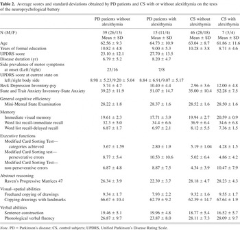
Table 2 reports demographic variables, depression and anxiety scores as well as average performance scores of PD and CS with or without alexithymia on tests of the neuropsychological battery. For the PD patients, UPDRS scores and side prevalence of motor symptoms are also reported. The four groups were comparable for age, level of formal education, and general cognitive efficiency (i.e., Mini-Mental State Examination score; F consistently < 1.5). Also, there was no difference in gender distribution in alexithymic and nonalexithymic subjects in either the PD (χ2 = 0.22) or the CS (χ2 = 0.81) group. With regard to severity of depression, the Group effect was not significant [F(1,103) = 0.31, p > .50], while the Alexithymia effect was [F(1,103) = 41.28, p < .001]. Indeed, subjects with alexithymia had higher average scores on the BDI-psy than those without alexithymia (11.2 and 4.3, respectively). The Group * Alexithymia interaction was also significant [F(1,103) = 4.23, p = .042]. Post hoc analyses revealed that, although the BDI-psy score did not differ between PD patients and CS with alexithymia (p > .10), among patients without alexithymia, PD patients were more depressed than CS (p = .003). With regard to severity of anxiety, only the main effect of Alexithymia was significant [F(1,103) = 24.84, p < .001]; indeed, subjects with alexithymia had higher average scores on the STAY-S than those without alexithymia (51.7 and 37.1, respectively). Furthermore, PD patients with and without alexithymia did not differ as to overall UPDRS score [F(1,52) = 1.47, p > .20], years of disease duration [F(1,52) = 0.82, p > .30], side prevalence of motor symptoms at disease onset (χ2 = 0.66), or current state (all F < 1). p. 20; lines 22 to 26 and p. 21; lines 1 to 8.
Average scores and standard deviations obtained by PD patients and CS with or without alexithymia on the tests of the neuropsychological battery

PD patients with unstable and stable therapeutic response did not differ with regard to the score achieved on any of the neuropsychological tests [F(1,68) range from 0.03 to 2.27, p > .10 in all cases] and on the BDI-psy [F(1,68) = 1.19, p > .20]. According to the TAS-20 total score, 5 patients suffering from LTTS and 10 patients with stable therapeutic response could be classified as alexithymic, while 23 patients with LTTS and 16 patients without LTTS did not present alexithymia; this distribution difference was not significant (χ2 = 2.85, df = 1, p > .09).
The assumption of antidepressant therapy was not related to the presence and severity of alexithymia. Indeed, the TAS-20 score did not differ between patients who were assuming or not antidepressant therapy [F(1,68) = 0.22, p > .60], and the number of PD patients that were administered antidepressant medication did not differ between the alexithymic (n = 4) and nonalexithymic (n = 11) groups (χ2 = 0.01). Finally, no difference between treated and untreated PD patients were found on the BDI-psy score [F(1,68) = 0.63, p > .40].
Comparison Between PD Patients and CS With and Without Alexithymia on Tests of the Neuropsychological Battery
Memory
Immediate visual memory. The Group effect was significant [F(1,101) = 5.20, p = .024], while the Alexithymia effect was not [F(1,101) = 0.12, p = .729]. Indeed, PD patients had lower average scores than CS (18.66 and 20.26, respectively). The Group * Alexithymia interaction was closer to the level of statistical significance [F(1,101) = 3.89, p = .051]. Post hoc analyses, performed to qualify this interaction, showed that PD patients with alexithymia obtained lower average scores than CS without alexithymia (p = .007, Cohen's d = 1.23). No other group comparisons approached statistical significance (p > .01).
Immediate Word-list recall. Statistical analyses did not reveal any significant effect of Group [F(1,101) = 2.25, p = .137] or Alexithymia [F(1,101) = 0.52, p = .474] in performances on this test. The Group * Alexithymia interaction also fell short of significance [F(1,101) = 1.37, p = .245].
Delayed Word-list recall. Also in this case, no significant effect was found [Group: F(1,101) = 0.62, p = .433; Alexithymia: F(1,101) = 0.001, p = .972; Group * Alexithymia interaction F(1,101) = 0.18, p = .674].
Executive functions
Modified Card Sorting Test: categories achieved. The Group effect result was significant [F(1,101) = 16.37, p < .001], documenting that PD patients obtained significantly lower average scores than CS (3.23 and 4.74, respectively). Otherwise, the Alexithymia effect and the interaction between the two factors did not approach statistical significance [F(1,101) = 1.78, p = .185 and F(1,101) = 0.03, p = .861, respectively].
Modified Card Sorting Test: perseverative errors. Also in this case, only the Group effect was significant [F(1,101) = 7.15, p = .008], documenting that, on average, PD patients made significantly more perseverative errors than CS (9.65 and 4.93, respectively). We did not find any significant effect of the Alexithymia factor [F(1,101 = 1.27,p = .263] or the Group * Alexithymia interaction [F(1,103) = 1.04, p = .309].
Modified Card Sorting Test: nonperseverative errors. Here, the Alexithymia effect approached statistical significance [F(1,101) = 3.43, p = .066], indicating that, in the overall sample, alexithymic subjects tended to make more errors than nonalexithymic subjects (9.66 and 5.60, respectively). Neither the Group factor nor the Group * Alexithymia interaction approached statistical significance [with F(1,101) = 0.07, p = .787 and F(1,101) = 1.71, p = .194, respectively].
Abstract reasoning
Raven's Progressive Matrices. The Group effect was significant [F(1,101) = 12.07, p < .001], indicating that PD patients' average scores were lower than those of CS (24.37 and 28.20, respectively), while the Alexithymia effect was not [F(1,101) = 0.36, p = .548]. The Group * Alexithymia interaction reached statistical significance [F(1,101) = 4.04, p = .047]. Post hoc analyses, performed to qualify this interaction, showed that PD patients with alexithymia had lower average scores than both PD patients without alexithymia (p = .003, Cohen's d = 2.01) and CS with or without alexithymia (p = .003, Cohen's d = 2.93 and p < .001, Cohen's d = 2.82, respectively). No other significant difference was found.
Visual–spatial abilities
Freehand copying of drawings. The Group effect approached statistical significance [F(1,101) = 3.00, p = .086], indicating that PD patients' average scores tended to be lower than those of CS (8.63 and 9.43), while the Alexithymia factor did not show any significant effect [F(1,101) = 0.14, p = .707]. The Group * Alexithymia interaction reached statistical significance [F(1,101) = 4.40, p = .038]. Post hoc LSD test revealed that PD patients with alexithymia had lower average scores than both PD patients without alexithymia (p = .009, Cohen's d = 1.01) and CS without alexithymia (p = .008, Cohen's d = 1.00). No other significant effect was found.
Copying drawings with landmarks. No significant effect emerged for this test [Group: F(1,101) = 0.003, p = .954; Alexithymia: F(1,101) = 0.33, p = .569; Group * Alexithymia interaction: F(1,101) = 2.66, p = .106].
It should be noted that differences in performance on copying drawings tests between alexithymic and nonalexithymic PD patients could be not accounted for by a different severity of right hand tremor. Indeed, as noted above, minor irregularities were not taken into account in the test scoring and, in any case, the two groups of patients did not differ in the UPDRS score on the right hand tremor item [F(1,52) = 2.61, p > .10].
Verbal abilities
Sentence construction. In this case, neither the main effects [Group: F(1,101) = 2.97, p = .087; Alexithymia: F(1,101) = 0.37, p = .545] nor the Group * Alexithymia interaction [F(1,103) = 0.50, p = .481] resulted significant.
Phonological verbal fluency. Also for this test, the MANCOVA documented a lack of significance for both the main effects [Group: F(1,101) = 1.48, p = .226; Alexithymia: F(1,101) = 0.07, p = .785] and the interaction [F(1,101) = 0.57, p = .451].
Because the results of the above reported analyses suggested that PD patients with alexithymia were particularly poor in performing neuropsychological tests involving visual–spatial data, in another MANCOVA, we directly contrasted the performance of PD patients and CS with and without alexithymia on two composite scores obtained by averaging Z scores on the verbal tasks (i.e., Immediate Word-list recall, Delayed Word-list recall, Sentence construction and Phonological verbal fluency) and on the visual–spatial tasks (i.e., Immediate visual memory, Raven's Progressive Matrices, Freehand copying of drawings, and Copying drawings with landmarks; see Carlesimo et al., 1996, for details about the two-factor clustering of tasks in the neuropsychological battery used here). Figure 1 reports the composite Z scores for the verbal and visual–spatial tasks in the four groups of subjects. Results of the three-way Group * Alexithymia * Task MANCOVA revealed a trend toward statistical significance for the Group effect [F(1,101) = 3.62, p = .056]. Indeed, average Z scores tended to be lower in the PD (mean = −2.5) than in the CS (mean = 0.02) group. Instead, the Alexithymia and Task main effects and the twofold interactions did not approach significance (F consistently < 2.0). However, the threefold Group * Alexithymia * Task interaction was highly significant [F(1,103) = 7.01, p = .009]. Post hoc analyses, made to qualify this interaction, revealed that the visual–spatial composite score was significantly lower in PD patients with alexithymia than in PD patients without alexithymia (p < .001, Cohen's d = 0.62) and in CS with and without alexithymia (p = .002, Cohen's d = 0.83 and p < .001; Cohen's d = 0.59, respectively). No other significant difference was detected (all p > .04).

Average Z scores obtained by Parkinson's disease (PD) patients and controls with and without alexithymia on the visual–spatial and verbal factors. Bars represent standard errors.
Psychopathological and Neuropsychological Factors Predicting Alexithymia
PD patients
Difficulty identifying feelings (F1). The first variable to enter the equation was BDI-psy [slope = 0.486, R2 = 0.130, t(68) = 3.4, p = .001]. This indicated an expected increase of 0.48 on the F1 subscale for each 1-point increment of BDI-psy. In the second step, Freehand copying of drawings contributed significantly to predicting the dependent variable (slope = −0.976, R2 change = 0.066, t(67) = −2.4, p = .021), indicating that subjects with worse performances in Freehand copying of drawings obtained higher scores on the F1 subscale. More precisely, for each unitary decrease in Freehand copying of drawings, an increase of 0.97 could be expected on the F1 subscale.
Difficulty describing feelings (F2). The only variable to enter the equation was STAY-S [slope = 0.137, R2 = 0.115, t(68) = 3.1, p = .002], indicating that higher scores on STAY-S significantly predicted higher scores on the F2 subscale.
Externally oriented thinking (F3). In this case, the only variable to enter the equation was BDI-psy [slope = 0.397, R2 = 0.1475, t(68) = 3.6; p = .001], showing that subjects with higher scores on BDI-psy obtained higher scores on the F3 subscale.
Control subjects
For all TAS-20 subscales, the only variable to enter the equation was BDI-psy, indicating that subjects with higher scores on this scale obtained higher scores on all TAS-20 subscales [F1 subscale: slope = 0.796, R2 = 0.409; t(68) = 6.9, p < .001; F2 subscale: slope = 0.458, R2 = 0.185; t(68) = 4.08; p < .001; F3 subscale: slope = 0.252, R2 = 0.044, t(68) = 2.0, p = .045, respectively).
To strengthen the finding of a specificity in the relationship between poor performances on Freehand copying of drawings and high scores on the F1 subscale in PD patients, the effect of Freehand copying of drawings on F1 scores was compared in the PD versus the CS group. For this purpose, an ANCOVA was performed with F1 as dependent variable, Group as between-subjects factor, and BDI-psy and Freehand copying of drawings as covariates. More precisely, Freehand copying of drawings was entered as both main effect and interactive term Freehand copying of drawings * Group. Other than the strongly significant effect of BDI (p < .001), the only significant term was the Freehand copying of drawings*Group interaction (p = .037). As shown in Figure 2, no effect was found in controls (R2 = 0.6%), but a clear dependence of F1 on Freehand copying of drawings was documented in PD patients (R2 = 13.6%).

Relationship between scores obtained on the Freehand copying of drawings and F1 subscale (i.e., difficulty identifying feelings) of the Toronto Alexithymia Scale in Parkinson's disease (PD) patients and controls. R2 (Rsq) values of analysis of variance are also reported (see text).
DISCUSSION
The present study investigated the relationship between alexithymia and neuropsychological deficits in a group of PD patients without dementia compared with a control group of patients suffering from orthopedic or peripheral nervous system diseases. Although PD patients (as well as CS) were investigated while hospitalized for a period of intensive motor rehabilitation, they appeared to be representative of the overall PD population. Indeed, clinical assessment, as well as neuroradiological investigation, ruled out any neurological and/or severe systemic disease other than PD that might interfere with cognitive functioning. Moreover, average UPDRS as well as Hoehn and Yahr scores were indicative of a mild to moderate motor disability, thus, documenting that PD patients recruited in the present study suffered from a mild to moderate form of the disease. Consistent with a large body of literature (e.g., Burn, 2002; Green et al., 2002; Janvin et al., 2003; Muslimovic et al., 2005; Weintraub et al., 2005), the PD patients who participated in the present study were more depressed and anxious than the CS, and displayed a neuropsychological profile mainly characterized by impaired executive functions (i.e., set-maintaining and -shifting) and visual–spatial abilities. With regard to the main issue of the present study, our data suggest a specific association between alexithymia and impaired visual–spatial abilities in PD patients. Indeed, PD patients classified as alexithymic, based on their TAS-20 scores, performed significantly worse than both PD patients without alexithymia and CS with or without alexithymia on several tasks requiring the elaboration of visual–spatial stimuli (i.e., Raven's Progressive Matrices, Freehand copying of drawings, and Immediate visual memory). However, no difference among groups was detected on measures of language abilities. Further strengthening the finding of a specific association between visual–spatial dysfunction and the presence of alexithymia in PD, the analysis that directly contrasted the composite Z scores on the four visual–spatial and the four verbal tasks showed significantly worse performances in the PD patients with alexithymia than in the other three groups, specifically in the visual–spatial tasks. Results of multiple regression analyses further qualified our finding of an association between alexithymia and visual–spatial abilities in PD patients. In particular, we found that performances on a visual–spatial task only predicted a specific manifestation of alexithymia, that is, difficulty in identifying emotions (F1 subscale of TAS-20). Instead, difficulty in describing feelings and reduced introspection (F2 and F3 subscales of TAS-20) were predicted only by anxiety and depression scores, respectively. Differently, in CS, neuropsychological variables failed to predict alexithymia. In fact, partial scores on all three individual subscales of TAS-20 were strongly predicted only by BDI-psy scores. An ANCOVA comparing the effect of Freehand copying of drawings on the F1 subscale in PD patients and CS, controlling for the effect of depression, confirmed that the association between low scores on the constructional praxis test and reduced ability to identify feelings is specific to PD patients.
The finding that, in our group of PD patients, high TAS-20 scores (specifically on the F1 subscale) were predicted by low scores on tasks investigating visual–spatial abilities suggests an association in these patients between alexithymia and right hemisphere dysfunction. Indeed, it is generally held that the right hemisphere is specifically involved in processing visual–spatial information (Hamsher et al., 1992; Haxby et al., 1993; Nichelli, 1996; Warrington & Rabin, 1970). Moreover, although some behavioral and neuroimaging data seem to suggest that visual–constructional praxis is underlined by a distributed network involving both cerebral hemispheres (Makuuchi et al., 2003; Trojano et al., 2004), constructional praxis alterations are reported to be more frequent and severe in patients with right brain damage (Arrigoni & De Renzi, 1964; Binder, 1982; Carlesimo et al., 1996; Piercy et al., 1960). Finally, in a previous study, aimed at validating the neuropsychological battery used in the present study in a large sample of healthy controls, we reported that Raven's Progressive Matrices, Freehand copying of drawings, and Immediate visual memory tests loaded in a unique visual–spatial factor and showed a good level of reliability in differentiating patients affected by focal brain damage of the left hemisphere from patients with right hemisphere lesions. Indeed, the latter group was particularly impaired on these tasks (Carlesimo et al., 1996).
The relationship we found in our PD patients between alexithymia and right hemisphere functioning could reflect a more basic association between the right hemisphere and emotional processing. In fact, a right hemisphere advantage in the elaboration of emotional stimuli has been variously demonstrated in both healthy subjects and neurological patients with and without PD (Borod et al., 1996; Caltagirone et al., 1989; Jacobs et al., 1995; Mandal et al., 1999; Troisi et al., 2002). For example, a deficit in the perception and recognition of emotions from both facial and vocal expressions has been frequently reported as a consequence of right hemisphere dysfunction (Adolphs et al., 1996; Hornak et al., 1996; Mandal et al., 1999; Ross & Mesulam, 1979). Moreover, behavioral and event-related potentials studies have also suggested the presence of a dysregulation of right hemisphere regions in patients with major depression (Alhaj et al., 2007; Miller et al., 1995; Min & Oh, 1992). The fact that, in our group of PD patients, the relationship between alexithymia and visual–spatial functioning was particularly related to the ability to identify one's own emotional response (i.e., subjective feeling) is relevant in the context of a neurobiological model that explains alexithymia as the result of an alteration of interhemispheric communication involving the corpus callosum (see Larsen et al., 2003, for a review). In this view, alexithymia is defined as difficulty in integrating affective experiences, which are processed by the right hemisphere, with the ability to communicate these same experiences to others, a function mediated by the left hemisphere (Parker et al., 1999). The results of the present study are coherent with this assumption. They indirectly suggest the possible involvement of the right hemisphere in modulating processes that allow the correct perception and recognition of the affective experience but not the ability to express emotions, which could be a more specific function of the left hemisphere.
Our data did not reveal a relationship between the lateralization of motor signs and alexithymia in PD patients. Although we cannot exclude that the effect of the dopaminergic therapy may have masked possible differences between the two subgroups as for the side of prevalent motor involvement, it should be noted that a failure to demonstrate a relationship between severity and/or lateralization of motor symptoms and tasks investigating cognitive and affective functioning has been previously reported (Huber et al., 1989; St. Clair et al., 1998). This finding supports the view that extrastriatal dopamine systems or nondopaminergic mechanisms may subserve cognitive/affective disturbance in PD (Cooper et al., 1991; Owen, 2004). For instance, recent investigations showed that the loss of dopamine terminals not only occurs in the striatum but also in the limbic system, even in the early stages of the disease (Ouchi et al., 1999). Moreover, it has been shown that the amygdala is involved in the detection and recognition of affectively salient stimuli (Anderson & Phelps, 2001), and the dopaminergic neurotransmission at this level has been reported to modulate cognitive as well as emotional processes in PD patients (Tessitore et al., 2002).
Based on the above-mentioned behavioral, neuroimaging, and EEG data from healthy subjects (Aftanas & Varlamov, 2004, 2007; Jessimer & Markham 1997; Kano et al., 2003), as well as behavioral findings in unilateral stroke patients (Spalletta et al., 2001), the relationship we found between alexithymia and poor performance on cognitive tests underlain by the right hemisphere is not unexpected. It remains to be explained why such an association was not found in our CS group. In our opinion, there are two factors that could account for this negative finding. First, the group of CS with alexithymia was very small (n = 7), and the variability of data in a particularly reduced number of patients could have made it difficult to detect a specific pattern of neuropsychological impairment in CS with alexithymia; second, the visual–spatial tasks comprising the neuropsychological battery used in the present study were validated in brain-damaged populations to highlight specific cognitive impairments resulting from brain injury. Therefore, they might be not sensitive enough to detect visual–spatial alterations in healthy subjects. Further studies on larger samples of healthy subjects with and without alexithymia and using experimental paradigms more suited for revealing mild cognitive dysfunctioning in people without brain damage are needed to confirm our hypothesis.
A somewhat unexpected result of this study was that measures of executive functioning substantially failed to predict alexithymia in PD patients. Indeed, statistical comparisons did not reveal more pronounced executive deficits in alexithymic than in nonalexithymic subjects when they were evaluated using indices of the Modified Card Sorting Test (i.e., categories achieved and perseverative errors). However, neuroimaging studies have reported convincing evidence of a relationship between frontal lobe dysregulation and alexithymia. In particular, both structural (Gundel et al., 2004) and functional (Berthoz et al., 2002) alterations, involving anterior cingulate and orbito-frontal cortices, have been reported to be strictly related to alexithymia. Furthermore, in a recent behavioral study in which alexithymia was investigated in patients with traumatic brain injury, a relationship was also found between frontal-related cognitive functions, as measured on fluency tasks, and some alexithymic characteristics (i.e., difficulty in identifying feelings; Henry et al., 2006). Although our data appear to be at variance with this evidence, it cannot be declared that frontal lobe alterations do not contribute to alexithymic expressions in PD patients. In fact, on one side there are claims that visual–spatial alterations in PD patients may be related to executive dysfunctioning (Crucian & Okun, 2003; Dubois & Pillon, 1997) and, on the other side, our failure to detect an association between some executive abilities and alexithymia in PD patients may be because the set of neuropsychological tests used in the present study did not completely cover frontal lobe-related cognitive functions. In fact, we did not adopt measures that strongly tap planning and switching abilities, monitoring capacities in the context of working memory or the ability to inhibit the effect of interference.
In conclusion, this is the first study that investigates the relationship between neuropsychological deficits and alexithymia in PD, controlling for the effect of other psychopathological variables (i.e., depression and anxiety). Our data, which indicate an association between alexithymia and right hemisphere-related cognitive functions in PD, are necessarily preliminary. Further behavioral and neuroimaging studies are clearly needed to clarify the neuropsychological and neurobiological correlates of alexithymia in this clinical population.
ACKNOWLEDGMENTS
For the present manuscript, financial or other relationships that could be interpreted as a conflict of interest are not present. Moreover, no financial interests or other relationships that include such things as grants or research support, or serving as an employee, consultant, major stock holder, or member of a speakers bureau of a corporation are present. Finally, the information in this manuscript and the manuscript itself has never been published either electronically or in print.