INTRODUCTION
The exudative lesion, which is a clinical feature of sheep scab, appears rapidly after Psoroptes ovis mites are applied to the skin of the ovine host and is the result of pro-inflammatory factors derived from the parasite (van den Broek and Huntley, 2003). The continued presence of the sheep scab mites on the skin, and their feeding, excretion and secretion of immunogenic compounds, provokes both intradermal inflammatory responses and the generation of mite-specific IgE and IgG antibodies (van den Broek et al. 2000, 2003). P. ovis allergens have therefore been implicated both in the pathogenesis and immunity to infection (Huntley et al. 2004). As a result, these allergens may offer novel targets for diagnosis, therapies and vaccine development (Nisbet and Huntley, 2006).
Lee et al. (2002) described the isolation of a cDNA representing a truncated form of Pso o 1 from P. ovis that encoded a cysteine proteinase with 54% amino acid (aa) identity (69% similarity) to the major house dust mite allergen Der f 1 from Dermatophagoides farinae. The group I allergens, which include Der f 1, Der p 1 and Eur m 1 from D. farinae, D. pteronyssinus and Euroglyphus maynei, respectively (Chua et al. 1988; Dilworth et al. 1991; Kent et al. 1992) belong to the papain-superfamily of cysteine proteinases, and approximately 90% of humans allergic to house dust mites possess IgE reactivity to these allergens (Arlian and Platts-Mills, 2001). Using a bacterially-expressed recombinant truncated Pso o 1, Lee et al. (2002) demonstrated that sheep developed a pronounced IgG response to this proteinase within the first 3 weeks of a primary P. ovis infestation and that some sheep also developed a weak, specific IgE response.
Der p 1 is synthesized as a prepro-enzyme of 320 amino acids (aa), composed of an 18-aa signal peptide and 80-aa N-terminal pro-sequence (Chua et al. 1988; Dilworth et al. 1991). Autocatalytic cleavage of the pro-region activates the mature enzyme, and studies using the specific and irreversible Der p 1 proteinase inhibitor PTL11028 have shown that the prevention of cleavage of the low-affinity receptor for IgE from human B cells by Der p 1 reduced significantly house dust mite allergen-induced hypersensitivity-related pathogenesis (John et al. 2000). This information indicates that the enzymatic activity of the cysteine proteinase is important or essential for the development of an allergic response. In addition, the correct tertiary structure of proteins is often critical for IgE binding. The previously characterized, truncated recombinant Pso o 1 protein lacked one of the C-terminal catalytic site residues characteristic for cysteine proteinases (Asn191 in the mature form of Der p 1) and was therefore unlikely to be enzymatically active (Lee et al. 2002). Recent reports (e.g. Takai et al. 2002) have demonstrated the success of producing mature recombinant Der p 1 with enzymatic activity and IgE binding-capacity in the yeast Pichia pastoris, by expressing the protein either as a prepro-enzyme or a pro-enzyme and allowing self-activation. In addition, expressed sequence tag (EST) analysis of P. ovis has shown the presence of a number of transcripts encoding proteins with a significant homology to both Pso o 1 and Der p 1 (Kenyon et al. 2003). The aims of the current study were to use data from the EST project to determine the entire coding sequence of Pso o 1, to express the recombinant protein in a eukaryotic system in order to increase the likelihood of obtaining an enzymatically active recombinant protein and to determine the tissue distribution of the native enzyme to infer its potential biological role.
MATERIALS AND METHODS
Molecular cloning and expression of Pso o 1
A contiguous sequence was formed of the known P. ovis sequences and expressed sequence tags (ESTs) encoding Der p 1 homologues (NCBI Accession AF495854 and EMBL Accession BQ834765). Oligonucleotide primers were designed to the contig, to amplify by the polymerase chain reaction (PCR) the putative 966 bp ORF of the gene encoding prepro-Pso o 1. The sequences of these primers were: 5′-ATGAAATTTGTTTTGGCCATCGCCTC-3′ (ppPsoo1Bf) and 5′-CTCAAAGCATTGCAGCCAATGGG-3′ (ppPsoo1Br).
The PCR was performed using the Advantage®2 PCR system (BD Biosciences) according to the manufacturer's instructions, employing a P. ovis cDNA library lysate (diluted 1[ratio ]100) as the template (Kenyon et al. 2003) and employing the following cycling parameters: 95 °C 1 min (1 cycle), 95 °C 15 sec, 68 °C 1 min (30 cycles). The resultant amplicon was column-purified (QIAquick® PCR purification kit, Qiagen), diluted 1[ratio ]100 and used as a template to amplify cDNAs encoding prepro-Pso o 1 and pro-Pso o 1 (without a signal peptide) while simultaneously incorporating restriction sites for the enzymes Xho1 and Not1 at the 5′ and 3′ ends of the coding sequence, respectively. The forward primer for prepro-Pso o 1 was thus Xho1Psoo1B; 5′-GTA-TCTCTCGAGAAGAGAATGAAATTTGTTT-TGG-3′ and that for pro-Pso o 1 was XhoXPsoo1B; 5′-GTATCTCTCGAGAAGAGAGCTTACCCA-TCAG-3′ (Xho1 cleavage sites are underlined). The latter (XhoXPsoo1B) was designed to amplify from the putative cleavage site of the signal peptide from the mature molecule (between Ala18 and Tyr19, see Fig. 1). The reverse primer in these reactions was Not1Psoo1B; 5′-AAAGCTGGCGGCCGCTCAA-AGCATT-3′ (Not1 restriction site underlined); the cycling parameters were 95 °C 1 min (1 cycle), 95 °C 15 sec, 53 °C 30 sec, 68 °C 1 min (30 cycles), 70 °C 10 min (1 cycle), using the Advantage®2 PCR system. The resultant amplicons were digested with the relevant restriction enzymes and ligated into the vector pPICZαC (Invitrogen) in frame with the secretion signal of the vector. The constructs were transformed into Escherichia coli JM109 (Promega) cultured in low-salt Luria Bertani medium (LB) prior to selection on low-salt LB agar containing Zeocin™ (25 μg/ml). Colonies with plasmids containing the cDNA inserts were isolated and plasmids extracted, using Wizard® Plus SV Minipreps (Promega), after overnight liquid culture in low-salt LB containing Zeocin™ (25 μg/ml). Automated sequencing using ET terminator chemistry on a MegaBACE DNA analyser confirmed the frame and sequence of each construct. Plasmids were linearised using Pme1 (New England BioLabs), column-purified (QIAquick® PCR purification kit, Qiagen), precipitated, and used to transform Pichia pastoris (Km71 strain) by electroporation, as described by the supplier (Invitrogen). Transformants, isolated by Zeocin™ selection on yeast peptone dextrose (YPD) agar plates, were grown in 5 ml of buffered complex medium with 1% glycerol (BMGY) for 16 h at 30 °C before centrifugation (1500 g for 10 min) and resuspended in the same medium containing 0·5% methanol (BMMY). Protein expression was induced by adding methanol to a final concentration of 0·5% at 24 h intervals thereafter for 96 h. For large-scale cultures, transformants were grown for 30 h at 30 °C in 500 ml BMGY in 2 l capacity baffled flasks with vigorous shaking, centrifuged and resuspended in 50 ml of BMMY in 250 ml flasks. Protein expression was induced as above for 72 h. Culture fluid was collected at 24 h intervals and centrifuged (13000 g for 10 min) to separate soluble proteins from the insoluble fraction. Pro-Pso o 1 was purified from the culture supernatant by gel filtration using a Sepharose S300 column, equilibrated and eluted with phosphate-buffered saline, pH 7·4 (PBS).

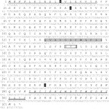
Fig. 1. Nucleotide and deduced amino acid sequence of prepro-Pso o 1. The putative signal peptide is underlined and amino acid residues differing from the deduced amino acid sequence derived from a truncated version of Pso o 1 (Lee et al. 2002) are shown as white text in black boxes. N-terminal and C-terminal residues not previously determined (Lee et al. 2002) are shown in italics. A potential N-glycosylation site is shown boxed. The consensus thiol proteinase cysteine active site (prosite motif PS00139) is grey shaded and that of the thiol proteinase asparagine active site (prosite motif PS00640) is double underlined.
Verification of Pso o 1 identity by Western blot and matrix-assisted laser desorption/ionization time of flight (MALDI-ToF) mass spectrometric analysis
Culture supernatant was used for electrophoresis on a NuPAGE® Bis-Tris 4–12% gel (Invitrogen) under reducing conditions. The gel was stained with SimplyBlue™, according to the manufacturer's instructions (Invitrogen), and the band detected (see Fig. 2) was excised, destained and reductively alkylated using dithiothreitol and iodoacetamide. The gel pieces were then digested overnight with trypsin or chymotrypsin at 37 °C. Digests were analysed using an Ultraflex II MALDI-ToF mass spectrometer (Bruker Daltonics), scanning the 600–5000 Dalton (Da) region in the reflectron mode producing monoisotopic resolution. The spectra generated were mass-calibrated using known standards and the peaks deisotoped. Databases were searched with the masses obtained using the MASCOT search engine (Swiss-Prot database) and a 50 ppm mass tolerance window. Significant matches from the Peptide Mass Fingerprint data were confirmed by MS/MS analysis using the search criteria described and an MS/MS-tolerance window of 0·5 Da.

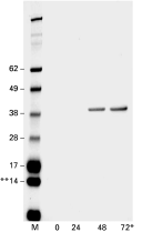
Fig. 2. Expression of Psoo1 in Pichia pastoris. Culture supernatant, removed from a pro-Pso o 1 culture 0–72 h after the induction of protein expression, was used for electrophoresis on a NuPAGE® Novex Bis-Tris 12–4% gel under reducing conditions and stained with SimplyBlue™ according to the manufacturer's instructions (Invitrogen). * Hours after induction of protein synthesis, ** Molecular mass (kDa).
For Western blots, culture supernatant, removed from a pro-Pso o 1 culture 72 h after the induction of protein expression, or purified Pso o 1 (before and after induction of proteinase activity), were subjected to electrophoresis in NuPAGE® (Invitrogen) Bis-Tris 4–12% gels under reducing conditions employing NuPAGE® MES SDS running buffer (Invitrogen). Proteins were transferred to a nitrocellulose membrane according to the manufacturer's instructions (Invitrogen). After transfer, the membranes were washed briefly in Tris-buffered saline, pH 7·4 (TBS; 25 mM Tris, 137 mM NaCl, 2·7 mM KCl) containing 0·1% (v/v) Tween 20 (TBST) and then incubated in TBST containing 5% (w/v) skimmed milk powder for 4 h at room temperature (RT; 22–24 °C) to block non-specific protein adsorption. To detect Pso o 1, the blotted proteins were incubated, for 1 h at RT, with serum produced in sheep against bacterially-expressed truncated Pso o 1 (Lee et al. 2002). Control blots were performed by omitting the test serum during this primary incubation step. Following extensive washing in TBST, membranes were incubated in affinity-purified donkey anti-sheep IgG horse-radish peroxidase (HRP) conjugate (Sigma) in TBST containing 5% (w/v) skimmed milk powder. Following incubation at RT for 1 h, and extensive washing, peroxidase activity was revealed using 3,3′-diaminobenzidine (DAB) as a substrate.
Measurement of enzymatic activity of recombinant Pso o 1
Activation of the recombinant pro-enzyme was attempted using each of the following methods. Dialysis against 100 mM acetate buffer (pH 4) for 72 h at 4 °C or at RT (Yasuhara et al. 2001; Takai et al. 2002); deglycosylation with endoglycosidase H (van Oort et al. 2002); exogenous activation with native Der p 1 (van Oort et al. 2002); heating to 60 °C in the presence of 20 mM cysteine (Jacquet et al. 2000; van Oort et al. 2002). In addition, the recombinant protein was incubated, for 10 min at 37 °C, with soluble extracts of P. ovis (0·02–0·4 μg protein, prepared as described previously (Nisbet and Billingsley, 2000) in 100 mM phosphate buffer, pH 5·8, containing cysteine (10 mM) prior to the testing in the enzyme assay. The enzymatic activity was assessed using the substrates butyloxycarbonyl-Val-Leu-Lys-MCA (Sasaki et al. 1984) and Z-Phe-Arg-MCA (Bando et al. 1986). Positive control reactions were performed for both substrates using a soluble extract of P. ovis and/or purified native Der p 1 as an enzyme source. Native Der p 1 was a generous gift from Dr Colin R. A. Hewitt, University of Leicester, UK.
Immunolocalization of Pso o 1
Mites (adults, nymphs and larvae) were collected from infested donor sheep (× Suffolk sheep, aged 1–2 years), fixed in Carnoy's fluid and stored at 4 °C until processing. Fixative was removed and mites were pelleted in 1% molten agarose and left to solidify before processing into paraffin wax. Sections (5 μm) were dried at 40 °C, dewaxed in xylene and rehydrated. Sections were treated to remove endogenous peroxidase (EnVision Kit™, DAKO) and blocked in 25% normal goat serum in TBS, pH 7·6, for 30 min before an overnight incubation at 4 °C in serum raised in rabbits against recombinant pro-Pso o 1 (1 in 2000 dilution in PBS). Following washing and incubation in goat anti-rabbit HRP conjugate (1[ratio ]1000 dilution) (DakoCytomation) for 1 h at RT, the sections were washed, and DAB substrate (Sigma) was used to detect the goat antibody conjugate. Negative controls included were a ‘pre-bleed’ serum from rabbits prior to injection with Pso o 1 (1 in 2000 dilution in PBS) and a ‘conjugate only’ control. Sections were washed in water, counterstained in haematoxylin for a few sec, washed in water, dehydrated, cleared and mounted in a resin-based mountant (Nisbet et al. 2006).
RESULTS
Molecular cloning and expression of Pso o 1
The PCR product obtained by amplification using primers ppPsoo1Bf and ppPsoo1Br was sequenced, and shown to contain an open reading frame (ORF) of 966 bp (nucleotide sequence data available in the EMBL, GenBank™ and DDJB databases under Accession number AM269885). This gene encoded a 322 aa predicted protein with 64% identity (80% similarity) to the major house dust mite faecal allergen Der f 1 (prepro-enzyme, Accession number BAC53948). The predicted protein, Pso o 1, also shared 99% identity over 263 aa residues with the previously characterized, truncated form (Lee et al. 2002). However, the asparagine residue involved in the catalytic activity of cysteine proteinases (Asn290 in the prepro-enzyme sequence) was present in the C-terminus of the full-length Pso o 1 described herein (Fig. 1).
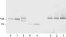
Pro-Pso o 1 was expressed efficiently in yeast within 48 h of induction (Fig. 2), but the expression of prepro-Pso o 1 was unsuccessful in this system (results not shown). The apparent molecular mass of the recombinant protein was 40 kDa, ~6 kDa larger than that based on the cDNA sequence. The recombinant protein reacted strongly with sheep anti-truncated Pso o 1 serum on Western blot (Fig. 3, lane 1); both the chymotrypsin and trypsin digests and MALDI-ToF analysis of the expressed protein confirmed that the recombinant protein was Pso o 1.

Fig. 3. Western blot of Pso o 1 and its derivatives. Lanes on blot are as follows: (1) pro-Pso o 1 culture supernatant (CS); (2) pro-Pso o 1 CS dialysed against acidic (pH 4) buffer for 72 h; (3) pro-Pso o 1 purified from CS by gel filtration; (4) purified native Der p 1; (5) soluble P. ovis extract containing native Pso o 1; (6) pro-Pso o 1 CS incubated at 60 °C for10 min; (7) pro-Pso o 1 CS treated with 12·5 mu endoglycosidase H; (8) purified pro-Pso o 1 treated with 12·5 mu endoglycosidase H. To detect Pso o 1, Der p 1 and derivatives, the blotted proteins were incubated with serum raised in sheep against bacterially-expressed truncated Pso o 1 before being incubated in donkey anti-sheep IgG horse radish peroxidase (HRP) conjugate and peroxidase activity was revealed using DAB as a substrate. * Molecular mass (kDa).
Measurement of enzymatic activity of recombinant Pso o 1
Large-scale expression of pro-Pso o 1 was performed, and attempts were made to activate the recombinant protein to a mature active enzyme. Enzymatic activity was assessed using the fluorogenic substrates butyloxycarbonyl-Val-Leu-Lys-MCA or Z-Phe-Arg-MCA. Activation of the recombinant enzyme was attempted employing each of the following treatments: dialysis against acidic buffer for 72 h; deglycosylation; activation with mite proteinases and heating to 60 °C. With the exception of the dialysis, each treatment reduced the apparent molecular mass of the major band by 2–4 kDa (Fig. 3), but none of the treatments resulted in a protein of the same molecular mass as native Pso o 1 or Der p 1 (Fig. 3 lanes 4 and 5). Soluble extract of P. ovis and native Der p 1 positive controls efficiently hydrolysed both substrates, but none of the treatments produced active enzyme from Pso o 1. In addition, treatment of the recombinant protein with a range of concentrations of a soluble extract of P. ovis failed to produce active enzyme.
Immunolocalization of Pso o 1
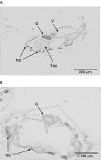
Pso o 1 was localized specifically to the gut in sections of adult P. ovis (Fig. 4A). The labelling of Pso o 1 was most intense near the luminal surface of the gut. Minor non-specific reactivity of the secondary antibody (goat anti-rabbit IgG) and/or the substrate was observed in the tissues underlying the cuticle in both the negative controls and the sections treated with rabbit anti-Pso o 1 (Fig. 4). Negative controls were identical when primary antibody was omitted (Fig. 4B) and when rabbit ‘pre-bleed’ serum was used in the primary incubation step (not shown).

Fig. 4. Immunolocalization of Pso o 1 in sections of adult Pichia ovis. (A) Incubated with rabbit anti-Pso o1 (1 in 2000 dilution) as primary antibody; (B) primary antibody omitted. Immunoreactivity was visualized using goat anti-rabbit HRP conjugate followed by incubation in DAB substrate. (B) Conjugate-only control. Areas annotated with ‘G’ show sections through gut; ‘V’, ventriculus; ‘NS’ denotes non-specific staining resulting from binding of the secondary antibody and/or reactivity of the substrate.
DISCUSSION
The entire coding sequence of Pso o 1 was determined to augment the truncated sequence which had already been published (Lee et al. 2002). The newly determined sequence encoded a protein with a predicted signal peptide which is cleaved between Ala18 and Tyr19 [predicted using SignalP 3.0 (Dyrløv Bendtsen et al. 2004)] and possessed significant homology to the house dust mite group 1 allergens Der f 1, Der p 1 and Eur m 1, suggesting that Pso o 1 is secretory in nature and that it may also act as an allergenic component in the pathogenesis of sheep scab disease. Lee et al. (2002) noted that Pso o 1 possessed a high degree of amino acid homology within the residues surrounding the propeptide cleavage site involved in the maturation of active enzyme in house dust mite group 1 allergens and suggested that this area may also be involved in the maturation of Pso o 1 cysteine proteinase activity. These residues are present in the full-length Pso o 1 molecule described herein. Interestingly, only 50% of the 12 residues surrounding the proposed cleavage site (between Ala98 and Gly99 in Fig. 1) are identical between Der p 1 and Pso o 1, compared with >80% identity in the same region between the different house dust mite allergens. This information may explain the failure of autocatalytic processing of the recombinant Pso o 1, suggesting that alternative processing may be required, though endogenous proteinase activity in soluble P. ovis extracts also failed to activate the recombinant enzyme. The ~6 kDa discrepancy between the observed and calculated masses for pro-Pso o 1 suggests hyperglycosylation of Pso o 1 in the yeast expression system, a phenomenon which has also been reported from a recent expression study of the group 1 house dust mite allergens (Takai et al. 2002).
Cysteine proteinase activity is abundant in the soluble extracts of a number of mite species, including P. ovis (see Nisbet and Billingsley, 2000). These enzymes are involved in fundamental processes in the pathogenesis of sheep scab, including the degradation of connective tissue and extracellular matrix molecules and the catalysis of immunoglobulins (Kenyon and Knox, 2002). By analogy to dust mite allergens, cysteine proteinases and Pso o 1, in particular, may also be involved in allergen-induced hypersensitivity-related effects (John et al. 2000). Studies using both storage mites (Acarus farris) and dust mites (D. farinae), fed on diets containing cysteine proteinase inhibitors, have also shown that the inhibition of cysteine proteinase activity in the guts of mites retarded their development and reduced survival, suggesting critical roles in the digestive process (Pernas et al. 1998; Sánchez-Ramos et al. 2004).
In addition, Der p 1 has been localized in the oral cavity, gut epithelium, gut contents and faecal pellets of D. pteronyssinus and is thought to be synthesized by cells lining the gastrointestinal tract (Rees et al. 1992). The present study demonstrated that Pso o 1 is localized within the gut of the P. ovis, making it potentially accessible to the immunoglobulin component of the exudates which have been shown to be imbibed by sheep scab mites feeding on the host (Pettit et al. 2000). Thus, an attractive possibility is that vaccination with gut cysteine proteinases, such as Pso o 1, will induce the production of ‘blocking antibodies’ which inhibit cysteine proteinase activity in the mite gut, resulting in a retardation of mite development and/or survival (Nisbet and Huntley, 2006). By further analogy with Der p 1, another possible beneficial effect of vaccination with Pso o 1 may be a down–regulation of the allergic inflammatory Th2 response involved in the pathogenesis of sheep scab disease (John et al. 2000).
Vaccination with cysteine proteinases has been shown to be effective against a number of other parasites. For example, the vaccination of dogs with Ac-CP-2, a recombinant cysteine proteinase derived from the hookworm Ancylostoma caninum, resulted in high antibody responses, the stunting of adult worms and a marked reduction in faecal egg output (Loukas et al. 2004). Also, IgG purified from the serum of vaccinated dogs inhibited the enzymatic activity of Ac-CP-2 by 73% (Loukas et al. 2004). Although enzymatic activity of recombinant proteins may be desirable for enhanced protection, it is not always essential. For example, vaccination with inactive recombinant cysteine proteinases (hmcp1, 4 and 6) from Haemonchus contortus resulted in a significant reduction in the intensity of infection in sheep (Redmond and Knox, 2004, 2006). Thus, although the recombinant Pso o 1 expressed in yeast herein was inactive as a cysteine proteinase, it may represent a vaccine candidate. Future studies will focus on this possibility.
A.J.N. and J.F.H. gratefully acknowledge funding by the Department for Environment Food and Rural Affairs, UK Research Contract OD0544 and J.F.H., A.McK and K.McL acknowledge funding by the Scottish Executive Environment and Rural Affairs Department. The authors are grateful to George Newlands, Jim Redmond and Karina Reinhardt, Moredun Research Institute, for their assistance in the expression, purification of recombinant enzymes and DNA analyses, respectively, and to Toshiro Takai, Juntendo University School of Medicine, Japan, for advice and useful discussions during this project. We are grateful to Dr Colin R. A. Hewitt, University of Leicester, for the gift of native Der p 1.