Acquired aortocardiac shunts are extremely rare paediatric heart lesions. Reference Rojas, Amat-Santos and Cortés1–Reference Nagi Haddad, Ly, Iserin and Malekzadeh-Milani4 Transcatheter closure is an attractive treatment modality, yet the anatomic complexity and morphological variability of these anomalies make them challenging to close using device occluders not originally designed for this purpose. Reference Nakamura, Passeri and Inglessis-Azuaje2–Reference Pate, Al Zubaidi, Chandavimol, Thompson, Munt and Webb5
Case presentation
An 8.5-year-old female patient (27 kg/129 cm) was referred to our institution for a progressively worsening aortic regurgitation that has been monitored since her last heart surgery during regular outpatient follow-up. The patient had type IIc tricuspid atresia with aortic coarctation that has been initially palliated with pulmonary artery banding and aortic repair in the neonatal period, and finally with a fenestrated total cavopulmonary connection and bulboventricular foramen enlargement (through right ventriculotomy) at the age of 6 years.
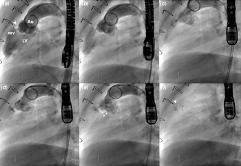
Upon admission to our unit, the patient had NYHA functional class II and physical examination showed a diastolic murmur of grade 4/6 along the left parasternal region. Chest X-ray and basic blood tests were normal. Baseline ultrasound showed a mild-to-severe eccentric aortic regurgitation (PHT of 450 ms, end-diastolic flow velocity of 0.35 m/second in the aortic isthmus). The ultrasound study was unable to accurately grade the regurgitation and depict its precise mechanism motivating a cardiac MRI that showed an anteriorly located aortic regurgitation and graded it as mild (regurgitation fraction of 25%). However, invasive diagnostic aortogram revealed normal aortic valve functioning and delineated an abnormal tunnel-like communication of 5.0 mm long× 2.6 mm large between the right anterior non-coronary aortic sinus and the rudimentary right ventricular cavity (Fig 1a). A complimentary pre-intervention planning CT angiogram confirmed catheterisation findings and excluded coronary arteries involvement. Full infective endocarditis workup came negative and the lesion was thereby considered as post-operatively acquired.
Diagnostic aortogram delineating a tunnel-like shunt (white pointed arrow) from the right anterior non-coronary aortic sinus to the rudimentary right ventricle (width: 2.61 mm; depth: 5.05 mm) ( a ). Consecutive sequences of the interventional closure of the shunt using a KONAR-MFO 5 × 3 mm connected from its right side ( b – d ). Control angiography before release showing complete shunt closure and normal aortic valve functioning (white stars) ( e ). Stable device after release (white pointed arrow) ( f ). Ao = aorta; LV = left ventricle; RRV = rudimentary right ventricle.
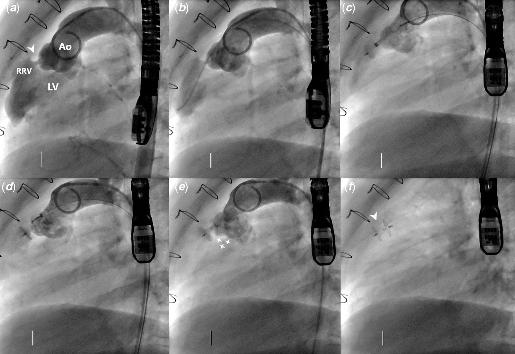
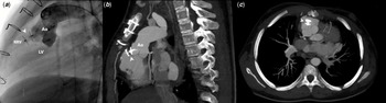
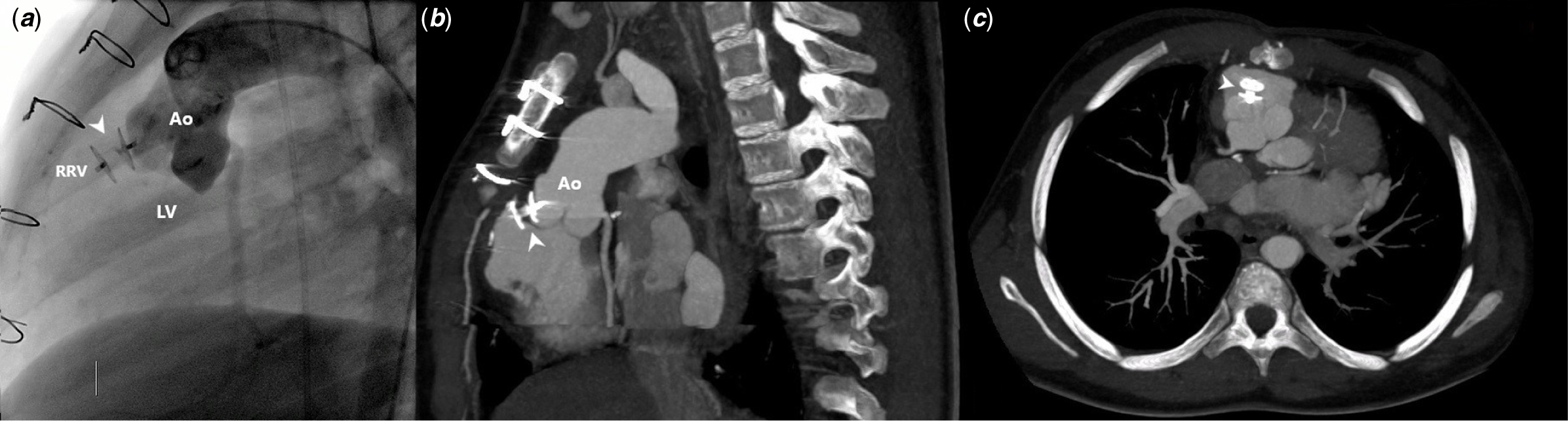
Appropriate informed consent was obtained for attempted device closure. The procedure was performed under general anaesthesia, biplane fluoroscopy, and transesophageal echocardiography. At the time of the procedure, the patient was under Warfarin therapy with INR ranging between 2 and 3. Antibiotic prophylaxis and systemic heparinisation were given as per institutional protocol. A bilateral 5-Fr arterial femoral access was obtained. Biplane aortogram was performed using a 5-Fr Pigtail diagnostic catheter (Cordis Corp., FL, United States of America) to relocate the defect that was easily crossed using a 5-Fr Judkins right coronary diagnostic catheter (Cordis Corp., FL, United States of America) in combination with an 0.032-inch/180 cm-long J-tip Radifocus® Hydrophilic Guidewire M (Terumo Corp., Tokyo, Japan). The catheter was then advanced into the rudimentary right ventricle and the wire was upgraded to a 0.035-inch/260 cm-long J-tip Stiff-type Radifocus® Hydrophilic Guidewire M (Terumo Corp., Tokyo, Japan). A 5 × 3 mm KONAR-MFO was selected and was connected to the delivery cable from its right side. The catheter was exchanged with a 5-Fr/80 cm SteerEase™ delivery sheath (Lifetech, Shenzhen, China) through which the device was advanced and sequentially deployed under fluoroscopy guidance (Fig 1b–d). Device stability, shunt closure, and normal aortic valve function were checked on ultrasound and control angiography before releasing the device (Fig 1e and f). No early complications were observed. The procedure required 25 minutes of fluoroscopy. The patient received 48 hours of therapeutic heparin and was safely discharged with no residual shunt or aortic regurgitation. The patient was prescribed to continue warfarin with oral aspirin therapy. One year follow-up confirmed excellent results with complete shunt closure and preserved aortic valve competence (Fig 2).
One-year follow-up control imaging with ascending aortogram ( a ) and multi-planar modified CT-scan reconstructions ( b , c ). Note device stability, complete shunt closure, and preserved aortic valve competence (white pointed arrow). Ao = aorta; LV = left ventricle; RRV = rudimentary right ventricle.
Discussion
Aortocardiac shunts have been previously described. Reference Rojas, Amat-Santos and Cortés1–Reference Pate, Al Zubaidi, Chandavimol, Thompson, Munt and Webb5 Aetiologies are numerous and treatment strategy remains debatable. Reference Rojas, Amat-Santos and Cortés1–Reference Kerkar, Lanjewar, Mishra, Nyayadhish and Mammen6 Surgery is most commonly considered as the primary treatment option, yet interventional device closure has been repetitively reported as a safe and effective alternative with attractive advantages. Multiple devices have been implanted and the majority of the results are encouraging. Reference Rojas, Amat-Santos and Cortés1–Reference Kerkar, Lanjewar, Mishra, Nyayadhish and Mammen6 The recently introduced KONAR-MFO has been smartly designed, combining technical features of previous occluder devices, to tackle encountered difficulties. Reference Boukaram, Haddad, Daou and Saliba7–Reference Haddad and Saliba9 Emerging reports are consecutively positive and clinical applications have been mainly limited to ventricular septal defects closure. Reference Haddad and Saliba9 This is the first use of the KONAR-MFO to percutaneously treat an iatrogenic aorta to right ventricle shunt occurring in a complex child following intracardiac repair.
Iatrogenic aorta to right ventricle fistulas has been commonly described following transcatheter or surgical aortic valve replacements and ventricular septal defect repairs. Reference Rojas, Amat-Santos and Cortés1–Reference Pate, Al Zubaidi, Chandavimol, Thompson, Munt and Webb5 The most commonly suggested mechanisms of this complication are ischaemic necrosis, aneurysm rupture, aggressive surgical debulking, and erosion between the implanted material and the aortic wall in late-onset perforation of the aortic wall. Reference Rojas, Amat-Santos and Cortés1–Reference Kerkar, Lanjewar, Mishra, Nyayadhish and Mammen6 Our patient underwent an opening of the accessory cavity beneath the aortic valve to enlarge the bulboventricular foramen. Based on the time frame sequence of the events and the aseptic status of the patient, we retrospectively advocate that an iatrogenic surgical injury to the area of contact between the right anterior non-coronary aortic sinus and accessory cavity could have been the likely cause for such a particular fistulous-like communication in our patient. Our case showed the utility of the aortogram as a complimentary exam to non-invasive diagnostic tools in the straightforward mapping of the aortic valve region. Diagnostic aortogram was very useful in adjusting the diagnosis and in providing the required information for the planning of the intervention. Reference Nagi Haddad, Ly, Iserin and Malekzadeh-Milani4,Reference Alam10 Multi-modality imaging and fusion technology can also be helpful for pre-procedural planning, device choice, and pre-procedural guidance, yet these technologies are not always necessary to achieve the intended outcomes. Reference Nagi Haddad, Ly, Iserin and Malekzadeh-Milani4
We chose KONAR-MFO as the most suitable device for this case. Indeed, the central disk perfectly fitted across the tunnel achieving immediate and stable complete shunt closure. The articulating soft right retention disk conformed to the aortic sinus curvature without interfering with the aortic valve function or the nearby coronary arteries. This is the first report of closure of this rare cardiac lesion using this particular device. Reference Haddad and Saliba9 The procedure was technically smooth. The tunnel was easily accessed from the aorta using proper wires and the device was promoted directly into position. The retrograde approach allowed straightforward delivery avoiding technical complications of the arterio-venous circuit and reducing irradiation exposure. Reference Haddad, Rizk, Saliba and Farah11 The anatomic complexity of these rare lesions makes them sometimes challenging to close using devices not originally designed for this purpose, yet we set additional proof of the efficacy of transcatheter treatment.
Conclusion
Retrograde transcatheter closure of acquired aorta to right ventricle shunt is a feasible and safe procedure with very good outcomes. The new KONAR-MultifunctionalTM VSD Occluder offers high anatomical conformability and procedural flexibility to effectively treat this complex type of heart lesion.
Acknowledgements
None.
Author contributions
RH collected clinical data, designed illustrative material, and took the lead in writing and revising the entire manuscript. All authors have read and approved the final version of the manuscript.
Financial support
This research received no specific grant from any funding agency, commercial, or not-for-profit sectors.
Conflicts of interest
None.
Ethical standards
The authors assert that all procedures contributing to this work comply with the ethical standards of the relevant national guidelines on human experimentation and with the Helsinki Declaration of 1975, as revised in 2008. The patient’s legal guardians signed informed consent for the reported procedures and this publication.