Introduction
Currently, the Microscaphidioidea Looss, 1900 is a superfamily including a group of gastrointestinal trematodes found in marine and freshwater chelonians and teleosts (Blair, Reference Blair, Gibson, Jones and Bray2005). In this superfamily, the oral sucker is absent and functionally replaced by the pharynx, the ventral sucker is absent, the oesophagus is long and mostly ends in a muscular oesophageal bulb, the two testes are inter- or post-caecal, the cirrus sac is absent and the seminal vesicle is free in the parenchyma, a hermaphroditic duct is present, the ovary is inter- or post-testicular, a uterine seminal receptacle is usually present, the uterus is mainly inter-caecal, the vitelline follicles are profuse or restricted and the excretory pore is terminal or postero-terminal. This superfamily includes two closely similar families, the Microscaphidiidae Looss, 1900 and the Mesometridae Poche, 1926. The essential difference between them is the ventral body surface. This surface is not modified in the former, but in the latter, it is modified (entirely concave or concave only anteriorly) to form an accessory attachment organ. Phylogenetic and molecular phylogenetic studies (e.g. Brooks et al., Reference Brooks, O'Grady and Glen1985; Olson et al., Reference Olson, Cribb, Tkach, Bray and Littlewood2003) have strongly suggested that the two families are paraphyletic. Presently, the Microscaphidiidae includes 22 genera, whereas the Mesometridae includes only five genera (Blair, Reference Blair, Gibson, Jones and Bray2005; Tkach and Snyder, Reference Tkach and Snyder2006). To date, very little is known about the life cycles of members of these families. Lotz & Corkum (Reference Lotz and Corkum1984) is the only known study on microscaphidiid life cycles, where the redia and cercaria of Dictyangium chelydrae Stunkard, 1943 from the snail Amnicola (Cincinnatid) peracuta and the metacercaria were briefly described. Similarly, Jousson & Bartoli (Reference Jousson and Bartoli1999) is the only known study on mesometrid life cycles, where the rediae and cercariae of three species, namely Elstia stossichianum Monticelli, 1892, Wardula capitellata (Rudolphi, 1819) Poche, 1926 and Centroderma spinosissima (Stossich, 1886) Lühe, 1901, from the snails Vermetus triqueter, Barleeia rubra and Rissoa ventricosa, respectively, and also the metacercariae, were described briefly. Prior to this study, Jousson et al. (Reference Jousson, Bartoli, Zaninetti and Pawlowski1998) used the internal transcribed spacer (ITS) rDNA for distinguishing the cercariae of these three mesometrid species from each other. Despite these observations, a complete life cycle is still unknown.
Currently (Al-Jahdali, Reference Al-Jahdali2013), the microscaphidiid genus Hexangium Goto & Ozaki, 1929 includes three valid species, all found in the intestine of siganid fishes: Hexangium sigani Goto & Ozaki, 1929 (type species) (from Siganus fuscescens off Misaki and Takamatsu, Japan), Hexangium brayi Hassanine & Gibson, Reference Hassanine and Gibson2005 (from Siganus luridus in the Red Sea off Sharm El-Sheikh, Egypt) and Hexangium saudii Al-Jahdali, Reference Al-Jahdali2013 (from Siganus rivulatus in the Red Sea off Rabigh, Saudi Arabia). The first is a well-known and widely distributed species, recorded from numerous different siganid fishes by several authors (e.g. Velasquez, Reference Velasquez1961; Fischthal & Kuntz, Reference Fischthal and Kuntz1964, Reference Fischthal and Kuntz1965; Al-Yamani & Nahhas, Reference Al-Yamani and Nahhas1981; Shen, Reference Shen1990; Martens & Moens, Reference Martens and Moens1995; Geets & Ollevier, Reference Geets and Ollevier1996; Dzikowski et al., Reference Dzikowski, Paperna and Diamant2003; Kardousha, Reference Kardousha2003; Sey et al., Reference Sey, Nahhas, Uch and Vang2003; El-Labadi et al., Reference El-Labadi, Ismail and Khalaf2006), whereas the other two species are known only from their original description and may be endemic to the Red Sea (Hassanine & Gibson, Reference Hassanine and Gibson2005; Al-Jahdali, Reference Al-Jahdali2013).
In the present study, we describe the life cycle of H. sigani under natural conditions. This represents the first complete life cycle for a member of the superfamily Microscaphidioidea. A knowledge of life cycles has proved important for many recent phylogenetic studies, since molecular phylogenetic studies on digenean groups have mostly used molecular data combined with a knowledge of life cycles (e.g. Olson et al., Reference Olson, Cribb, Tkach, Bray and Littlewood2003; Bray et al., Reference Bray, Waeschenbach, Cribb, Weedall, Dyal and Littlewood2009). The absence of such data forces researchers to make assumptions that have led to glaring gaps in their studies (Cribb et al. Reference Cribb, Bray, Olson and Littlewood2003).
Materials and methods
Collection and examination of fish and snails
In a small lagoon (c. 75 m in diameter, 2 m in depth) within mangrove swamps found on the Egyptian coast of the Gulf of Aqaba (between 28°7′N and 28°18′N), the rabbitfish S. rivulatus Forsskål & Niebuhr, 1775 (Teleostei: Siganidae) is permanently resident and highly parasitized by the intestinal trematode H. sigani Goto & Ozaki, 1929 (Digenea: Microscaphidiidae), the only trematode species found in this lagoon. Life-history stages (mother sporocysts, rediae and cercariae) of this parasite were found only in the gonads and digestive gland of Nassarius pullus Linnaeus, 1758 (Gastropoda: Nassariidae), a common snail in the same lagoon; other fishes and molluscs were completely free of trematodes.
Fish identifications were based on Randall (Reference Randall1983) and their names follow Froese & Pauly (Reference Froese and Pauly2014). Mollusc identifications were based on Sharabati (Reference Sharabati1984). Standard parasitological techniques were used to examine the alimentary canal of the fish and the different organs of the molluscs. Trematodes were removed from their fish or mollusc host under a stereomicroscope and observed live under a compound microscope; the examination of larval trematodes was facilitated by the use of neutral red as a vital stain, since the stain accumulated rapidly in their bodies.
Eight random samples, each containing 30–45 specimens of the snail N. pullus, were collected from the coast of Jubayl (160 km away from of the mangrove swamps), and examined carefully to make sure that they were not naturally infected with any trematode larvae. All of these specimens were uninfected. Therefore, 700 specimens of this snail were transferred live during March 2014, to special aquaria near the lagoon to follow their experimental infection with H. sigani from its beginning. Aquaria were maintained at the same water temperature as in the lagoon (20–26°C). Similarly, eight random samples, each containing 15–20 specimens of the fish S. rivulatus from the same location, were found to be uninfected with any trematode species. Therefore, 500 specimens of this fish were tagged and transferred live to the lagoon in order to allow their natural infection with H. sigani to occur, and to estimate the approximate time taken by this trematode to reach maturity in its definitive host. To prevent fish from escaping from the lagoon, a plastic net with a narrow mesh size was used as a barrier at the lagoon mouth; the lagoon can therefore be considered as a natural aquarium. Some uninfected fish were kept alive in aquaria during the study.
Experimental infections
Fully gravid individuals of H. sigani were taken alive from the intestine of naturally infected S. rivulatus inhabiting the lagoon, placed in a small Petri dish containing filtered sea water and, when gently ruptured with a fine needle, eggs were released from their uteri. To detect egg hatching in the sea water, numerous eggs were carefully concentrated in filtered seawater, placed in transparent embryo cups and observed for more than 3 weeks at 20–25°C. Also, attempts were made to obtain miracidia from the eggs in several ways, such as changing water temperature, exposure to light or changing osmotic conditions. To detect egg hatching in the snail, the contents of the Petri dish were gently poured into one of the aquaria containing the uninfected snails (transferred from Jubayl). Ten snails were taken from this aquarium at 12-h intervals, and their digestive glands and gonads were examined microscopically, searching for newly formed trematode larvae; also, snail faecal balls from the aquarium were examined carefully. To elucidate the successive development of larval stages (sporocysts, rediae and cercariae), ten snails were taken from the aquarium every week and their digestive glands and gonads were examined carefully. To determine whether the cercariae were released from the snails or not, some snails were placed singly in small glass bowls filled with filtered seawater and observed; the addition of a drop of neutral red to the bowls was helpful. The behaviour of cercariae was observed in a Petri dish under the microscope.
To estimate the approximate time taken by H. sigani to reach maturity in its definitive fish host, 1- to 4-day-old metacercariae encysted on filamentous algae were fed to uninfected S. rivulatus (from those transferred from Jubayl and kept alive in the aquarium). However, six tagged fish from those transferred to the lagoon (to allow their natural infection with H. sigani to occur) were taken every week to examine the different developmental stages of this parasite found in their intestines.
Intra-molluscan and sexually mature stages of H. sigani
The most accurate drawings were obtained by killing the intra-molluscan stages of the parasite, especially the cercariae, in whirling hot water prior to fixation in hot 5% formalin without pressure. This procedure gives specimens of a uniform size and shape. Some mature worms were fixed in alcohol–formalin–acetic acid (AFA) under a very slight coverslip pressure and preserved in 75% ethyl alcohol. Whole mounts were stained with alum carmine, cleared in terpineol and mounted in Canada balsam. Specimens were examined using a fully motorized Nikon Eclipse 90i microscope equipped with differential interference contrast optics (Nomarski), a DS-5M Nikon digital camera, Nikon ACT-2U measuring software and a drawing tube attachment (Nikon, Florida, USA). Measurements are quoted as the range, with the mean in parentheses, and are given in micrometres.
Results
Hexangium sigani Goto & Ozaki, 1929
-
Final host. Siganus rivulatus Forsskål & Niebuhr, 1775 (Siganidae). Site: intestine.
-
Molluscan host. Nassarius pullus Linnaeus, 1758 (Gastropoda: Nassariidae). Site: gonads and digestive gland.
-
Locality. Mangrove swamps on the Egyptian coast of the Gulf of Aqaba.
Eggs
Eggs oval, yellowish, thick-walled, non-operculate, moderately large, 70–76 × 38–46 (73 × 42). Eggs incubated in seawater at 20–25°C for more than 3 weeks did not hatch. Also, attempts to obtain miracidia from eggs by changing water temperature, exposure to light or changing osmotic conditions were unsuccessful. However, eggs of the same shape and size were observed in the digestive tracts of many infected individuals of the snail N. pullus, and empty eggshells were recovered from their faecal balls.
Mother sporocyst
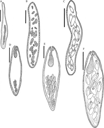
Young mother sporocyst (1 week post-infection; based on 20 specimens) (fig. 1A): body elongate claviform, opaque white, thin-walled, 373–445 × 44–58 (409 × 51). Germinal balls longitudinally aggregated in wider half of body; narrower half appeared to be transparent and its narrowness seemed temporary following penetration of snail tissue.
Stages in the life cycle of Hexangium sigani Goto & Ozaki, 1929 from the nassariid gastropod Nassarius pullus, Red Sea: (A) young mother sporocyst; (B) moderate-sized mother sporocyst; (C) fully developed mother sporocyst; (D) young redia; (E) moderate-sized redia; (F) fully developed redia. Scale bars: 100 μm (A, D); 200 μm (B, E); 500 μm (C, F).

Moderate-sized mother sporocyst (3–4 weeks post-infection; based on 20 specimens) (fig. 1B): body elongate, 952–1142 × 168–206 (1047 × 187), contained 16–20 redial embryos; smallest 65 × 28, largest 86 × 42. Few germinal balls remained scattered in body.
Fully developed mother sporocyst (5–7 weeks post-infection; based on 20 specimens) (fig. 1C): body elongate, slightly curved, 1840–2224 × 298–368 (2032 × 333), contained 16–20 rediae, which are usually at different stages of development; smallest 170 × 60, largest 340 × 102. Germinal balls rarely observed at this stage. Lateral birth-pore observed at 306–352 (329) from anterior end. Walls of sporocyst are capable of contraction and expansion, but no movement was observed.
Redia
Young redia (8–10 weeks post-infection; based on 20 specimens) (fig. 1D): weakly contractile. Body spindle-shaped, with thick muscular wall, opaque white, without appendages, 316–378 (374) in length, 70–102 (86) in maximum width at its middle. Oral opening antero-terminal. Pharynx well developed, globular, 17–23 × 20–26 (20 × 23), connected directly to short saccular caecum restricted to anterior quarter of body; contains yellowish, finely granular material. Germinal balls aggregated in elongated cluster extending to posterior third of body.
Moderate-sized redia (11–13 weeks post-infection; based on 20 specimens) (fig. 1E): weakly contractile. Body spindle-shaped, 972–1162 (1067) in length, 268–310 (289) in maximum width at its middle. Oral opening antero-terminal. Pharynx well developed, globular, 50–62 × 58–70 (56 × 64), connected directly to relatively long saccular caecum restricted to anterior third of body and filled with yellowish droplets (probably ingested from snail tissue). Cercarial embryos (16–22/redia) aggregated in cluster extending into posterior half of body. Few germinal balls remained undifferentiated near posterior end of body.
Fully developed redia (16–17 weeks post-infection; based on 20 specimens) (fig. 1F): immobile. Body spindle-shaped, 1960–2326 (2143) in length, 522–642 (582) in maximum width at its middle. Birthpore just postero-lateral to pharynx and easily seen during emergence of cercariae. Oral opening anteroterminal. Pharynx globular, 83–105 × 106–118 (94 × 112), connected directly to relatively long saccular caecum not reaching into middle third of body, and filled with yellowish droplets and amorphous white material (probably ingested from snail tissue). Differentiated cercariae 18–26 in number, at different stages of development, usually crowded together in posterior half of body. Germinal balls and cercarial embryos completely absent.
Cercaria
After expulsion from the redia, cercariae remain for about 2 weeks within the gonad of the snail before their emergence, which occurs in night.
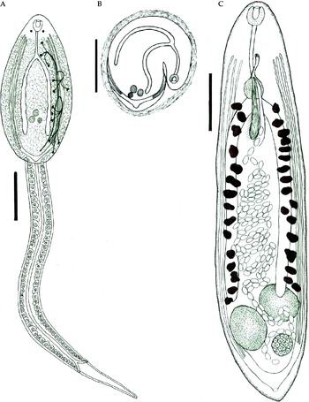
Fully developed cercaria (emerges from snail 18–19 weeks post-infection; based on 20 specimens) (fig. 2A): cercaria monostome. Body elongate, oval, 292–312 (302) in length, 110–136 (123) in maximum at its middle. Tegument smooth and unarmed. Two lateral eyespots present on dorsal surface posterior to pharynx. Tail simple, long, cylindrical, without fins and cystogenous cells, 1.95–2.2 (2.07) times as long as body, capable of extension and contraction; it attaches to body dorso-subterminally. Suckers absent. Oral opening surrounded directly by subterminal barrel-shaped muscular pharynx measuring 18–24 × 16–20 (21 × 18). Oesophagus moderately long, narrow, 38–46 (42) in length, thin-walled; wall slightly thicker at end of oesophagus, with some transverse muscular fibres; this thickening corresponds to oesophageal bulb of adult stage. Intestinal caeca simple, relatively wide, slightly sinuous, extending to near posterior end of body. Cystogenous gland cells dorsal, relatively large, ovoid, extending from eyespots to posterior end of body; these are negative to neutral red. Penetration glands absent. Testes: two, round, diagonal, inter-caecal, in middle of posterior half of body, subequal, 11–15 (13) in diameter. Anlagen of ovary sinistrally submedian, immediately posterior to testes; testes and ovary arranged as in adult. Genital ducts not visible. Excretory system consists of a thin-walled V-shaped excretory vesicle, situated between testes and posterior end of body; lateral arm on each side divides into three long collecting ducts extending anteriorly to near pharynx. Flame-cell formula 2[(3+3)+(3+3)] = 24. Excretory pore dorso-subterminal. Axial duct of tail bifurcates into two short arms in middle of posterior third of tail; these open to exterior via two lateral primary excretory pores.
Stages in the life cycle of Hexangium sigani Goto & Ozaki, 1929: (A) fully developed cercaria from the nassariid gastropod Nassarius pullus, Red Sea; (B) metacercaria; (C) fully gravid specimen (ventral view) from Siganus rivulatus, Red Sea. Scale bars: 100 μm (A, B); 500 μm (C).

Metacercaria
(The formation of the metacercaria (fig. 2B) was observed in a Petri dish filled with filtered seawater and containing small pieces of washed filamentous algae and other aquatic vegetation on its bottom.) After leaving the snail, the cercaria appeared to be at rest for only a few seconds, and then suddenly became very active for few seconds, swimming through the water using its long tail. After which, the cercaria swam down to reach the substratum, and when it encountered an algal thallus it started a series of thrusting and revolving movements, which resulted in the detachment of the tail and triggered the emission of secretions from the cystogenous glands. The transparent material emitted formed a flexible membranous cyst around the metacercaria; some of this cyst material was anchored to the substratum. One or 2 min later, the cercaria began a series of complex oscillatory movements inside the unfinished cyst, apparently in order to detach its body wall from the cyst wall.
The metacercaria measured 205–233 (219) in diameter, remained alive for about 1 week; no noticeable progenesis was observed during this period. Generally, it is closely similar to the cercaria, only differing in having a bent body within the cyst and more turgid intestinal caeca. The lagoon bottom is extremely muddy and, consequently, metacercarial cysts cannot be observed on the aquatic vegetation, which is usually covered by a muddy layer. Two-day-old metacercariae encysted on filamentous algae fed to uninfected S. rivulatus developed into fully gravid worms 5–6 weeks post-infection. Juvenile worms of H. sigani were also found in the intestine of many tagged fish 2 days after being transferred to the lagoon, and developed naturally into mature, fully gravid worms after 5-6 weeks. Thus, the complete life cycle of H. sigani extends for 23–25 (24) weeks.
Sexually adult stage (marita) from S. rivulatus
Adult stage (fig. 2C) (based on 30 fully gravid specimens): body elongate or lanceolate, stout, somewhat dorso-ventrally flattened, 2057–4285 (3171) in length, 628–942 (785) in maximum width at its middle. Tegument armed with fine sharp spines (often lost during fixation). Suckers absent; oral opening sub-terminal. Pharynx well developed, globular, muscular, directly abuts oral opening, 151–201 × 148–176 (176 × 162). Oesophagus moderately long, 394–570 (482), provided with round oesophageal bulb at its posterior end; bulb small, 140–194 (167) in diameter. Intestinal bifurcation at middle of anterior half of body; caeca simple, long, wide, extend to middle of posterior half of body. Testes: two, usually oval, sometimes round, oblique, immediately post-caecal, slightly separated or contiguous, subequal, 360–510 × 270–350 (435 × 310). Cirrus sac absent. Seminal vesicle claviform, surrounded by weakly developed fibres, extends from short distance posterior to intestinal bifurcation to near genital pore, 484–680 (582) in length; distal end of vesicle unites with uterus to form relatively short hermaphroditic duct. Genital pore situated mid-way between pharynx and intestinal bifurcation. Ovary round, usually sinistral to posterior testis, 175–237 (206) in diameter. Seminal receptacle and Laurer's canal absent; uterine seminal receptacle present. Uterus relatively long, mainly inter-caecal; few uterine coils extend to near posterior end of body. Eggs oval, yellowish, thick-shelled, moderately large, 70–76 × 38–46 (73 × 42). Vitelline follicles moderately large, irregular, extend in lateral fields between testes and intestinal bifurcation. Excretory vesicle V-shaped; excretory arms short, divide at level of ovary into three long collecting ducts extending anteriorly to level of genital pore; the medial pair of these ducts may represent a lymphatic system. Excretory pore dorso-subterminal.
Discussion
Very little has been known about microscaphidiid life cycles. Our knowledge was restricted to the only known study by Lotz & Corkum (Reference Lotz and Corkum1984), in which the rediae and cercariae of D. chelydrae Stunkard, 1943 from the snail A. (C.) peracuta and also its metacercariae were briefly described. Nevertheless, the known life cycle of this microscaphidiid is still incomplete. Nothing is known of the hatching of the eggs, the sporocysts and the duration of each life-cycle stage. In the present study, all attempts to obtain miracidia from the eggs of H. sigani were unsuccessful. It is, therefore, safe to assume that the eggs of this species do not normally hatch in seawater and that the snail N. pullus becomes infected by ingesting them. This was confirmed when eggs of the same shape and size were observed in the digestive tracts of many infected snails, and empty eggshells were recovered from their faecal balls. Consequently, there is almost certainly no free-swimming miracidium in the life cycle of H. sigani and, indeed, other microscaphidiids.
Sporocysts of D. chelydrae were not observed by Lotz & Corkum (Reference Lotz and Corkum1984), whereas the mother sporocyst of H. sigani was readily found in the gonads of N. pullus. The redia of D. chelydrae has no procruscula or appendages, a birth pore was not observed and the number of cercarial embryos in its lumen was not mentioned (Lotz & Corkum Reference Lotz and Corkum1984); this is possibly because its description was based on young or not fully developed specimens. Similarly, the redia of H. sigani has no procruscula or appendages, its birth pore is just postero-lateral to the pharynx and it contains 18–26 differentiated cercarial embryos.
The cercaria of D. chelydrae is monostome, suckerless and without penetration glands. It has ten combined characteristics (data from Lotz & Corkum Reference Lotz and Corkum1984): (1) two eyespots; (2) unarmed tegument; (3) cystogenous cells occupying almost the entire body; (4) short oesophagus; (5) inconspicuous oesophageal bulb; (6) caeca not reaching the posterior end of the body; (7) inter-caecal genital primordia; (8) excretory system consisting of a thin-walled vesicle, receiving on each side a short duct formed by the confluence of two secondary ducts (the medial pair of secondary ducts may represent a lymphatic system); (9) tail long, without alae; and (10) axial excretory duct of tail with two primary excretory pores in the middle of the posterior third of the tail. A few minutes after emergence from the snail, the cercariae of D. chelydrae rapidly encyst as metacercariae on aquatic vegetation. So their existence in water is very brief and transitory, and they have no chance to find a second intermediate host. Cercariae of H. sigani are closely similar to these cercariae and typically share all of their characteristics and behaviour. As in the case of D. chelydrae, the metacercariae of H. sigani have a round cyst and are not progenetic. Consequently, the intra-molluscan stages and metacercariae of H. sigani are substantially similar to those of D. chelydrae as described by Lotz & Corkum (Reference Lotz and Corkum1984).
Characteristics of the life-history stages, especially cercarial morphology, are of great importance in studying the phylogenetic relationships between the groups and subgroups of digenean trematodes (Bozhkov, Reference Bozhkov1982; Brooks et al., Reference Brooks, O'Grady and Glen1985; Gibson, Reference Gibson1987; Cribb et al., Reference Cribb, Bray, Olson and Littlewood2003; Bray et al., Reference Bray, Waeschenbach, Cribb, Weedall, Dyal and Littlewood2009). Although little is known about microscaphidiid and mesometrid life cycles, phylogenetic and molecular phylogenetic studies (e.g. Brooks et al., Reference Brooks, O'Grady and Glen1985; Olson et al., Reference Olson, Cribb, Tkach, Bray and Littlewood2003) have strongly suggested that Microscaphidiidae and Mesometridae are paraphyletic families.
To date, Jousson & Bartoli (Reference Jousson and Bartoli1999) is the only known study on the mesometrid life cycles. In this study, rediae and cercariae of three species, namely E. stossichianum Monticelli, 1892, W. capitellata (Rudolphi, 1819) Poche, 1926 and C. spinosissima (Stossich, 1886) Lühe, 1901 from the snails V. triqueter, B. rubra and R. ventricosa, respectively, and their metacercariae were described briefly, but the life cycles of these mesometrids are incompletely known. Nothing is known of the hatching of the eggs, the sporocysts and the duration of each stage in the life cycle. Mesometrid rediae have no procruscula or appendages, and are closely similar to those of the microscaphidiids. Like those of microscaphidiids, mesometrid cercariae are monostome, suckerless, lack penetration glands, and have two eyespots, cystogenous cells occupying almost the entire body, a long tail without alae and a tail axial duct with two primary excretory pores. Unlike those of microscaphidiids, mesometrid cercariae have a spinous tegument, a long oesophagus, a conspicuous oesophageal bulb, post-caecal genital primordia, an excretory system consisting of a paranephridial network of anastomosed ducts, and a tail axial duct with two primary excretory pores near the end of the tail. However, mesometrid cercariae have the same behaviour as microscaphidiid cercariae, i.e. a few minutes after emergence from the snail, they rapidly encyst as metacercariae on aquatic vegetation, and the metacercariae have a round cyst and are not progenetic. So, apart from a few morphological features of the cercariae, the characteristics of the intra-molluscan stages of mesometrids are similar to those of microscaphidiids. This and the close similarity of their adult characters (Blair, Reference Blair, Gibson, Jones and Bray2005), implicitly support a phylogenetic relationship between these two families, as inferred from molecular phylogenetic studies. Microscaphidiids and mesometrids appear to have a similar two-host life cycle, in which eggs are ingested by a snail (there is no free-swimming miracidium); monostome cercariae, produced in rediae, emerge from the snail and encyst in the open as metacercariae on a potential food source of the definitive host (herbivorous or benthic grazing fish or turtle).
In retrospect, the life cycle of H. sigani is the first complete life cycle of a member of the Microscaphidiidae and, indeed, the superfamily Microscaphidioidea. A knowledge of life cycles is necessary for most modern phylogenetic studies on digenean groups, since these studies mostly use molecular data combined with a knowledge of life cycles (e.g. Olson et al., Reference Olson, Cribb, Tkach, Bray and Littlewood2003; Bray et al., Reference Bray, Waeschenbach, Cribb, Weedall, Dyal and Littlewood2009). The absence of knowledge of life cycles has forced some researchers to make assumptions that have led to glaring errors or omissions in their studies (Cribb et al., Reference Cribb, Bray, Olson and Littlewood2003).
Acknowledgements
We should like to extend our appreciation to the staff of Rabigh-Faculty of Science and Arts, King Abdulaziz University, for their help during the collection of the material.
Financial support
We are very grateful to King Abdulaziz University, Saudi Arabia, for continued encouragement and support.
Conflict of interest
None.