Ventricular septal defect is the most common form of CHD. It accounts for 17 to 37% of all types of congestive heart disease. Large ventricular septal defect with significant left-to-right shunt lesions will cause an increase in pulmonary blood flow. This condition will eventually cause hyperkinetic pulmonary hypertension. Reference Park and Salamat1 Pulmonary hypertension is characterised by a progressive increase in the pulmonary vascular resistance and pulmonary artery pressure, which may lead to right heart failure and death, ultimately. Reference Gu, Zhang and Wei2
Vitamin D is a fat-soluble vitamin and hormone precursor that classically is known to contribute in bone mineralisation and blood calcium homoeostasis. The role of vitamin D has been extended in pathogenesis of various diseases such as autoimmune disease cancer, metabolic syndrome, and CVD. In the last 15 years, the immunomodulatory and anti-inflammatory properties of vitamin D have been dicovered through multiple pathways. Reference Wimalawansa3 There are two functional vitamin D; vitamin D2 (Ergocalciferol) is present in food such as livestock product and vitamin D3 (Cholecalciferol) is synthesised in the skin with the help of sunlight exposure. Despite the essential role of vitamin D that has been well appreciated, hypovitaminosis D is still a global problem of all age and gender groups. Southeast Asian Nutrition Survey stated that 40–50% of children experienced vitamin D insufficiency. It was also stated that adequate level of vitamin D was found in only 5% of Indonesian children, despite the country having adequate sunlight all year round. Reference Poh, Rojroongwasinkul and Nguyen4 Previous study had reported associations between vitamin D deficiency, the increased risk of respiratory infections, and the severity of the disease. Reference Haugen, Basnet and Hardang5 Previous studies through various mechanisms showed that vitamin D deficiency could lead to CVD in adult patients. However, the results of the studies done regarding this topic were still inconsistent. Reference Kheiri, Abdalla, Osman, Ahmed, Hassan and Bachuwa6,Reference Wang, Sun and Ge7
Previous study showed that children with congestive heart disease has lower serum vitamin D level compared with healthy controls and even lower in congestive heart disease children with cyanotic. Reference Noori, Nakhaee-Moghadam, Teimouri, Pakravan and Boryri8 The evidence of vitamin D deficiency in adult patients with pulmonary hypertension has been well established, and vitamin D supplementation to increase vitamin D level has been shown to improve the clinical condition in these patients. Reference Tanaka, Kataoka and Isobe9 Vitamin D deficiency is very common in adult patients with pulmonary hypertension, and lower vitamin levels are associated with worse prognosis and worse response to some treatments. Reference Tanaka, Kataoka and Isobe9–Reference Callejo, Blanco, Barberá and Perez-Vizcaino11 The causal relationship between vitamin D deficiency and pulmonary vascular disease has been demonstrated in animal models. Reference Callejo, Mondejar-Parreño and Morales-Cano12 However, the serum level of vitamin D in pulmonary hypertension as complication of congestive heart disease in children has not been established. Thus, our study aimed to investigate the serum vitamin D level in congestive heart disease children with pulmonary hypertension.
Materials and methods
A cross-sectional study was conducted from January to June, 2019 in the Department of Child Health, Dr Hasan Sadikin General Hospital, Bandung, Indonesia. Children aged 0–18 years with isolated ventricular septal defect were included. Subjects were taken from Dr Hasan Sadikin General Hospital Pediatric Cardiology Outpatient.The exclusion criteria were the presence of any other congenital abnormalities or syndromes, children who had received vitamin D supplementation or drugs that affected calcium and bone metabolism, or children who suffered from kidney, liver, skin, gastrointestinal and hormonal diseases, and obesity. Exclusion was done when information regarding the aforementioned criteria were obtained through history taking, physical examination, or additional examinations, such as laboratory, X-ray, chromosome examination, supported the exclusion criteria. Consent was given to the parents of the study subjects prior to data collection. Subjects’ characteristics including sex, age, body weight, and height were collected.
Nutritional status was determined using World Health Organization growth charts and was classified as normal, mild, moderate, and severe malnutrition. Reference Bouma13 The presence of heart failure was evaluated according to the modified Ross grading score for heart failure diagnosis in paediatric patients. Reference Ross14
Blood samples were collected from eligible subjects. The blood was taken by well-trained laboratory assistant. The laboratory assessment included total serum hydroxy-vitamin D 25-hydroxyvitamin D measurement by electrochemiluminescence method based on manufacture protocol. Vitamin D deficiency was defined as 25-hydroxyvitamin D values below 20 ng/mL, while vitamin D insufficiency was defined as 25-hydroxyvitamin D values between 21 and 29 ng/mL. Reference Holick, Binkley and Bischoff-ferrari15
Type and size of ventricular septal defect, left ventricular function, and pulmonary artery pressure were measured by echocardiography performed by a paediatric cardiologist. Ventricular septal defect was classified as perimembranous, subaortic doubly committed, muscular, and inlet according to the location. Two-dimensional echocardiography was used to determine the size of the defect and was recorded as the maximal ventricular septal defect diameter in multiple planes. The defect size was classified into three groups: (a) small defect was defined as a defect less than 3 mm, (b) moderate defect was defined as a defect between 3 and 5 mm, and (c) large defect was defined as a more than 5 mm defect. Reference Cresti, Giordano and Koestenberger16 Pulmonary hypertension was defined as mean pulmonary artery systolic pressure >20 mm Hg for children >3 months of age at sea level. Reference Hansmann, Koestenberger and Alastalo17 Pulmonary arterial pressure was the approximated right ventricular systolic pressure, which was the derivative of the tricuspid regurgitant jet velocity and modified Bernoulli equation2 × 4). Reference Sciomer, Magrì and Badagliacca18 Left ventricular function was measured by echocardiography. Reference Park and Salamat1
Basic descriptive statistics were reviewed for all variables. For comparing patients with vitamin D levels that were deficient/insufficient and patients with levels that were sufficient, Chi square and alternative Fisher exact test were used for binary variables and unpaired T-test or alternative Mann Whitney test were used for continuous variables. Statistical analysis was done to find out about the correlations between type and size of ventricular septal defect, left ventricular function, nutritional status against vitamin D level, and pulmonary artery pressure using Rank Spearman’s correlation. History of pneumonia, heart failure against vitamin D level, and pulmonary artery pressure were analysed using Point Biserial correlation. Statistical significance was present if p < 0.05. The data were analysed using SPSS ver. 23. This study was approved by the Health Research Ethics Committee of the Faculty of Medicine, Universitas Padjadjaran/Dr Hasan Sadikin General Hospital LB.02.01/X.6.5/162/2019 and did not receive any external funding.
Results
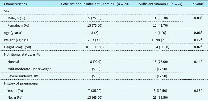
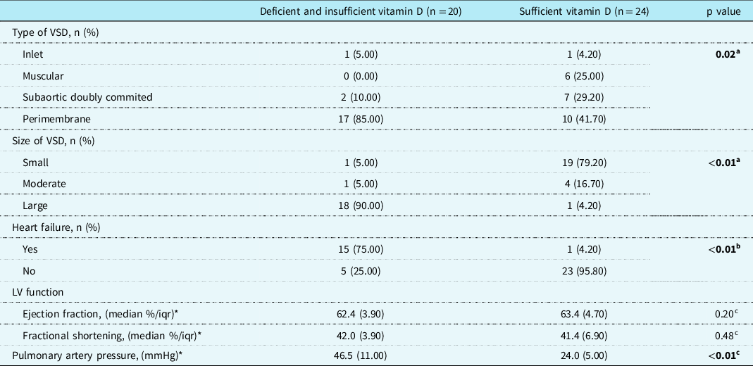
During the study period, 44 subjects who met the inclusion criteria were enrolled. Table 1 showed the characteristics of the subjects, and Table 2 showed the result of the study. The majority of the study subjects had normal nutritional status (82% of total subjects) and were diagnosed with perimembranous ventricular septal defect (61% of total subjects). We took blood sample from all subjects (44 samples) from the outpatient clinic to determine the vitamin D level.
Basic characteristics of the subjects

**Mean (standard deviation) for data with normal distribution.
* Median (interquartile range) for data with skewed distribution.
a Chi-square;
b Fisher Exact;
c Mann Whitney;
d Unpaired t-test, p < 0.05 significance.
Comparison of clinical characteristics among subjects with deficient and sufficient vitamin D

VSD: ventricular septal defect; LV: left ventricle.
* Median (iqr).
a Chi-square;
b Fisher Exact;
c Mann Whitney, p < 0.05 significance.
Associations between vitamin D level and type and size of ventricular septal defect, nutritional status, heart failure occurrence, left ventricular function, and pulmonary arterial pressure were shown in Table 2. This study found that vitamin D level was associated with pulmonary hypertension, type and size of ventricular septal defect, and heart failure (p < 0.05). There was no association between vitamin D level and nutritional status, pneumonia, and left ventricular function (p > 0.05).
Further analysis to find out about the prevalence ratio of pulmonary hypertension in samples with low vitamin D status was shown in Table 3. The result in Table 3 showed that patients with Vitamin D deficiency and insufficiency are at a higher risk of having pulmonary hypertension (prevalence ratio: 24.0).
Prevalence ratio for pulmonary hypertension among subjects with deficient and sufficient vitamin D

CI: confidence interval.
a Fisher Exact, p < 0.05 significance.
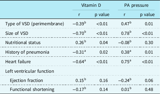
This study observed that higher vitamin D level was correlated with better nutritional status (r = 0.26, p = 0.05). Lower vitamin D level was correlated with perimembranous ventricular septal defect (r = −0.39, p = 0.005), bigger defect size (r = −0.70, p < 0.05), history of pneumonia (r = −0.31, p = 0.02), and existence of heart failure (r = −0.64, p < 0.001) (Table 4). Meanwhile, a higher pulmonary artery pressure was correlated with perimembranous ventricular septal defect (r = 0.47, p = <0.05), bigger defect size (r = 0.78, p < 0.05), history of pneumonia (r = 0.38, p = <0.05), and existence of heart failure (r = 0.75, p < 0.05) (Table 4).
Correlation between, type and size of VSD, nutritional status, heart failure, left ventricular function, and vitamin D and PA pressure

VSD: ventricular septal defect; PA: pulmonary artery.
a Point biserial correlation;
b Rank Spearman correlation, p < 0.05 significance.
Discussion
Hypovitaminosis D has become a worldwide problem found in all age and gender groups, especially in children. Reference Poh, Rojroongwasinkul and Nguyen4 The current evidence shows that vitamin D deficiency is a highly prevalent problem found in children with congestive heart disease. Reference McNally and Menon19 One study by Noori et al. reported that the level of vitamin D is significantly lower in children with congestive heart disease compared to their healthy controls. Reference Noori, Nakhaee-Moghadam, Teimouri, Pakravan and Boryri8 One review article that included the results of several studies reported that vitamin D levels in post-operative congestive heart disease patients demonstrated statistically significant associations between lower vitamin D levels and the increased need for cardiovascular support, cardiovascular function, and clinical outcomes such as Paediatric Risk of Mortality III scores, hypocalcaemia or calcium supplementation, fluid requirements, and the length of stay in Paediatric Intensive Care Unit. Reference McNally and Menon19
The average level of vitamin D found in our study is 33.16 ng/mL. All studies used 25-hydroxyvitamin D as vitamin D marker, as recommended by Endocrine Society Clinical Practice guideline. Reference Noori, Nakhaee-Moghadam, Teimouri, Pakravan and Boryri8,Reference Holick, Binkley and Bischoff-ferrari15
Vitamin D is a type of fat-soluble vitamin that is required for in many organs of the body to function well. Reference McNally, Menon and Chakraborty20 Identification of 25(OH)D receptors in several cells of the heart, such as endothelial cells, smooth muscle cells, and myocytes, has led to the hypothesis that 25-hydroxyvitamin D might be involved in the pathogenesis of CVD. Reference Swales and Wang21 Furthermore, a growing body of evidence suggests that low vitamin D levels may affect the cardiovascular function adversely and that there might be an association between vitamin D levels and cardiovascular health. Reference Ku22 In the current study, there is a strong negative correlation between the size of ventricular septal defect (r = −0.703; p < 0.001) and the presence of heart failure (r = −0.642; p < 0.001) with vitamin D. From this result, we can see that the greater the size of ventricular septal defect, the lower the vitamin D level in the blood will be. The presence of heart failure is also related to the lower level of vitamin D.
Large ventricular septal defect often causes heart failure as one of its complications, Reference Park and Salamat1 and in our study, heart failure is also correlated with the low level of vitamin D. This finding is in line with several studies in adults, which showed that vitamin D deficiency is associated with the progression of heart failure and having a worse prognosis. Reference Schierbeck, Jensen, Bang, Jensen, Køber and Jensen23–Reference Wang, Song and Manson26
Vitamin D has a role as the negative regulator of the renin-angiotensin-aldosterone system, and it also affects the myocardial extracellular matrix turnover. Studies on vitamin D receptors in mice showed that low vitamin D levels will cause cardiac hypertrophy, increased water intake, sodium retention, and increased metalloprotease activity. Eventually, myocardial tissue will be destructed and the ventricular remodelling will happen. Zhao et al. performed a meta-analysis on randomised controlled trial studies to evaluate the benefit of vitamin D supplementation on ventricular remodelling in patients with heart failure and found that in patients with heart failure, vitamin D supplementation inhibited myocardial remodelling, and improved cardiac function. Reference Zhao, Jia, Dong, Zhao, Li and Zhang27
In accordance with that, this study found that vitamin D level is associated with pulmonary hypertension, type and size of ventricular septal defect, and heart failure. Lower vitamin D status is found in children with perimembranous type ventricular septal defect, larger defect size, and heart failure.
Many studies have also reported associations between vitamin D deficiency and the increased risk of the development of respiratory infections and the increased severity of the disease. Vitamin D was reported as an independent risk factor for treatment failure and the longer duration of severe lower respiratory infections in children. Reference Haugen, Basnet and Hardang5 Recent meta-analysis study showed that vitamin D has an important protective role to prevent the exacerbations of severe asthma and also to prevent acute viral infections from happening. Reference Cepeda, Zenteno, Fuentes and Bustos28
In the current study, normal children cannot be used as a control; thus, the comparison of the result of Vitamin D value in the sample of this study with the value found in normal children cannot be done. Normal pulmonary artery systolic pressure of children and adults is 30 mmHg or less, and the mean pulmonary artery pressure is 25 mmHg or less. Pulmonary hypertension is diagnosed when the mean pulmonary artery pressure is 20 mmHg or greater in resting individual older than 3 months, and the golden standard is usually done using cardiac catheterisation. Reference Hansmann, Koestenberger and Alastalo17 However, in the current study, due to the limitation of the resource, we could only use echocardiography as the diagnostic tool; thus, the confirmation of pulmonary hypertension using cardiac catheterisation was not done. In the future, we suggest to doing further study regarding this field by comparing other congestive heart disease and using normal children as control.
It is possible that vitamin D deficiency might also have a causal effect relationship with other manifestations that are found in the current study, such as pulmonary hypertension, pneumonia, heart failure, and also nutritional problems. Ideally, multivariate analysis should have been done on those variables too. However, due to the limited number of data available, it is not possible to do multivariate analysis in this study. Thus, we recommend that in the future, researches with similar topic with our study should include more samples, so that multivariate analysis and the causal effect of vitamin D deficiency and its outcomes can be described more clearly.
As the conclusion, in children with ventricular septal defect, pulmonary arterial pressure is correlated with the type of ventricular septal defect, the defect size, the history of pneumonia, and heart failure. In addition, vitamin D deficiency is identified as a risk factor of pulmonary hypertension.
Acknowledgement
None.
Financial support
This research received no specific grant from any funding agency, commercial, or non profit sectors.
Conflicts of interest
None.
Ethical standards
The authors assert that all procedures contributing to this work comply with the ethical standards with the Helsinki Declaration and has been approved by the institutional committees of Universitas Padjadjaran.